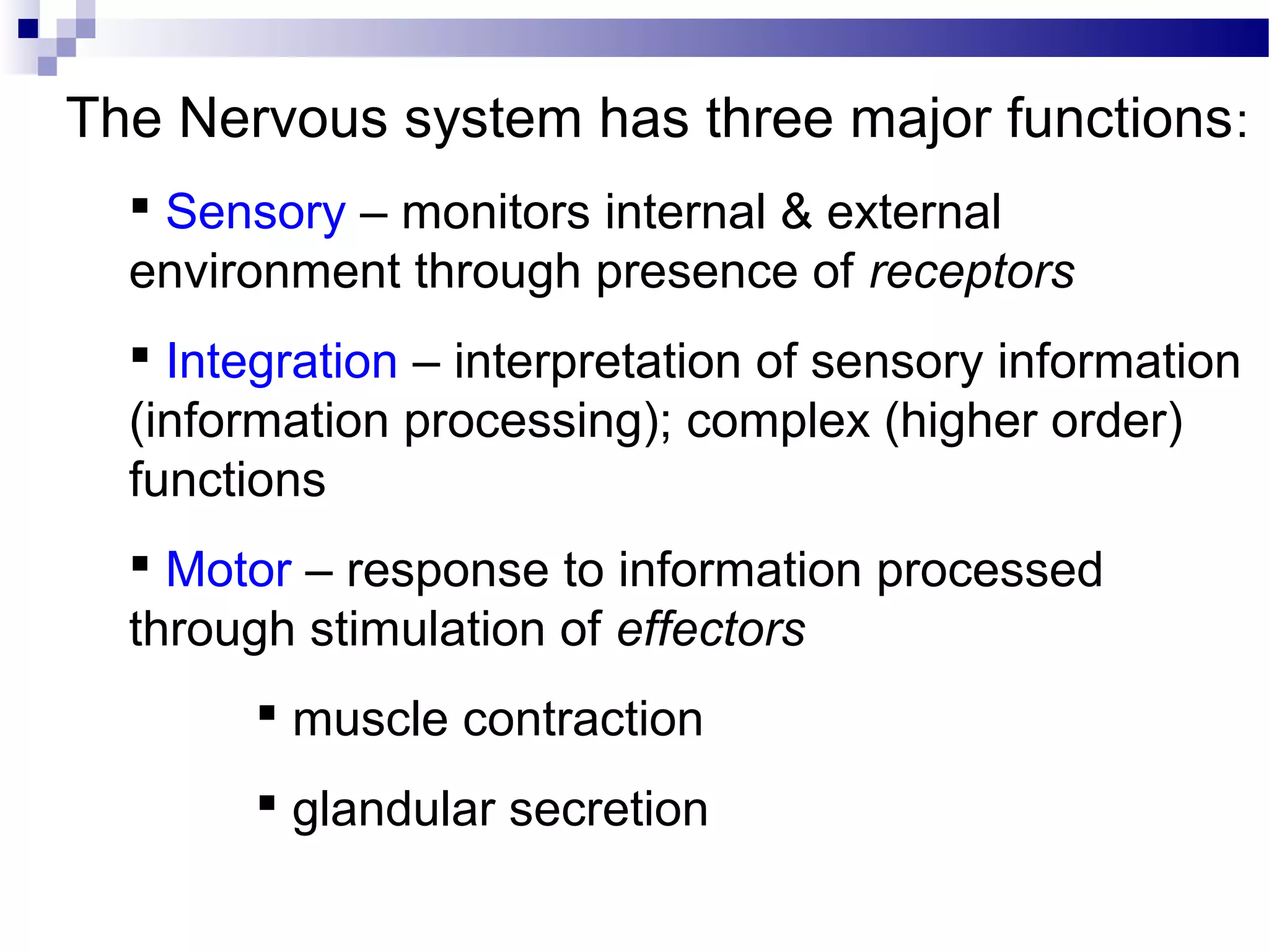
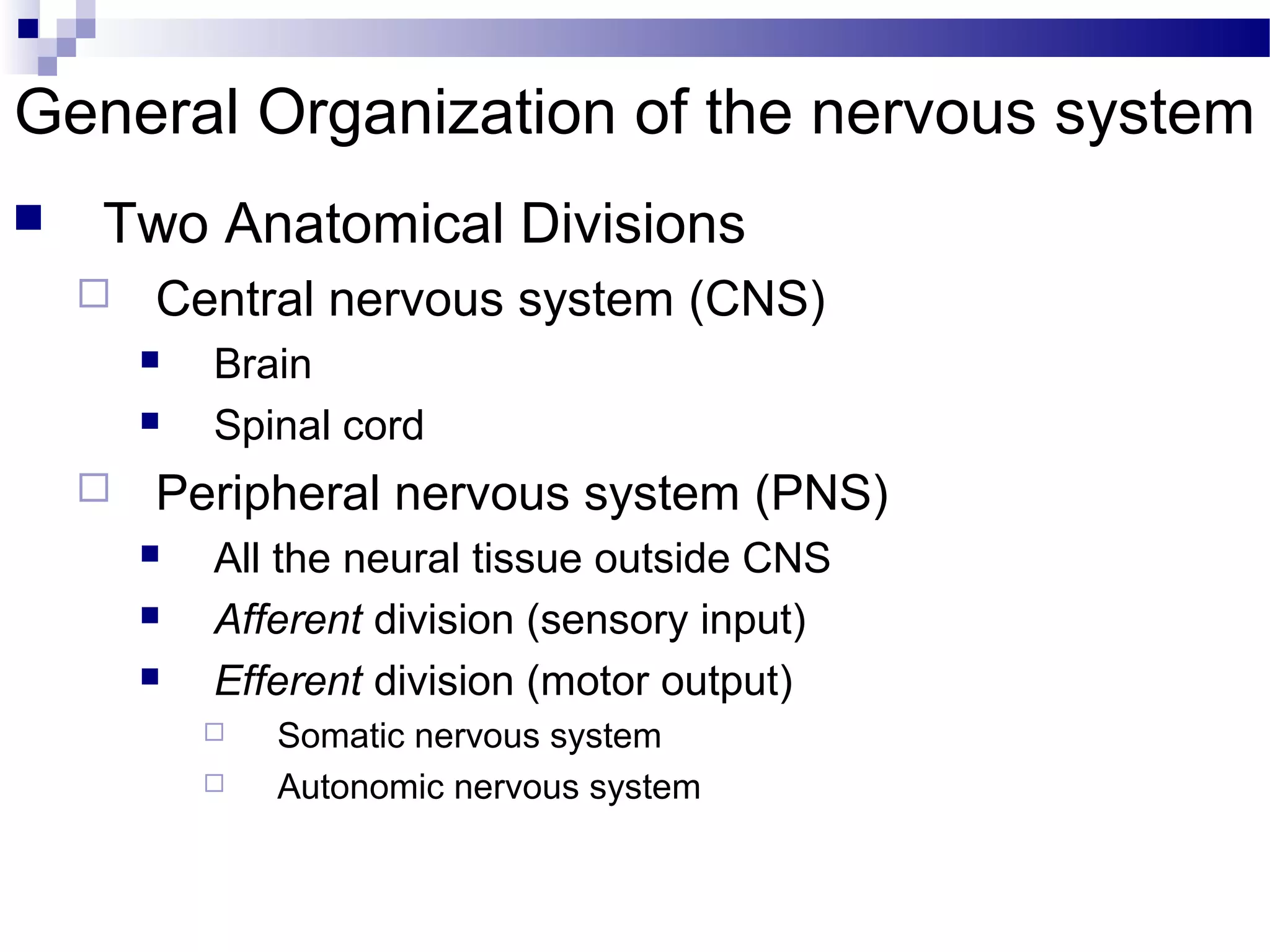
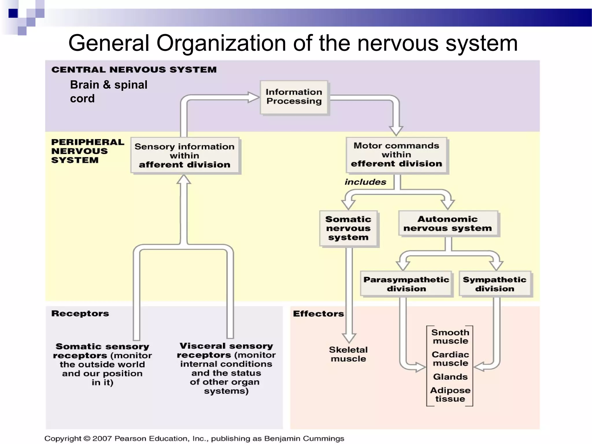
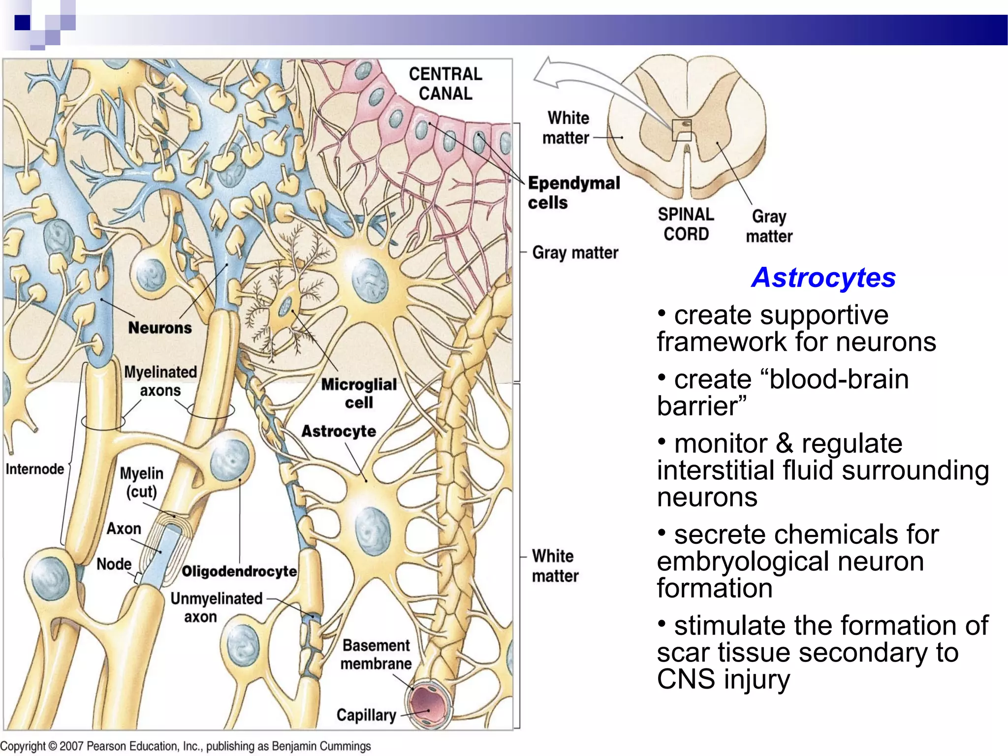
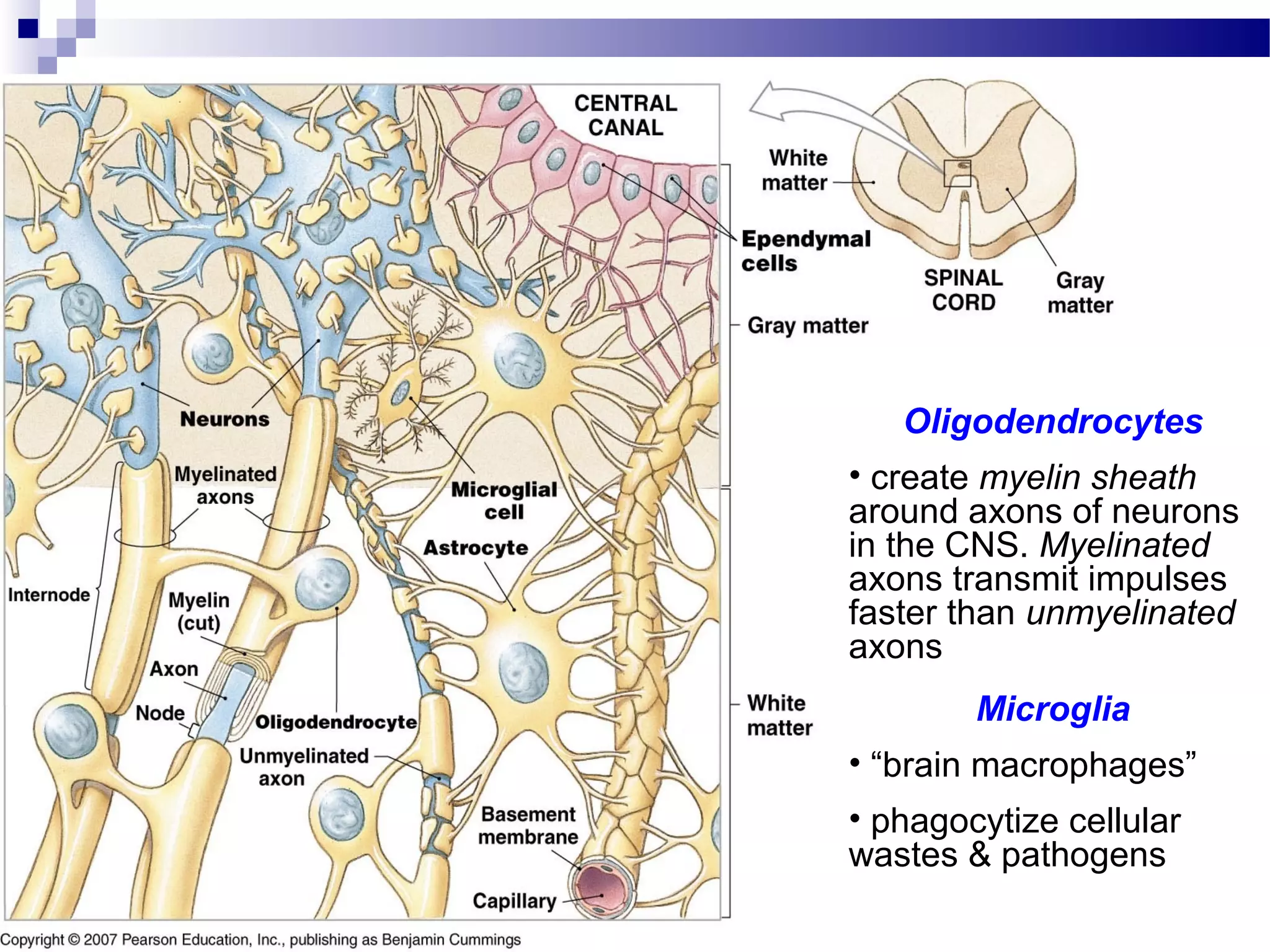
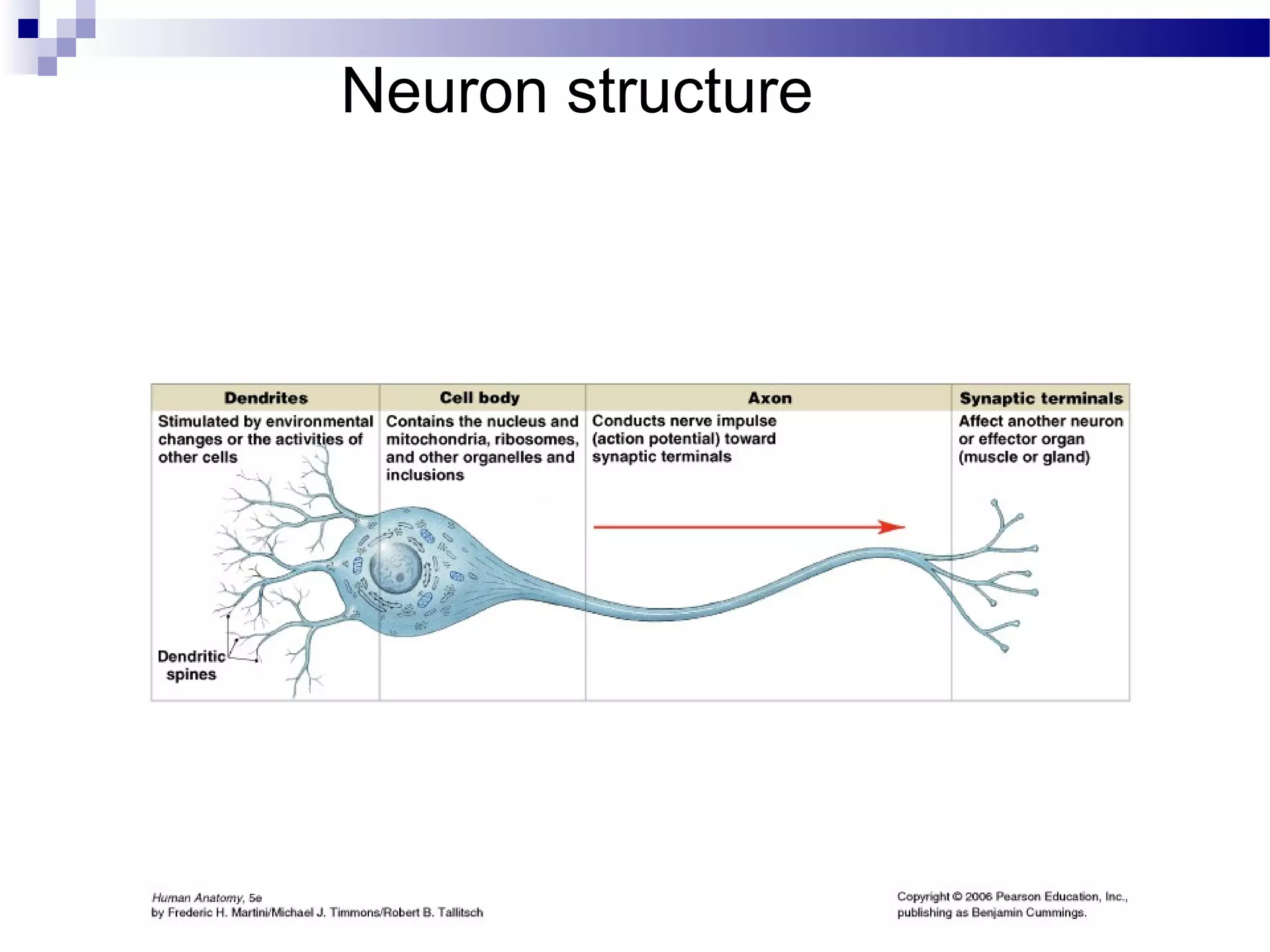
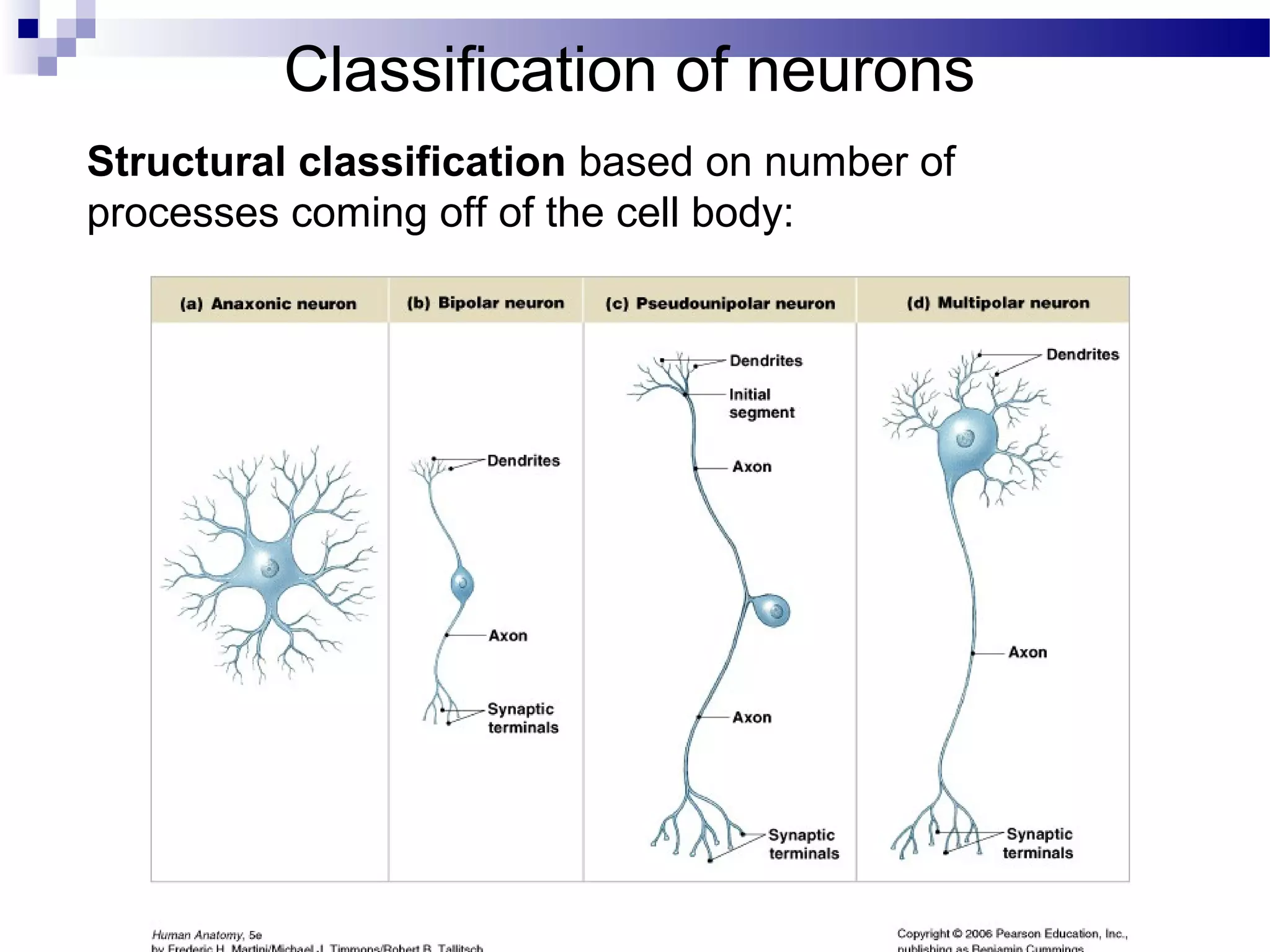
The Nervous System- NervousThe Nervous System- Nervous
TissueTissue
Chapter 13Chapter 13
The Nervous system has three major functions:
 Sensory – monitors internal & external
environment through presence of receptors
 Integration – interpretation of sensory information
(information processing); complex (higher order)
functions
 Motor – response to information processed
through stimulation of effectors
 muscle contraction
 glandular secretion
 Two Anatomical Divisions
 Central nervous system (CNS)
 Brain
 Spinal cord
 Peripheral nervous system (PNS)
 All the neural tissue outside CNS
 Afferent division (sensory input)
 Efferent division (motor output)
 Somatic nervous system
 Autonomic nervous system
General Organization of the nervous system
Brain & spinal
cord
Histology of neural tissue
Two types of neural cells in the nervous system:
 Neurons - For processing, transfer, and storage
of information
 Neuroglia – For support, regulation & protection
of neurons
Neuroglia (glial cells)
CNS neuroglia:
• astrocytes
• oligodendrocytes
• microglia
• ependymal cells
PNS neuroglia:
• Schwann cells (neurolemmocytes)
• satellite cells
Astrocytes
• create supportive
framework for neurons
• create “blood-brain
barrier”
• monitor & regulate
interstitial fluid surrounding
neurons
• secrete chemicals for
embryological neuron
formation
• stimulate the formation of
scar tissue secondary to
CNS injury
Oligodendrocytes
• create myelin sheath
around axons of neurons
in the CNS. Myelinated
axons transmit impulses
faster than unmyelinated
axons
Microglia
• “brain macrophages”
• phagocytize cellular
wastes & pathogens
Ependymal cells
• line ventricles of brain &
central canal of spinal cord
• produce, monitor & help
circulate CSF
(cerebrospinal fluid)
Schwann cells
• surround all axons of neurons in
the PNS creating a neurilemma
around them. Neurilemma allows
for potential regeneration of
damaged axons
• creates myelin sheath around
most axons of PNS
Satellite cells
• support groups of cell bodies
of neurons within ganglia of the
PNS
Neuron structure
•Most axons of the nervous system are
surrounded by a myelin sheath
(myelinated axons)
•The presence of myelin speeds up
the transmission of action potentials
along the axon
•Myelin will get laid down in segments
(internodes) along the axon, leaving
unmyelinated gaps known as “nodes
of Ranvier”
•Regions of the nervous system
containing groupings of myelinated
axons make up the “white matter”
•“gray matter” is mainly comprised of
groups of neuron cell bodies, dendrites
& synapses (connections between
neurons)
of Ranvier
Classification of neurons
Structural classification based on number of
processes coming off of the cell body:
Anaxonic neurons
• no anatomical clues to
determine axons from
dendrites
• functions unknown
Multipolar neuron
• multiple dendrites & single
axon
• most common type
Bipolar neuron
• two processes coming off
cell body – one dendrite &
one axon
• only found in eye, ear &
nose
Unipolar (pseudounipolar)
neuron
• single process coming
off cell body, giving rise
to dendrites (at one end)
& axon (making up rest of
process)
Classification of neurons
Functional classification based on type of information &
direction of information transmission:
• Sensory (afferent) neurons –
• transmit sensory information from receptors of PNS towards the CNS
• most sensory neurons are unipolar, a few are bipolar
• Motor (efferent) neurons –
• transmit motor information from the CNS to effectors
(muscles/glands/adipose tissue) in the periphery of the body
• all are multipolar
• Association (interneurons) –
• transmit information between neurons within the CNS; analyze inputs,
coordinate outputs
• are the most common type of neuron (20 billion)
• are all multipolar
Conduction across synapses
Most synapses within the nervous system are
chemical synapses, & involve the release of a
neurotransmitter
In order for neural control to occur, “information”
must not only be conducted along nerve cells, but
must also be transferred from one nerve cell to
another across a synapse
The Structure of a Typical Synapse
Neuronal Pools
Anatomical organization of neurons
Neurons of the nervous system tend to group together into
organized bundles
The axons of neurons are bundled together to form nerves in
the PNS & tracts/pathways in the CNS. Most axons are
myelinated so these structures will be part of “white matter”
The cell bodies of neurons are clustered together into
ganglia in the PNS & nuclei/centers in the CNS. These are
unmyelinated structures and will be part of “gray matter”
Neural Tissue Organization
Anatomical structure of Nerves
Fig. 14.6

Anatomy nervous tissue

  • 1.
    The Nervous System-NervousThe Nervous System- Nervous TissueTissue Chapter 13Chapter 13
  • 2.
    The Nervous systemhas three major functions:  Sensory – monitors internal & external environment through presence of receptors  Integration – interpretation of sensory information (information processing); complex (higher order) functions  Motor – response to information processed through stimulation of effectors  muscle contraction  glandular secretion
  • 3.
     Two AnatomicalDivisions  Central nervous system (CNS)  Brain  Spinal cord  Peripheral nervous system (PNS)  All the neural tissue outside CNS  Afferent division (sensory input)  Efferent division (motor output)  Somatic nervous system  Autonomic nervous system General Organization of the nervous system
  • 4.
    General Organization ofthe nervous system Brain & spinal cord
  • 5.
    Histology of neuraltissue Two types of neural cells in the nervous system:  Neurons - For processing, transfer, and storage of information  Neuroglia – For support, regulation & protection of neurons
  • 6.
    Neuroglia (glial cells) CNSneuroglia: • astrocytes • oligodendrocytes • microglia • ependymal cells PNS neuroglia: • Schwann cells (neurolemmocytes) • satellite cells
  • 7.
    Astrocytes • create supportive frameworkfor neurons • create “blood-brain barrier” • monitor & regulate interstitial fluid surrounding neurons • secrete chemicals for embryological neuron formation • stimulate the formation of scar tissue secondary to CNS injury
  • 8.
    Oligodendrocytes • create myelinsheath around axons of neurons in the CNS. Myelinated axons transmit impulses faster than unmyelinated axons Microglia • “brain macrophages” • phagocytize cellular wastes & pathogens
  • 9.
    Ependymal cells • lineventricles of brain & central canal of spinal cord • produce, monitor & help circulate CSF (cerebrospinal fluid)
  • 10.
    Schwann cells • surroundall axons of neurons in the PNS creating a neurilemma around them. Neurilemma allows for potential regeneration of damaged axons • creates myelin sheath around most axons of PNS Satellite cells • support groups of cell bodies of neurons within ganglia of the PNS
  • 11.
  • 12.
    •Most axons ofthe nervous system are surrounded by a myelin sheath (myelinated axons) •The presence of myelin speeds up the transmission of action potentials along the axon •Myelin will get laid down in segments (internodes) along the axon, leaving unmyelinated gaps known as “nodes of Ranvier” •Regions of the nervous system containing groupings of myelinated axons make up the “white matter” •“gray matter” is mainly comprised of groups of neuron cell bodies, dendrites & synapses (connections between neurons) of Ranvier
  • 13.
    Classification of neurons Structuralclassification based on number of processes coming off of the cell body:
  • 14.
    Anaxonic neurons • noanatomical clues to determine axons from dendrites • functions unknown
  • 15.
    Multipolar neuron • multipledendrites & single axon • most common type
  • 16.
    Bipolar neuron • twoprocesses coming off cell body – one dendrite & one axon • only found in eye, ear & nose
  • 17.
    Unipolar (pseudounipolar) neuron • singleprocess coming off cell body, giving rise to dendrites (at one end) & axon (making up rest of process)
  • 18.
    Classification of neurons Functionalclassification based on type of information & direction of information transmission: • Sensory (afferent) neurons – • transmit sensory information from receptors of PNS towards the CNS • most sensory neurons are unipolar, a few are bipolar • Motor (efferent) neurons – • transmit motor information from the CNS to effectors (muscles/glands/adipose tissue) in the periphery of the body • all are multipolar • Association (interneurons) – • transmit information between neurons within the CNS; analyze inputs, coordinate outputs • are the most common type of neuron (20 billion) • are all multipolar
  • 20.
    Conduction across synapses Mostsynapses within the nervous system are chemical synapses, & involve the release of a neurotransmitter In order for neural control to occur, “information” must not only be conducted along nerve cells, but must also be transferred from one nerve cell to another across a synapse
  • 21.
    The Structure ofa Typical Synapse
  • 22.
  • 23.
    Anatomical organization ofneurons Neurons of the nervous system tend to group together into organized bundles The axons of neurons are bundled together to form nerves in the PNS & tracts/pathways in the CNS. Most axons are myelinated so these structures will be part of “white matter” The cell bodies of neurons are clustered together into ganglia in the PNS & nuclei/centers in the CNS. These are unmyelinated structures and will be part of “gray matter”
  • 24.
  • 25.
    Anatomical structure ofNerves Fig. 14.6