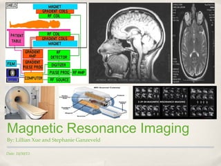
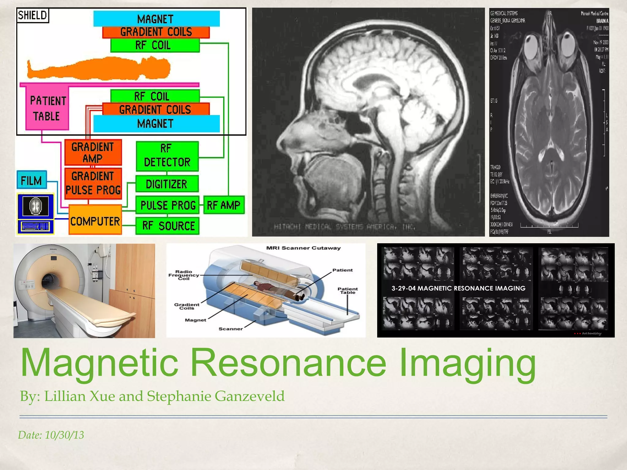
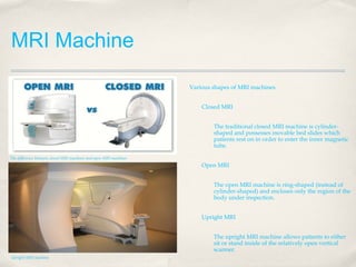
Magnetic Resonance Imaging
By: Lillian Xue and Stephanie Ganzeveld
Date: 10/30/13
MRI Machine
Various shapes of MRI machines
Closed MRI
The traditional closed MRI machine is cylindershaped and possesses movable bed slides which
patients rest on in order to enter the inner magnetic
tube.
The difference between closed MRI machines and open MRI machines

Open MRI
The open MRI machine is ring-shaped (instead of
cylinder-shaped) and encloses only the region of the
body under inspection.
Upright MRI
The upright MRI machine allows patients to either
sit or stand inside of the relatively open vertical
scanner.
Upright MRI machine
Practical Uses
Diagnosis of brain tumors
Identification of hemorrhage
Recognition of abnormal brain development
Discernment of lesions connected with multiple sclerosis
Diagnosis of hormonal instability

Possible locations for malignant brain tumors

Abnormal brain development in young children

Identification of hemorrhage
Method
To begin, MRI machines focus primarily on the nuclei of hydrogen
atoms in polar water molecules.
Hydrogen protons, under normal circumstances, spin
arbitrarily with their “axises” pointing in various directions.
When the body is placed under a magnetic current (as issued by the
MRI Machine), all of the protons line up to face in the same
direction.
The alignment of positive subatomic particles creates a
magnetic vector which is consequently identified by the
MRI scanner.

Protons of Hydrogen spinning in arbitrary directions

Radio waves are then added in concurrence with the magnetic
current to deflect the original magnetic vector.
When the radio waves are turned off, the magnetic vector returns to
its initial state, causing another radio wave signal to be produced.
This signal is what MRI machines translate to produce accurate
scans.

Protons of Hydrogen spinning in the same direction
Method Continued
Receiver coils are placed around the part of the body
being inspected to improve signal emission
How long it takes protons to return to their resting state
can be evaluated in two ways:
Time taken for the magnetic vector to return to
its initial state (T1 relaxation)
Time needed for the axial spin to return to its
resting state (T2 relaxation)
Receiver coils in MRI machines

In essence, MRI scans are composed of a series of pulse
patterns.
Since different tissues have different relaxation
states they can thus be distinguished from one
another.
In this respect, diseases can be identified by increased
water content.
T1 and T2 relaxation
Advantages of MRI
Does not involve x- or exposure to radiation and is not invasive.
Provides clear images of soft tissue structures such as grey matter.
Provides images of the brain at any direction, or slice.
Provides information on the blood moving through areas of the body.

The difference between white and grey matter

Various brain slice angles
Disadvantages of MRI
Can be very expensive and unaffordable
Can be dangerous for patients with metallic devices placed within the body such as pacemakers
Performed in small spaces which will be difficult to be performed on claustrophobic patients
Movement during scanning may cause blurry images, so the patient must lie still during the scan.
RF transmitters can cause severe burns if misused or mishandled.

Claustrophobia as a result of closed MRI machines

Pacemakers and other metallic devices are not permitted

RF (radio frequency) power should be used with caution
Advantages of fMRI
non-invasive and effective
no exposure to radiation, unlike CT, X-Rays, and PET scans
high resolution images (1mm)
signals can be recorded from all regions in the brain, unlike EEG scans
more precise than PET scans

Example of functional MRI scans

High resolution images

fMRI scans are able to show the brain from all regions
Disadvantages of fMRI
can only examine the blood flow in the brain (cannot detect activities of neurons)
results may be difficult to interpret (cannot specify emotions or feelings)

Unfortunately, fMRI scans can only examine blood flow in the brain

Results may be difficult to interpret or analuze
Difference between MRI and fMRI
Magnetic Resonance Imaging (MRI)
views anatomical structure
focuses on protons in hydrogen nuclei
high, spatial resolution
utilized for experimental purposes
MRI scan

Functional Magnetic Resonance Imaging (fMRI)
views metabolic function
calculates oxygen levels
long-distance resolution
utilized for diagnostic purposes
fMRI scan
Maguire and MRI
Used fMRI to locate blood flow in the brain
"FMRI can tell you which brain areas are involved in memory but you are never sure which ones are really
necessary. That is where the study of patients comes in."
Used to see which areas were active when participants were trying to locate a specific region from spatial
memory
The hippocampus was not the only active region of the brain
Used MRI to compare the structures of the brain

Hippocampus

Hippocampus as shown on the fMRI scan

Identification of spatial navigation
References (Sources)
Tilakaratna, P. (n.d.). Magnetic Resonance Imaging : Basic principles of how it works. Retrieved November 4, 2013 from
http://www.howequipmentworks.com/physics/medical_imaging/mri/magnetic_resonance_imaging.html

Berger, A. (January, 2002). Magnetic Resonance Imaging. Retrieved November 2, 2013 from: http://www.ncbi.nlm.nih.gov/pmc/articles/PMC1121941/#!po=16.6667

Blake, L. (2013). What does MRI equipment look like? Retrieved November 4, 2013 from: http://www.ehow.com/facts_5499464_mri-equipment-look-like.html

Smith, A. (2013). Reasons for an MRI of the brain. Retrieved November 4, 2013 from: http://www.ehow.com/facts_5499464_mri-equipment-look-like.html

Kalapurayil, M. (2013, July). What is MRI? How does MRI work? Retrieved November 4, 2013 from: http://www.medicalnewstoday.com/articles/146309.php

Seven Counties Services Inc. (n.d.). Functional Magnetic Resonance Imaging. Retrieved November 4, 2013 from http://www.sevencounties.org/poc/view_doc.php?type=doc&id=8947&cn=18

CancerQuest (2011, October). Benefits and Disadvantages of MRI. Retrieved November 4, 2013, from http://www.cancerquest.org/mri-advantages-and-disadvantages.html

Schwartz., A. (n.d.). MRI: Advantages and Disadvantages. MRI: Advantages and Disadvantages. Retrieved November 4, 2013, from
http://www.anapolschwartz.com/practices/gadolinium-nsf/blog/mri-side-effects.html

Health information you can trust. (n.d.). MRI scan. Retrieved November 4, 2013, from http://www.nhsinform.com/health-library/articles/m/mri-scan/advantages

Chudler, E. (2013). Brain imaging. Retrieved November 4, 2013 from: http://faculty.washington.edu/chudler/image.html
References (Images)
http://www.magnet.fsu.edu/education/tutorials/magnetacademy/mri/fullarticle.html

http://occlusionconnections.com/TMJ/wp-content/uploads/2012/03/MRI-TMJ-Occlusion-Connections-Clayton-A.-Chan-DDS-9.png

http://classes.soe.ucsc.edu/cmps161/Winter10/projects/projects/upopov/images/general/whiteGreyMatterT1-1.jpg

http://lcni.uoregon.edu/~dow/Space_software/Space_documentation/Volume_segmentation/Inner_ear_MRI/renderings/L_inner_ear_T2_slices.png

http://www.magnet.fsu.edu/education/tutorials/magnetacademy/mri/images/mri-brainslices.jpg

http://info.blockimaging.com/Portals/21795/images/H--Alicia%20Wiklund-HubSpot-Blogs-Open_MRI_vs_Closed_MRI.jpg

http://www.tribbleassociates.com/Images/Upright%20MRI%202.JPG

http://www.ehow.com/list_6106146_reasons-mri-brain.html

www.yalemedicalgroup.org

http://medcastaway.edublogs.org/files/2011/03/cerebral-hemorrhage-mjkqx4.JPG
References (Images) Continued
http://medcastaway.edublogs.org/files/2011/03/cerebral-hemorrhage-mjkqx4.JPG

http://www.mcva.com/mcaimages/pacemaker.jpg

http://www.mysafetysign.com/img/lg/L/RF-Power-Warning-Safety-Label-LB-0102.gif

http://www.nih.gov/news/pr/dec2005/nimh_07.jpg

http://www.brianjogrady.com/images/fmri.jpg

http://www.med.nyu.edu/thesenlab/group/images/fmri1.jpg

http://faculty.washington.edu/chudler/vessel.gif

http://lifebyexperimentation.com/wp-content/uploads/2011/12/fmri.h2.gif

http://www.bannerhealth.com/NR/rdonlyres/7062ADED-D701-4BEC-A3D4-0856EACC21CC/18190/Picture2.gif
References (Images) Continued
http://www.nih.gov/news/pr/dec2005/nimh_07.jpg

http://www.brianjogrady.com/images/fmri.jpg

http://www.med.nyu.edu/thesenlab/group/images/fmri1.jpg

http://faculty.washington.edu/chudler/vessel.gif

http://lifebyexperimentation.com/wp-content/uploads/2011/12/fmri.h2.gif

http://www.bannerhealth.com/NR/rdonlyres/7062ADED-D701-4BEC-A3D4-0856EACC21CC/18190/Picture2.gif

http://morphonix.com/software/education/science/brain/game/specimens/images/hippocampus.gif

http://www.neuro24.de/hip0006.jpg

MRI Presentation Final