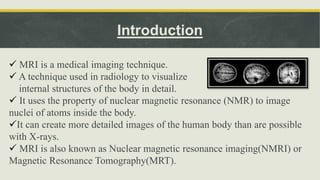
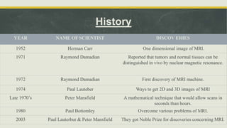
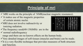
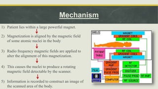
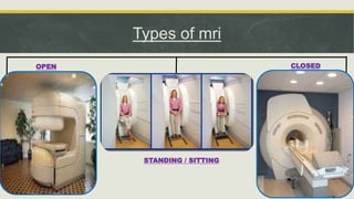
Magnetic Resonance Imaging (MRI) is a medical imaging technique that uses nuclear magnetic resonance to visualize internal structures of the body. MRI works by aligning the magnetization of atomic nuclei when placed in a magnetic field and exposing them to radio frequencies, allowing for detailed images of tissues. The history of MRI involved early developments in one-dimensional imaging in the 1950s and the first MRI machine in the 1970s. MRI can be open or closed, and used for standing/sitting or lying down. Applications of MRI include detecting diseases, tumors, strokes and musculoskeletal and neurological conditions.
basic and brief but informative knowledge about how MRI works and what are its components ... easy to understand as well as presenting during lectures and in classes . share it
this slide sharer contents are basic principle of CT fluoroscopy , software and hardware parts of equipment and image aqua cation and radiation dose comparison and videos related to equipment .
MRI is an imaging technique that uses strong magnetic fields and radio waves to create detailed images of the human body. It is primarily used in medical settings to get high-resolution images of the inside of the human body. MRI is also used to monitor the effectiveness of certain medical treatments, such as surgery and radiation therapy.
There are many different imaging techniques used in medicine and other fields. Some examples include X-ray imaging, magnetic resonance imaging (MRI), computed tomography (CT) scans, ultrasound, and positron emission tomography (PET) scans. These techniques all work in different ways to produce images of the inside of the body or other objects. For example, X-rays use radiation to create images, while MRI and CT scans use powerful magnets and computers to produce detailed images of the body's internal structures. Ultrasound uses sound waves to create images, and PET scans use radioactive tracers to create images of the body's metabolic activity. These imaging techniques are often used in conjunction with one another to provide a comprehensive view of a patient's condition.
MRI is a powerful imaging technique that can be used to diagnose a variety of medical conditions.
MRI is a non-invasive and painless procedure, but it can be time-consuming and expensive.
Patients who are considering an MRI scan should talk to their doctor to see if it is the right imaging procedure for them.
This presentation discusees a brief history of the MRI, it's mechanism of action, applications in dentistry and recent advancements in its technology. Also it's advantages and disadvantages in comparison with the CT scan
Magnetic Resonance Imaging or MRI Scanning as we commonly call it is a process where strong magnetic fields and radio waves are used to produce detailed images of the inside of a human body.Contact Us: Open MRI of Orlando 668 N. Orlando Avenue, Suite 1005 Maitland, FL 32751., Tel No: (407) 740-8848, Fax: 407-740-0324, Email: NewPatient@OpenMRIofOrlando.com
Imagine an MRI experience that is comfortable, stress-free, and completely reliable. Sounds too good to be true? It isn’t. Upright MRI of Deerfield is recognized as the world-leader in open MRI innovation, offering patients the peace-of-mind they deserve, and the most accurate diagnoses possible.
The term isolation refers to the separation of a strain from a natural, mixed population of living microbes, as present in the environment. It becomes necessary to maintain the viability and purity of the microorganism by keeping the pure culture free from contamination.
THE IMPORTANCE OF MARTIAN ATMOSPHERE SAMPLE RETURN.Sérgio Sacani
The return of a sample of near-surface atmosphere from Mars would facilitate answers to several first-order science questions surrounding the formation and evolution of the planet. One of the important aspects of terrestrial planet formation in general is the role that primary atmospheres played in influencing the chemistry and structure of the planets and their antecedents. Studies of the martian atmosphere can be used to investigate the role of a primary atmosphere in its history. Atmosphere samples would also inform our understanding of the near-surface chemistry of the planet, and ultimately the prospects for life. High-precision isotopic analyses of constituent gases are needed to address these questions, requiring that the analyses are made on returned samples rather than in situ.
(May 29th, 2024) Advancements in Intravital Microscopy- Insights for Preclini...Scintica Instrumentation
Intravital microscopy (IVM) is a powerful tool utilized to study cellular behavior over time and space in vivo. Much of our understanding of cell biology has been accomplished using various in vitro and ex vivo methods; however, these studies do not necessarily reflect the natural dynamics of biological processes. Unlike traditional cell culture or fixed tissue imaging, IVM allows for the ultra-fast high-resolution imaging of cellular processes over time and space and were studied in its natural environment. Real-time visualization of biological processes in the context of an intact organism helps maintain physiological relevance and provide insights into the progression of disease, response to treatments or developmental processes.
In this webinar we give an overview of advanced applications of the IVM system in preclinical research. IVIM technology is a provider of all-in-one intravital microscopy systems and solutions optimized for in vivo imaging of live animal models at sub-micron resolution. The system’s unique features and user-friendly software enables researchers to probe fast dynamic biological processes such as immune cell tracking, cell-cell interaction as well as vascularization and tumor metastasis with exceptional detail. This webinar will also give an overview of IVM being utilized in drug development, offering a view into the intricate interaction between drugs/nanoparticles and tissues in vivo and allows for the evaluation of therapeutic intervention in a variety of tissues and organs. This interdisciplinary collaboration continues to drive the advancements of novel therapeutic strategies.
Richard's entangled aventures in wonderlandRichard Gill
Since the loophole-free Bell experiments of 2020 and the Nobel prizes in physics of 2022, critics of Bell's work have retreated to the fortress of super-determinism. Now, super-determinism is a derogatory word - it just means "determinism". Palmer, Hance and Hossenfelder argue that quantum mechanics and determinism are not incompatible, using a sophisticated mathematical construction based on a subtle thinning of allowed states and measurements in quantum mechanics, such that what is left appears to make Bell's argument fail, without altering the empirical predictions of quantum mechanics. I think however that it is a smoke screen, and the slogan "lost in math" comes to my mind. I will discuss some other recent disproofs of Bell's theorem using the language of causality based on causal graphs. Causal thinking is also central to law and justice. I will mention surprising connections to my work on serial killer nurse cases, in particular the Dutch case of Lucia de Berk and the current UK case of Lucy Letby.
Observation of Io’s Resurfacing via Plume Deposition Using Ground-based Adapt...Sérgio Sacani
Since volcanic activity was first discovered on Io from Voyager images in 1979, changes
on Io’s surface have been monitored from both spacecraft and ground-based telescopes.
Here, we present the highest spatial resolution images of Io ever obtained from a groundbased telescope. These images, acquired by the SHARK-VIS instrument on the Large
Binocular Telescope, show evidence of a major resurfacing event on Io’s trailing hemisphere. When compared to the most recent spacecraft images, the SHARK-VIS images
show that a plume deposit from a powerful eruption at Pillan Patera has covered part
of the long-lived Pele plume deposit. Although this type of resurfacing event may be common on Io, few have been detected due to the rarity of spacecraft visits and the previously low spatial resolution available from Earth-based telescopes. The SHARK-VIS instrument ushers in a new era of high resolution imaging of Io’s surface using adaptive
optics at visible wavelengths.
Cancer cell metabolism: special Reference to Lactate PathwayAADYARAJPANDEY1
Normal Cell Metabolism:
Cellular respiration describes the series of steps that cells use to break down sugar and other chemicals to get the energy we need to function.
Energy is stored in the bonds of glucose and when glucose is broken down, much of that energy is released.
Cell utilize energy in the form of ATP.
The first step of respiration is called glycolysis. In a series of steps, glycolysis breaks glucose into two smaller molecules - a chemical called pyruvate. A small amount of ATP is formed during this process.
Most healthy cells continue the breakdown in a second process, called the Kreb's cycle. The Kreb's cycle allows cells to “burn” the pyruvates made in glycolysis to get more ATP.
The last step in the breakdown of glucose is called oxidative phosphorylation (Ox-Phos).
It takes place in specialized cell structures called mitochondria. This process produces a large amount of ATP. Importantly, cells need oxygen to complete oxidative phosphorylation.
If a cell completes only glycolysis, only 2 molecules of ATP are made per glucose. However, if the cell completes the entire respiration process (glycolysis - Kreb's - oxidative phosphorylation), about 36 molecules of ATP are created, giving it much more energy to use.
IN CANCER CELL:
Unlike healthy cells that "burn" the entire molecule of sugar to capture a large amount of energy as ATP, cancer cells are wasteful.
Cancer cells only partially break down sugar molecules. They overuse the first step of respiration, glycolysis. They frequently do not complete the second step, oxidative phosphorylation.
This results in only 2 molecules of ATP per each glucose molecule instead of the 36 or so ATPs healthy cells gain. As a result, cancer cells need to use a lot more sugar molecules to get enough energy to survive.
introduction to WARBERG PHENOMENA:
WARBURG EFFECT Usually, cancer cells are highly glycolytic (glucose addiction) and take up more glucose than do normal cells from outside.
Otto Heinrich Warburg (; 8 October 1883 – 1 August 1970) In 1931 was awarded the Nobel Prize in Physiology for his "discovery of the nature and mode of action of the respiratory enzyme.
WARNBURG EFFECT : cancer cells under aerobic (well-oxygenated) conditions to metabolize glucose to lactate (aerobic glycolysis) is known as the Warburg effect. Warburg made the observation that tumor slices consume glucose and secrete lactate at a higher rate than normal tissues.
Multi-source connectivity as the driver of solar wind variability in the heli...Sérgio Sacani
The ambient solar wind that flls the heliosphere originates from multiple
sources in the solar corona and is highly structured. It is often described
as high-speed, relatively homogeneous, plasma streams from coronal
holes and slow-speed, highly variable, streams whose source regions are
under debate. A key goal of ESA/NASA’s Solar Orbiter mission is to identify
solar wind sources and understand what drives the complexity seen in the
heliosphere. By combining magnetic feld modelling and spectroscopic
techniques with high-resolution observations and measurements, we show
that the solar wind variability detected in situ by Solar Orbiter in March
2022 is driven by spatio-temporal changes in the magnetic connectivity to
multiple sources in the solar atmosphere. The magnetic feld footpoints
connected to the spacecraft moved from the boundaries of a coronal hole
to one active region (12961) and then across to another region (12957). This
is refected in the in situ measurements, which show the transition from fast
to highly Alfvénic then to slow solar wind that is disrupted by the arrival of
a coronal mass ejection. Our results describe solar wind variability at 0.5 au
but are applicable to near-Earth observatories.
Earliest Galaxies in the JADES Origins Field: Luminosity Function and Cosmic ...Sérgio Sacani
We characterize the earliest galaxy population in the JADES Origins Field (JOF), the deepest
imaging field observed with JWST. We make use of the ancillary Hubble optical images (5 filters
spanning 0.4−0.9µm) and novel JWST images with 14 filters spanning 0.8−5µm, including 7 mediumband filters, and reaching total exposure times of up to 46 hours per filter. We combine all our data
at > 2.3µm to construct an ultradeep image, reaching as deep as ≈ 31.4 AB mag in the stack and
30.3-31.0 AB mag (5σ, r = 0.1” circular aperture) in individual filters. We measure photometric
redshifts and use robust selection criteria to identify a sample of eight galaxy candidates at redshifts
z = 11.5 − 15. These objects show compact half-light radii of R1/2 ∼ 50 − 200pc, stellar masses of
M⋆ ∼ 107−108M⊙, and star-formation rates of SFR ∼ 0.1−1 M⊙ yr−1
. Our search finds no candidates
at 15 < z < 20, placing upper limits at these redshifts. We develop a forward modeling approach to
infer the properties of the evolving luminosity function without binning in redshift or luminosity that
marginalizes over the photometric redshift uncertainty of our candidate galaxies and incorporates the
impact of non-detections. We find a z = 12 luminosity function in good agreement with prior results,
and that the luminosity function normalization and UV luminosity density decline by a factor of ∼ 2.5
from z = 12 to z = 14. We discuss the possible implications of our results in the context of theoretical
models for evolution of the dark matter halo mass function.
Comparing Evolved Extractive Text Summary Scores of Bidirectional Encoder Rep...University of Maribor
Slides from:
11th International Conference on Electrical, Electronics and Computer Engineering (IcETRAN), Niš, 3-6 June 2024
Track: Artificial Intelligence
https://www.etran.rs/2024/en/home-english/
Slide 1: Title Slide
Extrachromosomal Inheritance
Slide 2: Introduction to Extrachromosomal Inheritance
Definition: Extrachromosomal inheritance refers to the transmission of genetic material that is not found within the nucleus.
Key Components: Involves genes located in mitochondria, chloroplasts, and plasmids.
Slide 3: Mitochondrial Inheritance
Mitochondria: Organelles responsible for energy production.
Mitochondrial DNA (mtDNA): Circular DNA molecule found in mitochondria.
Inheritance Pattern: Maternally inherited, meaning it is passed from mothers to all their offspring.
Diseases: Examples include Leber’s hereditary optic neuropathy (LHON) and mitochondrial myopathy.
Slide 4: Chloroplast Inheritance
Chloroplasts: Organelles responsible for photosynthesis in plants.
Chloroplast DNA (cpDNA): Circular DNA molecule found in chloroplasts.
Inheritance Pattern: Often maternally inherited in most plants, but can vary in some species.
Examples: Variegation in plants, where leaf color patterns are determined by chloroplast DNA.
Slide 5: Plasmid Inheritance
Plasmids: Small, circular DNA molecules found in bacteria and some eukaryotes.
Features: Can carry antibiotic resistance genes and can be transferred between cells through processes like conjugation.
Significance: Important in biotechnology for gene cloning and genetic engineering.
Slide 6: Mechanisms of Extrachromosomal Inheritance
Non-Mendelian Patterns: Do not follow Mendel’s laws of inheritance.
Cytoplasmic Segregation: During cell division, organelles like mitochondria and chloroplasts are randomly distributed to daughter cells.
Heteroplasmy: Presence of more than one type of organellar genome within a cell, leading to variation in expression.
Slide 7: Examples of Extrachromosomal Inheritance
Four O’clock Plant (Mirabilis jalapa): Shows variegated leaves due to different cpDNA in leaf cells.
Petite Mutants in Yeast: Result from mutations in mitochondrial DNA affecting respiration.
Slide 8: Importance of Extrachromosomal Inheritance
Evolution: Provides insight into the evolution of eukaryotic cells.
Medicine: Understanding mitochondrial inheritance helps in diagnosing and treating mitochondrial diseases.
Agriculture: Chloroplast inheritance can be used in plant breeding and genetic modification.
Slide 9: Recent Research and Advances
Gene Editing: Techniques like CRISPR-Cas9 are being used to edit mitochondrial and chloroplast DNA.
Therapies: Development of mitochondrial replacement therapy (MRT) for preventing mitochondrial diseases.
Slide 10: Conclusion
Summary: Extrachromosomal inheritance involves the transmission of genetic material outside the nucleus and plays a crucial role in genetics, medicine, and biotechnology.
Future Directions: Continued research and technological advancements hold promise for new treatments and applications.
Slide 11: Questions and Discussion
Invite Audience: Open the floor for any questions or further discussion on the topic.
3. Introduction
MRI is a medical imaging technique.
A technique used in radiology to visualize
internal structures of the body in detail.
It uses the property of nuclear magnetic resonance (NMR) to image
nuclei of atoms inside the body.
It can create more detailed images of the human body than are possible
with X-rays.
MRI is also known as Nuclear magnetic resonance imaging(NMRI) or
Magnetic Resonance Tomography(MRT).
4. History
YEAR NAME OF SCIENTIST DISCOV ERIES
1952 Herman Carr One dimensional image of MRI.
1971 Raymond Damadian Reported that tumors and normal tissues can be
distinguished in vivo by nuclear magnetic resonance.
1972 Raymond Damadian First discovery of MRI machine.
1974 Paul Lauteber Ways to get 2D and 3D images of MRI
Late 1970’s Peter Mansfield A mathematical technique that would allow scans in
seconds than hours.
1980 Paul Bottomley Overcome various problems of MRI.
2003 Paul Lauterbur & Peter Mansfield They got Noble Prize for discoveries concerning MRI.
5. Principle of mri
MRI works on the principle of NMR(nuclear magnetic resonance).
It makes use of the magnetic properties
of certain atomic nuclei.
MRI does not involve radioactivity or
ionizing radiation.
The frequency used(40-130)MHz are in the
normal radiofrequency
range and there are no known effects on the human body.
Very detailed images of soft tissues (muscles and brain) can be made.
It is a very flexible technique that provides measures of both structure
and function.
6. Mechanism
1) Patient lies within a large powerful magnet.
2) Magnetization is aligned by the magnetic field
of some atomic nuclei in the body
3) Radio frequency magnetic fields are applied to
alter the alignment of this magnetization.
4) This causes the nuclei to produce a rotating
magnetic field detectable by the scanner.
5) Information is recorded to construct an image of
the scanned area of the body.
7. Types of mri
OPEN CLOSEDSTANDING / SITTING
•This machine has a
larger opening than
standard MRI’S .
•It has magnets that do
not completely surround
your body.
•Open MRI’S is good for
those who get nervous.
• This machine is useful in
specific cases.
•This machine allows the
patient to sit or stand.
•Currently they do not
provide a good image
quality.(may improve in
future)
•Useful in specific cases.
•This machine scan
patients faster.
•It has high field strength
and are capable of
achieving greater
resolution and thinner
slices for viewing smaller
parts of the anatomy.
•Useful in detecting
stroke, sclerosis knee
defect etc.
OPEN
STANDING / SITTING
CLOSED
8. Types of mri scan
MRI OR MAGNETIC RESONANCE IMAGING
9. applications
MRI’s are administrated to patients suffering from the following:-
• Inflammation or infection in an organ
• Degenerative diseases.
• Strokes
• Muscoskeletal disorders
• Tumors
• Other irregularities that exist in tissue or organs in their body.
10. High-resolution images of the organs or any area of the body can be made without the need
for using X-rays because MRI’s use radiofrequency(RF) light. Since they use RF light, MRI’s
do not present any known health risks to the patients, however anyone with mental implants
could not receive a MRI. If a person’s nervous system needed to be studied, an MRI would be
the best imaging method to use, especially if the brain or spinal cord needed to be
investigated.
Functionally MRI’s are done to determine which parts of the brain have control over which
uses of the human body. These MRI’s are critical in determining motor imagery, speech
portions of the brain, and diagnosing which parts of the brain may be effected by a tumor.
Some operations are deferred because a portion of the brain that is vital may be removed, and
this is only determined via functional MRI’s.
applications
11. conclusion
•MRI are a relatively new technology to hit the medical world, and have completely
revolutionized medical imaging and the diagnosing process as we know it.
• In- vivo images can be taken of the human body, meaning that internal images can
be seen without making any incisions.
•Completely non-intrusive procedures are used, which makes MRI’s very effective,
but somewhat expensive, for doctors to use.