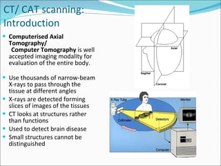
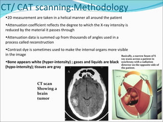
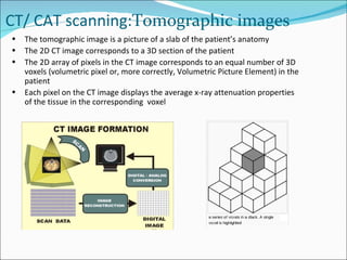
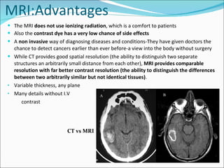
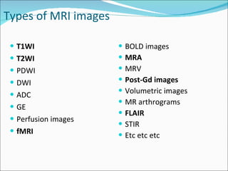
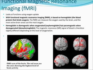
SlideShare a Scribd company logo
 
Lecture Outline  ,[object Object],[object Object],[object Object],[object Object],[object Object],[object Object],[object Object],[object Object],[object Object],[object Object],[object Object],[object Object],[object Object],[object Object],Sagittal Axial
 
CT/ CAT scanning: Introduction ,[object Object],[object Object],[object Object],[object Object],[object Object],[object Object],[object Object]
CT/ CAT scanning: Methodology ,[object Object],[object Object],[object Object],[object Object],[object Object],Basically, a narrow beam of X ray scans across a patient in synchrony with a radiation detector on the opposite side of the patient. CT scan  Showing a  brain tumor
CT/ CAT scanning: Tomographic images ,[object Object],[object Object],[object Object],[object Object]
 
What is a MRI? MRI stands for  magnetic resonance imaging. A MRI scanner has a magnetic field that is frequently up to  60,000  times as strong as Earth’s magnetic field! MRI equipment is expensive. 1.5 tesla scanners often cost between $1 million and $1.5 million USD. 3.0 tesla scanners often cost between $2 million and $2.3 million USD. Construction of MRI suites can cost up to $500,000 USD, or more, depending on project scope. Dangers of MRI's Video:  http://www.youtube.com/watch?v=_lBxYtkh4ts
MRI How does it work? The Basics ,[object Object],[object Object],[object Object],[object Object],[object Object],[object Object],[object Object],[object Object],[object Object],Molecules with magnetic charges align with the strong magnetic field. A radio frequency is used to tip these molecules over.
MRI How the image is made ,[object Object],[object Object],[object Object],[object Object],[object Object],[object Object],[object Object],Magnetic Resonance Imaging (MRI) image of the brain
MRI:Advantages ,[object Object],[object Object],[object Object],[object Object],[object Object],[object Object],[object Object],CT vs MRI
MRI the disadvantages ,[object Object],[object Object],[object Object],[object Object],[object Object],[object Object],[object Object],[object Object],[object Object],[object Object],[object Object],[object Object],[object Object],[object Object],[object Object]
Material to read latter- Before MRI, there is a checklist! ,[object Object],[object Object],[object Object],[object Object],[object Object],[object Object],[object Object],[object Object]
Material to read latter-MRI Further checklist! ,[object Object],[object Object],[object Object],[object Object],[object Object],[object Object]
Types of MRI images ,[object Object],[object Object],[object Object],[object Object],[object Object],[object Object],[object Object],[object Object],[object Object],[object Object],[object Object],[object Object],[object Object],[object Object],[object Object],[object Object],[object Object]
Types of MRI images: T1- T2-weighted ,[object Object]
Pooley, R. A. Radiographics 2005;25:1087-1099 T1-weighted contrast In the brain  T 1 -weighted scans provide good gray matter/white matter contrast , in other words put simply,  T1 Weighted Images highlights fat deposition. Types of MRI images: T1WI
Pooley, R. A. Radiographics 2005;25:1087-1099 T2-weighted contrast Types of MRI images: T2WI T2 images are particularly well suited to edema as they are sensitive to water content (edema is characterized by increased water content). In other words, put more simply,  T2 weighted images light up liquid on the images being visualized .
Magnetic Resonance Angiography (MRA)  is a group of techniques based on Magnetic Resonance Imaging (MRI) to image blood vessels . MRA  generates pictures of the arteries to evaluate them for  stenosis  (abnormal narrowing) or  aneurysms  (vessel wall dilatations, at risk of rupture). A variety of techniques can be used to generate the pictures, such as administration of a paramagnetic contrast agent ( gadolinium, Gd ). Types of MRI images: Magnetic resonance angiography (MRA) Magnetic Resonance Angiography: Maximum intensity projection of an MRA covering from the top of the heart to just below the circle of Willis MRA showing the circle of Willis in the brain.
Material to read latter-T 1  vs T 2  MRI: Tissue Appearance WT FAT H2O MUSC LIG BONE T1 B D I D D Proton  Density I I I D D T2 I B I D D
Material to read latter-T 1  vs T 2  MRI: Tissue Appearance
Spine imaging ,[object Object],[object Object],[object Object],[object Object]
 
Functional Magnetic Resonance Imaging (fMRI)  ,[object Object],[object Object],[object Object],fMRI scan of the brain. The red areas are the ones showing the highest level of brain activity
 
Electroencephalograph (EEG) ,[object Object],[object Object],[object Object],[object Object],[object Object]
 
Material to read latter-Magnetoencephalograph (MEG) ,[object Object]
 
PET ,[object Object],[object Object],[object Object],[object Object],[object Object],[object Object],[object Object]
 
Wilson’s disease Das SK and Ray K (2006) Wilson's disease: an update Nat Clin Pract  Neurol   2: 482 – 493 10.1038/ncpneuro0291 Hyperintensities   due to copper  deposition in the bilateral basal ganglia and thalami shown by T2-weighted MRI of the brain
Radiology:  Glioblastoma is usually seen as a  grossly heterogeneous mass . R ing enhancement surrounding a necrotic center  is the most common presentation, but there may be multiple rings. Characterized by  irregular ring-enhancement surrounding a central non-enhancing region of necrosis . Note the shaggy inner-margin of the ring, and the remarkable variation in its thickness. The small foci of internal enhancement represent islands of  living tumor within the regions of necrosis .  Surrounding vasogenic edema  can be impressive, and adds significantly to the mass effect.  Glioblastoma multiforme ( GBM) Axial Gd Enhanced T1W MRI Axial T2W MRI
MRI appearance two months after whole brain radiation (small lesions gone and large lesion much smaller) Metastatic brain tumors
[object Object],[object Object],[object Object],[object Object],[object Object],[object Object],[object Object],[object Object],[object Object],Basic Features of  Brain Lesions  Mass effect
 
Multiple sclerosis (MS) Axial Gd Enhanced T1W MR Axial T2W MR MRI imaging of the brain Gd enhanced helps diagnose MS. Typical MS white matter lesions are bright lesions on T2-weighted image (left image), especially in the corpus callosum and periventricular regions. T2W axial T2W sagittal
Exam prep-what to know! ,[object Object],[object Object]
Neuroimaging Lecture

More Related Content

What's hot

MRI BRAIN
MRI BRAINMRI BRAIN
MRI BRAIN
Shruti Shirke
 
Ct and mri interpretation
Ct and mri interpretationCt and mri interpretation
Ct and mri interpretation
Dr. Mohamed Maged Kharabish
 
how to read spine mri
 how to read spine mri how to read spine mri
how to read spine mri
FK UNCEN
 
Normalmribrain 150112010842-conversion-gate02
Normalmribrain 150112010842-conversion-gate02Normalmribrain 150112010842-conversion-gate02
Normalmribrain 150112010842-conversion-gate02
Dr Praman Kushwah
 
MRI Brain
MRI BrainMRI Brain
MRI Brain
Qamar Zaman
 
MRI basics - How to read and understand MRI sequences
MRI basics - How to read and understand MRI sequencesMRI basics - How to read and understand MRI sequences
MRI basics - How to read and understand MRI sequences
Ramesh Babu
 
Anatomy of hippocampus ( radiology )
Anatomy of hippocampus ( radiology )Anatomy of hippocampus ( radiology )
Anatomy of hippocampus ( radiology )Sajith Selvaganesan
 
MR spectroscopy
MR spectroscopyMR spectroscopy
MR spectroscopy
airwave12
 
Mri brain anatomy Dr Muhammad Bin Zulfiqar
Mri brain anatomy Dr Muhammad Bin ZulfiqarMri brain anatomy Dr Muhammad Bin Zulfiqar
Mri brain anatomy Dr Muhammad Bin Zulfiqar
Dr. Muhammad Bin Zulfiqar
 
Neuroradiology slideshare
Neuroradiology slideshareNeuroradiology slideshare
Neuroradiology slideshare
REKHAKHARE
 
Anatomy of normal ct brain
Anatomy of  normal ct brainAnatomy of  normal ct brain
Anatomy of normal ct brain
Maajid Mohi ud din
 
Anatomy of the middle cerebral artery (MCA)
Anatomy of the middle cerebral artery (MCA)Anatomy of the middle cerebral artery (MCA)
Anatomy of the middle cerebral artery (MCA)
Mohamed M.A. Zaitoun
 
MRI of Brain: Basics
MRI of Brain: BasicsMRI of Brain: Basics
Brain Tumors
Brain TumorsBrain Tumors
Brain Tumors
Drhardik Patel
 
MRI Procedure of Brain
MRI Procedure of BrainMRI Procedure of Brain
MRI Procedure of BrainSudil Paudyal
 
Normal CT BRAIN
Normal CT BRAINNormal CT BRAIN
Normal CT BRAIN
NeurologyKota
 
Craniovertebral anomalies
Craniovertebral anomaliesCraniovertebral anomalies
Craniovertebral anomalies
Shivshankar Badole
 

What's hot (20)

MRI BRAIN
MRI BRAINMRI BRAIN
MRI BRAIN
 
Ct and mri interpretation
Ct and mri interpretationCt and mri interpretation
Ct and mri interpretation
 
how to read spine mri
 how to read spine mri how to read spine mri
how to read spine mri
 
BRAIN CT SCAN
BRAIN CT SCANBRAIN CT SCAN
BRAIN CT SCAN
 
Normalmribrain 150112010842-conversion-gate02
Normalmribrain 150112010842-conversion-gate02Normalmribrain 150112010842-conversion-gate02
Normalmribrain 150112010842-conversion-gate02
 
MRI Brain
MRI BrainMRI Brain
MRI Brain
 
MRI basics - How to read and understand MRI sequences
MRI basics - How to read and understand MRI sequencesMRI basics - How to read and understand MRI sequences
MRI basics - How to read and understand MRI sequences
 
Anatomy of hippocampus ( radiology )
Anatomy of hippocampus ( radiology )Anatomy of hippocampus ( radiology )
Anatomy of hippocampus ( radiology )
 
Ppt mri brain
Ppt mri brainPpt mri brain
Ppt mri brain
 
MR spectroscopy
MR spectroscopyMR spectroscopy
MR spectroscopy
 
Mri brain anatomy Dr Muhammad Bin Zulfiqar
Mri brain anatomy Dr Muhammad Bin ZulfiqarMri brain anatomy Dr Muhammad Bin Zulfiqar
Mri brain anatomy Dr Muhammad Bin Zulfiqar
 
Neuroradiology slideshare
Neuroradiology slideshareNeuroradiology slideshare
Neuroradiology slideshare
 
Anatomy of normal ct brain
Anatomy of  normal ct brainAnatomy of  normal ct brain
Anatomy of normal ct brain
 
MRI sequences
MRI sequencesMRI sequences
MRI sequences
 
Anatomy of the middle cerebral artery (MCA)
Anatomy of the middle cerebral artery (MCA)Anatomy of the middle cerebral artery (MCA)
Anatomy of the middle cerebral artery (MCA)
 
MRI of Brain: Basics
MRI of Brain: BasicsMRI of Brain: Basics
MRI of Brain: Basics
 
Brain Tumors
Brain TumorsBrain Tumors
Brain Tumors
 
MRI Procedure of Brain
MRI Procedure of BrainMRI Procedure of Brain
MRI Procedure of Brain
 
Normal CT BRAIN
Normal CT BRAINNormal CT BRAIN
Normal CT BRAIN
 
Craniovertebral anomalies
Craniovertebral anomaliesCraniovertebral anomalies
Craniovertebral anomalies
 

Viewers also liked

Brain imaging.pdf
Brain imaging.pdf Brain imaging.pdf
Brain imaging.pdf coburgpsych
 
Neuroimaging
NeuroimagingNeuroimaging
Diagnostic Neuroradiology
Diagnostic NeuroradiologyDiagnostic Neuroradiology
Diagnostic NeuroradiologySumit Prajapati
 
Introduction to Neuroimaging
Introduction to NeuroimagingIntroduction to Neuroimaging
Introduction to Neuroimaging
Sunghyon Kyeong
 
Neuroimaging in psychiatry
Neuroimaging in psychiatryNeuroimaging in psychiatry
Neuroimaging in psychiatrySantanu Ghosh
 
Advances in neuroimaging techniques
Advances in neuroimaging techniquesAdvances in neuroimaging techniques
Advances in neuroimaging techniques
Sreenivasa Raju
 
fMRI preprocessing steps (in SPM8)
fMRI preprocessing steps (in SPM8)fMRI preprocessing steps (in SPM8)
fMRI preprocessing steps (in SPM8)
Sunghyon Kyeong
 
Introduction to fMRI
Introduction to fMRIIntroduction to fMRI
Introduction to fMRI
Katharina Seidl-Rathkopf
 
fMRI Presentation
fMRI PresentationfMRI Presentation
fMRI Presentation
ricksw78
 
Brain Anatomy
Brain AnatomyBrain Anatomy
Brain Anatomy
Lasitha vipin
 
Advances in neuroimaging
Advances in neuroimagingAdvances in neuroimaging
Advances in neuroimaging
fahad shafi
 
fMRI Brain Imaging
fMRI Brain ImagingfMRI Brain Imaging
fMRI Brain Imaging
Merrett
 
Brain tumor neuroimaging 4 17th may 02
Brain tumor neuroimaging   4 17th may 02Brain tumor neuroimaging   4 17th may 02
Brain tumor neuroimaging 4 17th may 02PS Deb
 
Diagnostic Imaging of Brain Tumors
Diagnostic Imaging of Brain TumorsDiagnostic Imaging of Brain Tumors
Diagnostic Imaging of Brain Tumors
Mohamed M.A. Zaitoun
 
PPT on BRAIN TUMOR detection in MRI images based on IMAGE SEGMENTATION
PPT on BRAIN TUMOR detection in MRI images based on  IMAGE SEGMENTATION PPT on BRAIN TUMOR detection in MRI images based on  IMAGE SEGMENTATION
PPT on BRAIN TUMOR detection in MRI images based on IMAGE SEGMENTATION
khanam22
 
Functional MRI
Functional MRI Functional MRI
Functional MRI
Nora Abdullah
 
fMRI Study Design
fMRI Study DesignfMRI Study Design
fMRI Study Design
Russell James
 
Radiological anatomy of the brain
Radiological anatomy of the brainRadiological anatomy of the brain
Radiological anatomy of the brain
Mohamed Shaaban
 

Viewers also liked (20)

Brain imaging.pdf
Brain imaging.pdf Brain imaging.pdf
Brain imaging.pdf
 
Neuroimaging
NeuroimagingNeuroimaging
Neuroimaging
 
Basics of neuroimaging
Basics of neuroimagingBasics of neuroimaging
Basics of neuroimaging
 
Diagnostic Neuroradiology
Diagnostic NeuroradiologyDiagnostic Neuroradiology
Diagnostic Neuroradiology
 
Introduction to Neuroimaging
Introduction to NeuroimagingIntroduction to Neuroimaging
Introduction to Neuroimaging
 
Neuroimaging in psychiatry
Neuroimaging in psychiatryNeuroimaging in psychiatry
Neuroimaging in psychiatry
 
Advances in neuroimaging techniques
Advances in neuroimaging techniquesAdvances in neuroimaging techniques
Advances in neuroimaging techniques
 
fMRI preprocessing steps (in SPM8)
fMRI preprocessing steps (in SPM8)fMRI preprocessing steps (in SPM8)
fMRI preprocessing steps (in SPM8)
 
Introduction to fMRI
Introduction to fMRIIntroduction to fMRI
Introduction to fMRI
 
fMRI Presentation
fMRI PresentationfMRI Presentation
fMRI Presentation
 
Brain Anatomy
Brain AnatomyBrain Anatomy
Brain Anatomy
 
Advances in neuroimaging
Advances in neuroimagingAdvances in neuroimaging
Advances in neuroimaging
 
Fmri overview
Fmri overviewFmri overview
Fmri overview
 
fMRI Brain Imaging
fMRI Brain ImagingfMRI Brain Imaging
fMRI Brain Imaging
 
Brain tumor neuroimaging 4 17th may 02
Brain tumor neuroimaging   4 17th may 02Brain tumor neuroimaging   4 17th may 02
Brain tumor neuroimaging 4 17th may 02
 
Diagnostic Imaging of Brain Tumors
Diagnostic Imaging of Brain TumorsDiagnostic Imaging of Brain Tumors
Diagnostic Imaging of Brain Tumors
 
PPT on BRAIN TUMOR detection in MRI images based on IMAGE SEGMENTATION
PPT on BRAIN TUMOR detection in MRI images based on  IMAGE SEGMENTATION PPT on BRAIN TUMOR detection in MRI images based on  IMAGE SEGMENTATION
PPT on BRAIN TUMOR detection in MRI images based on IMAGE SEGMENTATION
 
Functional MRI
Functional MRI Functional MRI
Functional MRI
 
fMRI Study Design
fMRI Study DesignfMRI Study Design
fMRI Study Design
 
Radiological anatomy of the brain
Radiological anatomy of the brainRadiological anatomy of the brain
Radiological anatomy of the brain
 

Similar to Neuroimaging Lecture

mri-170827044641.pdf
mri-170827044641.pdfmri-170827044641.pdf
mri-170827044641.pdf
JoehadaSalon
 
Magnetic Resonance Imaging - MRI
Magnetic Resonance Imaging - MRI Magnetic Resonance Imaging - MRI
Magnetic Resonance Imaging - MRI
Khalid Ibrahim
 
1. MRI interpretation.docx
1. MRI interpretation.docx1. MRI interpretation.docx
1. MRI interpretation.docx
ssuserc88386
 
Brain imaging in psychiatry
Brain imaging in psychiatryBrain imaging in psychiatry
Brain imaging in psychiatry
Dr. Subhendu Sekhar Dhar
 
COMPUTED TOMOGRAPHY
COMPUTED TOMOGRAPHY COMPUTED TOMOGRAPHY
COMPUTED TOMOGRAPHY
vamsi krishna
 
Neuroradiology lecture3
Neuroradiology lecture3Neuroradiology lecture3
Neuroradiology lecture3
REKHAKHARE
 
1 diagnostic imaging
1 diagnostic imaging1 diagnostic imaging
1 diagnostic imagingSimba Syed
 
Imaging In Orthopaedics.pptx
Imaging In Orthopaedics.pptxImaging In Orthopaedics.pptx
Imaging In Orthopaedics.pptx
Joydeep Tripathi
 
Project report on Diagnostic imaging.docx
Project report on Diagnostic imaging.docxProject report on Diagnostic imaging.docx
Project report on Diagnostic imaging.docx
UtsabPal
 
Project report on Diagnostic imaging.docx
Project report on Diagnostic imaging.docxProject report on Diagnostic imaging.docx
Project report on Diagnostic imaging.docx
TINKUGARAI1
 
Radiology & Medical Imaging
Radiology & Medical ImagingRadiology & Medical Imaging
Radiology & Medical Imaging
Madhuka Perera
 
MRI & CT scan
MRI & CT scanMRI & CT scan
MRI & CT scan
Dr.S.Selvaraj
 
Mri in ent
Mri in entMri in ent
Mri in ent
Ankit Choudhary
 
Magnetic resonance imaging
Magnetic resonance imagingMagnetic resonance imaging
Magnetic resonance imaging
vishal gupta
 
Basics Of MRI
Basics Of MRIBasics Of MRI
Basics Of MRI
Keshav Kulkarni
 
Biomedical instrumentation PPT
Biomedical instrumentation PPTBiomedical instrumentation PPT
Biomedical instrumentation PPT
abhi1802verma
 
Mrisequences 130118064505-phpapp02
Mrisequences 130118064505-phpapp02Mrisequences 130118064505-phpapp02
Mrisequences 130118064505-phpapp02
Hereward College
 

Similar to Neuroimaging Lecture (20)

mri-170827044641.pdf
mri-170827044641.pdfmri-170827044641.pdf
mri-170827044641.pdf
 
Magnetic Resonance Imaging - MRI
Magnetic Resonance Imaging - MRI Magnetic Resonance Imaging - MRI
Magnetic Resonance Imaging - MRI
 
1. MRI interpretation.docx
1. MRI interpretation.docx1. MRI interpretation.docx
1. MRI interpretation.docx
 
Brain imaging in psychiatry
Brain imaging in psychiatryBrain imaging in psychiatry
Brain imaging in psychiatry
 
COMPUTED TOMOGRAPHY
COMPUTED TOMOGRAPHY COMPUTED TOMOGRAPHY
COMPUTED TOMOGRAPHY
 
Neuroradiology lecture3
Neuroradiology lecture3Neuroradiology lecture3
Neuroradiology lecture3
 
1 diagnostic imaging
1 diagnostic imaging1 diagnostic imaging
1 diagnostic imaging
 
Imaging In Orthopaedics.pptx
Imaging In Orthopaedics.pptxImaging In Orthopaedics.pptx
Imaging In Orthopaedics.pptx
 
Project report on Diagnostic imaging.docx
Project report on Diagnostic imaging.docxProject report on Diagnostic imaging.docx
Project report on Diagnostic imaging.docx
 
Project report on Diagnostic imaging.docx
Project report on Diagnostic imaging.docxProject report on Diagnostic imaging.docx
Project report on Diagnostic imaging.docx
 
Radiology & Medical Imaging
Radiology & Medical ImagingRadiology & Medical Imaging
Radiology & Medical Imaging
 
Neruoimaging final
Neruoimaging finalNeruoimaging final
Neruoimaging final
 
MRI & CT scan
MRI & CT scanMRI & CT scan
MRI & CT scan
 
Mri in ent
Mri in entMri in ent
Mri in ent
 
Magnetic resonance imaging
Magnetic resonance imagingMagnetic resonance imaging
Magnetic resonance imaging
 
MRI Basics by Saleh Asad
MRI  Basics  by Saleh AsadMRI  Basics  by Saleh Asad
MRI Basics by Saleh Asad
 
Imaging of brain tumours
Imaging of brain tumoursImaging of brain tumours
Imaging of brain tumours
 
Basics Of MRI
Basics Of MRIBasics Of MRI
Basics Of MRI
 
Biomedical instrumentation PPT
Biomedical instrumentation PPTBiomedical instrumentation PPT
Biomedical instrumentation PPT
 
Mrisequences 130118064505-phpapp02
Mrisequences 130118064505-phpapp02Mrisequences 130118064505-phpapp02
Mrisequences 130118064505-phpapp02
 

More from test

Recombinant protein expression and purification Lecture
Recombinant protein expression and purification LectureRecombinant protein expression and purification Lecture
Recombinant protein expression and purification Lecture
test
 
Stem cell & therapeutic cloning Lecture
Stem cell & therapeutic cloning LectureStem cell & therapeutic cloning Lecture
Stem cell & therapeutic cloning Lecture
test
 
Short presentation version cns infections Lecture
Short presentation version cns infections LectureShort presentation version cns infections Lecture
Short presentation version cns infections Lecture
test
 
Cns infections Lecture
Cns infections LectureCns infections Lecture
Cns infections Lecture
test
 
Spinal tumors lecture
Spinal tumors lectureSpinal tumors lecture
Spinal tumors lecture
test
 
Headaches Lecture
Headaches LectureHeadaches Lecture
Headaches Lecture
test
 
Traumatic brain injury
Traumatic brain injuryTraumatic brain injury
Traumatic brain injury
test
 
Movement disorders lecture
Movement disorders lectureMovement disorders lecture
Movement disorders lecture
test
 
Parkinsons Disease
Parkinsons DiseaseParkinsons Disease
Parkinsons Disease
test
 
Cns tumors
Cns tumorsCns tumors
Cns tumors
test
 

More from test (10)

Recombinant protein expression and purification Lecture
Recombinant protein expression and purification LectureRecombinant protein expression and purification Lecture
Recombinant protein expression and purification Lecture
 
Stem cell & therapeutic cloning Lecture
Stem cell & therapeutic cloning LectureStem cell & therapeutic cloning Lecture
Stem cell & therapeutic cloning Lecture
 
Short presentation version cns infections Lecture
Short presentation version cns infections LectureShort presentation version cns infections Lecture
Short presentation version cns infections Lecture
 
Cns infections Lecture
Cns infections LectureCns infections Lecture
Cns infections Lecture
 
Spinal tumors lecture
Spinal tumors lectureSpinal tumors lecture
Spinal tumors lecture
 
Headaches Lecture
Headaches LectureHeadaches Lecture
Headaches Lecture
 
Traumatic brain injury
Traumatic brain injuryTraumatic brain injury
Traumatic brain injury
 
Movement disorders lecture
Movement disorders lectureMovement disorders lecture
Movement disorders lecture
 
Parkinsons Disease
Parkinsons DiseaseParkinsons Disease
Parkinsons Disease
 
Cns tumors
Cns tumorsCns tumors
Cns tumors
 

Neuroimaging Lecture

  • 1.  
  • 2.
  • 3.  
  • 4.
  • 5.
  • 6.
  • 7.  
  • 8. What is a MRI? MRI stands for magnetic resonance imaging. A MRI scanner has a magnetic field that is frequently up to 60,000 times as strong as Earth’s magnetic field! MRI equipment is expensive. 1.5 tesla scanners often cost between $1 million and $1.5 million USD. 3.0 tesla scanners often cost between $2 million and $2.3 million USD. Construction of MRI suites can cost up to $500,000 USD, or more, depending on project scope. Dangers of MRI's Video: http://www.youtube.com/watch?v=_lBxYtkh4ts
  • 9.
  • 10.
  • 11.
  • 12.
  • 13.
  • 14.
  • 15.
  • 16.
  • 17. Pooley, R. A. Radiographics 2005;25:1087-1099 T1-weighted contrast In the brain T 1 -weighted scans provide good gray matter/white matter contrast , in other words put simply, T1 Weighted Images highlights fat deposition. Types of MRI images: T1WI
  • 18. Pooley, R. A. Radiographics 2005;25:1087-1099 T2-weighted contrast Types of MRI images: T2WI T2 images are particularly well suited to edema as they are sensitive to water content (edema is characterized by increased water content). In other words, put more simply, T2 weighted images light up liquid on the images being visualized .
  • 19. Magnetic Resonance Angiography (MRA) is a group of techniques based on Magnetic Resonance Imaging (MRI) to image blood vessels . MRA generates pictures of the arteries to evaluate them for stenosis (abnormal narrowing) or aneurysms (vessel wall dilatations, at risk of rupture). A variety of techniques can be used to generate the pictures, such as administration of a paramagnetic contrast agent ( gadolinium, Gd ). Types of MRI images: Magnetic resonance angiography (MRA) Magnetic Resonance Angiography: Maximum intensity projection of an MRA covering from the top of the heart to just below the circle of Willis MRA showing the circle of Willis in the brain.
  • 20. Material to read latter-T 1 vs T 2 MRI: Tissue Appearance WT FAT H2O MUSC LIG BONE T1 B D I D D Proton Density I I I D D T2 I B I D D
  • 21. Material to read latter-T 1 vs T 2 MRI: Tissue Appearance
  • 22.
  • 23.  
  • 24.
  • 25.  
  • 26.
  • 27.  
  • 28.
  • 29.  
  • 30.
  • 31.  
  • 32. Wilson’s disease Das SK and Ray K (2006) Wilson's disease: an update Nat Clin Pract Neurol 2: 482 – 493 10.1038/ncpneuro0291 Hyperintensities due to copper deposition in the bilateral basal ganglia and thalami shown by T2-weighted MRI of the brain
  • 33. Radiology: Glioblastoma is usually seen as a grossly heterogeneous mass . R ing enhancement surrounding a necrotic center is the most common presentation, but there may be multiple rings. Characterized by irregular ring-enhancement surrounding a central non-enhancing region of necrosis . Note the shaggy inner-margin of the ring, and the remarkable variation in its thickness. The small foci of internal enhancement represent islands of living tumor within the regions of necrosis . Surrounding vasogenic edema can be impressive, and adds significantly to the mass effect. Glioblastoma multiforme ( GBM) Axial Gd Enhanced T1W MRI Axial T2W MRI
  • 34. MRI appearance two months after whole brain radiation (small lesions gone and large lesion much smaller) Metastatic brain tumors
  • 35.
  • 36.  
  • 37. Multiple sclerosis (MS) Axial Gd Enhanced T1W MR Axial T2W MR MRI imaging of the brain Gd enhanced helps diagnose MS. Typical MS white matter lesions are bright lesions on T2-weighted image (left image), especially in the corpus callosum and periventricular regions. T2W axial T2W sagittal
  • 38.

Editor's Notes

  1. Figure MRI of childs head
  2. Figure Left CT scan showing intracranial tumor
  3. Axial plane CT vs MRI of brain tumor, same subject-mri clearly has better contrast resolution.
  4. Dystonia caused by defect in copper excretion
  5. WHO Grade IV Cell of Origin: ASTROCYTE Synonyms: GBM, glioblastoma multiforme, spongioblastoma multiforme Common Locations: cerebral hemispheres, occasionally elsewhere (brainstem, cerebellum, cord) Demographics: peak from 45-60 years Histology: grossly heterogeneous, degeneration, necrosis and hemorrhage are common Special Stains: GFAP varies, often present in areas of better differentiation Progression : Can't get any worse. Radiology: Glioblastoma is usually seen as a grossly heterogeneous mass. Ring enhancement surrounding a necrotic center is the most common presentation, but there may be multiple rings. Surrounding vasogenic edema can be impressive, and adds significantly to the mass effect. Signs of recent (methemoglobin) and remote (hemosiderin) hemorrhage are common. Despite it’s apparent demarcation on enhanced scans, the lesion may diffusely infiltrate into the brain, crossing the corpus callosum in 50-75% of cases.