Downloaded 12,675 times


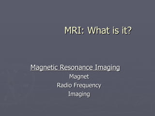

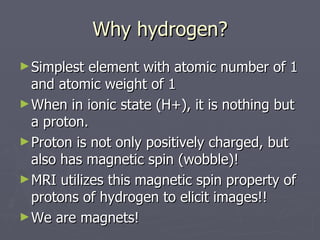
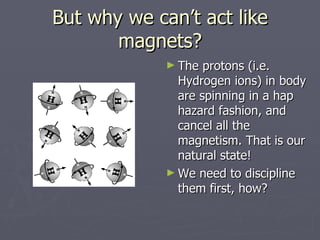
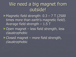
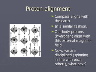
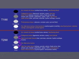

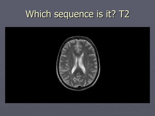
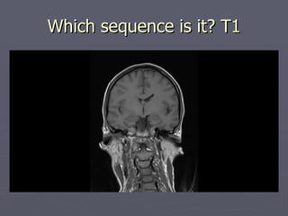
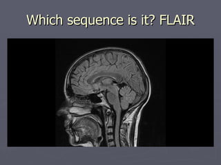
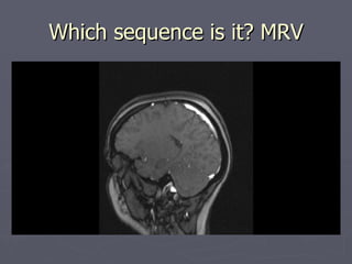
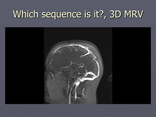
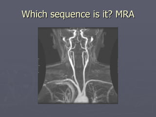
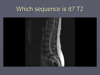
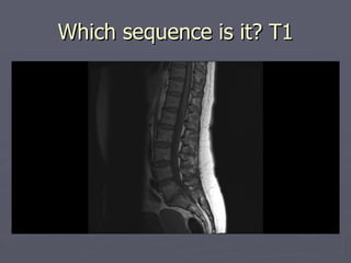
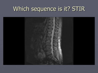
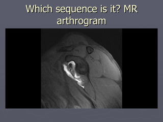


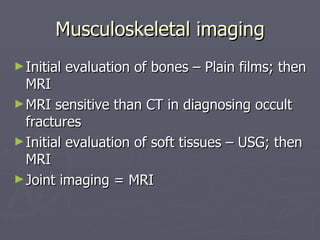
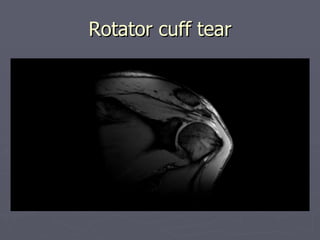
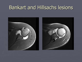
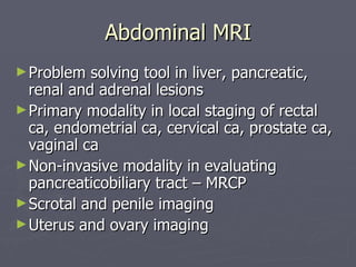
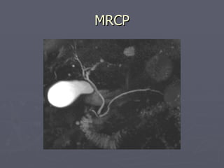


The document provides an overview of magnetic resonance imaging (MRI), including how it works, the types of images it can produce, and its applications in various parts of the body. It explains that MRI uses strong magnetic fields and radio waves to align hydrogen protons in the body and produce signals used to form images. Key applications mentioned include neuroimaging, musculoskeletal imaging, and evaluating diseases of the abdomen, blood vessels, heart, breast and fetus.


![MAGNETIC_RESONANCE.._IMAGING[MRI][1].pptx](https://cdn.slidesharecdn.com/ss_thumbnails/magneticresonanceimagingmri1-240903182728-4f857936-thumbnail.jpg?width=640&height=640&fit=bounds)


![ONFH[AVN HIP] -TRIPLE REGIME -A NOVAL SURGICAL CONCEPT .pptx](https://cdn.slidesharecdn.com/ss_thumbnails/onfhavnhip2026koaconcalicutdrgokuldevdrmashraf-260210064517-213ec005-thumbnail.jpg?width=640&height=640&fit=bounds)


