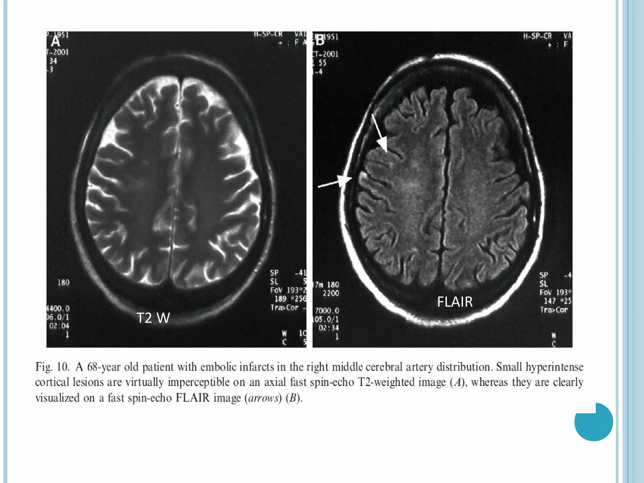
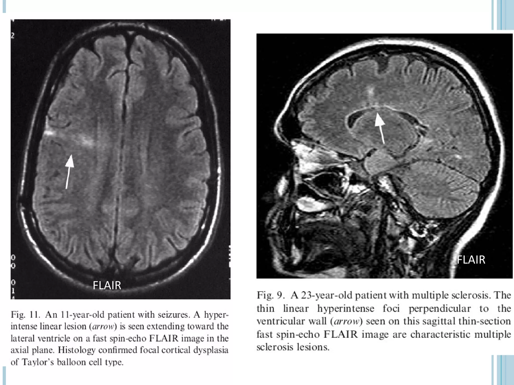
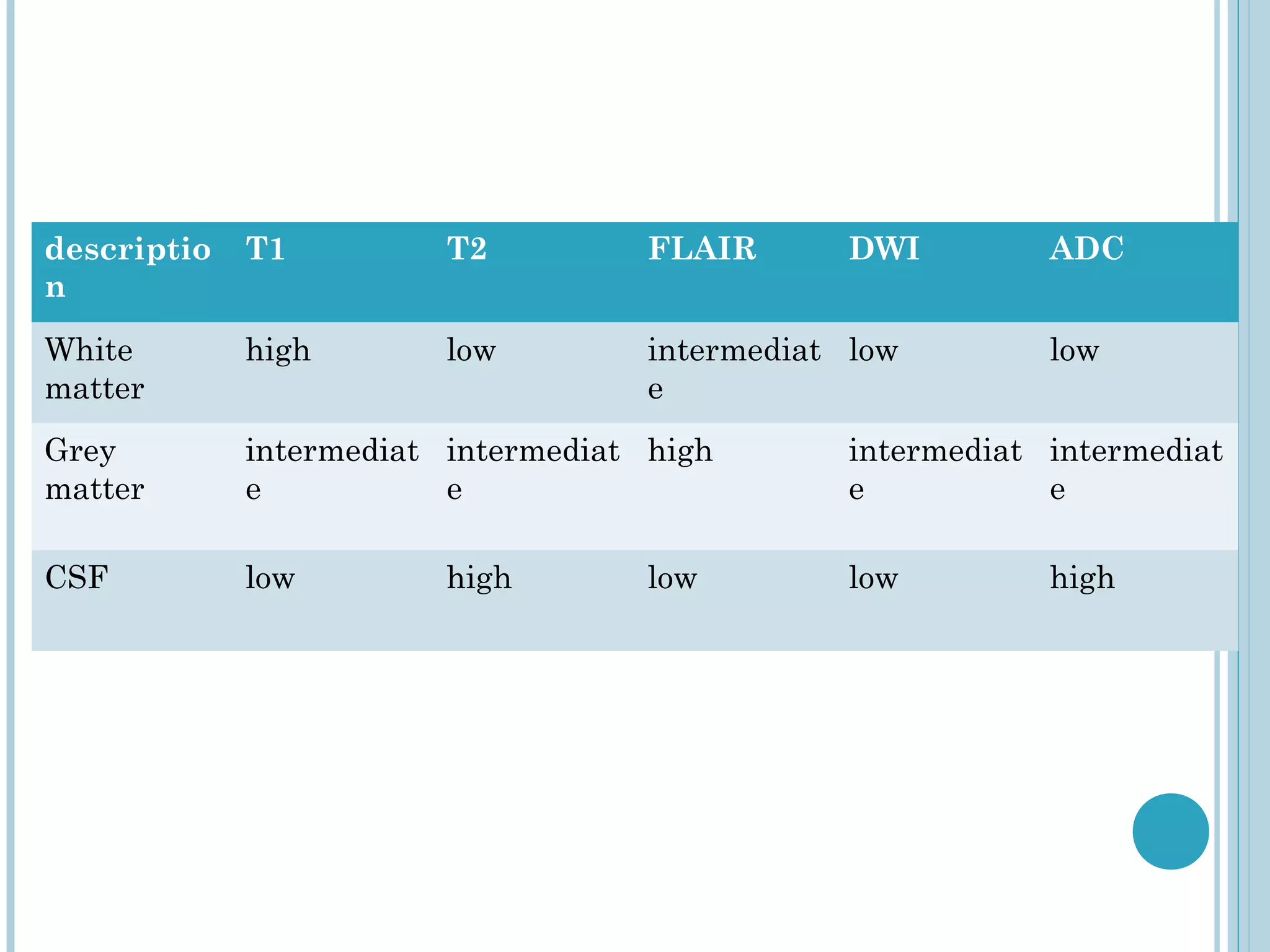
MRI sequences utilize the magnetic spin property of hydrogen protons to generate images. T1-weighted images highlight fat and tissues with slow-flowing blood while T2-weighted images highlight edema, inflammation and fluid. FLAIR sequences suppress the signal from cerebrospinal fluid to improve detection of lesions near CSF-containing spaces such as in multiple sclerosis or mesial temporal sclerosis. Choosing the appropriate sequence depends on the desired tissue contrast and abnormalities being evaluated.
SWI , high susceptibility for blood products, iron depositions, and calcifications
makes susceptibility-weighted imaging an important additional sequence for the diagnostic
workup of pediatric brain pathologic abnormalities. Compared with conventional MRI
sequences, susceptibility-weighted imaging may show lesions in better detail or with higher
sensitivity
Magnetic Resonance Angiography and VenographyAnjan Dangal
Introduction to MR Angiography and Venography Procedure of Brain . Includes Indication, MRI protocol, planning and anatomy as well as brief intoduction to physics behind MRA and MRV principle.
A review of advances in Brachytherapy treatment planning and delivery in last decade or so, with main focus on brachytherapy for Prostate cancer, Breast cancer and Cervical cancer
Magnetic resonance imaging (MRI) is an imaging technique used primarily in medical settings to produce high quality images of the soft tissues of the human body.
These lecture slides, by Dr Sidra Arshad, offer a quick overview of physiological basis of a normal electrocardiogram.
Learning objectives:
1. Define an electrocardiogram (ECG) and electrocardiography
2. Describe how dipoles generated by the heart produce the waveforms of the ECG
3. Describe the components of a normal electrocardiogram of a typical bipolar leads (limb II)
4. Differentiate between intervals and segments
5. Enlist some common indications for obtaining an ECG
Study Resources:
1. Chapter 11, Guyton and Hall Textbook of Medical Physiology, 14th edition
2. Chapter 9, Human Physiology - From Cells to Systems, Lauralee Sherwood, 9th edition
3. Chapter 29, Ganong’s Review of Medical Physiology, 26th edition
4. Electrocardiogram, StatPearls - https://www.ncbi.nlm.nih.gov/books/NBK549803/
5. ECG in Medical Practice by ABM Abdullah, 4th edition
6. ECG Basics, http://www.nataliescasebook.com/tag/e-c-g-basics
micro teaching on communication m.sc nursing.pdfAnurag Sharma
Microteaching is a unique model of practice teaching. It is a viable instrument for the. desired change in the teaching behavior or the behavior potential which, in specified types of real. classroom situations, tends to facilitate the achievement of specified types of objectives.
- Video recording of this lecture in English language: https://youtu.be/lK81BzxMqdo
- Video recording of this lecture in Arabic language: https://youtu.be/Ve4P0COk9OI
- Link to download the book free: https://nephrotube.blogspot.com/p/nephrotube-nephrology-books.html
- Link to NephroTube website: www.NephroTube.com
- Link to NephroTube social media accounts: https://nephrotube.blogspot.com/p/join-nephrotube-on-social-media.html
Anti ulcer drugs and their Advance pharmacology ||
Anti-ulcer drugs are medications used to prevent and treat ulcers in the stomach and upper part of the small intestine (duodenal ulcers). These ulcers are often caused by an imbalance between stomach acid and the mucosal lining, which protects the stomach lining.
||Scope: Overview of various classes of anti-ulcer drugs, their mechanisms of action, indications, side effects, and clinical considerations.
Report Back from SGO 2024: What’s the Latest in Cervical Cancer?bkling
Are you curious about what’s new in cervical cancer research or unsure what the findings mean? Join Dr. Emily Ko, a gynecologic oncologist at Penn Medicine, to learn about the latest updates from the Society of Gynecologic Oncology (SGO) 2024 Annual Meeting on Women’s Cancer. Dr. Ko will discuss what the research presented at the conference means for you and answer your questions about the new developments.
Lung Cancer: Artificial Intelligence, Synergetics, Complex System Analysis, S...Oleg Kshivets
RESULTS: Overall life span (LS) was 2252.1±1742.5 days and cumulative 5-year survival (5YS) reached 73.2%, 10 years – 64.8%, 20 years – 42.5%. 513 LCP lived more than 5 years (LS=3124.6±1525.6 days), 148 LCP – more than 10 years (LS=5054.4±1504.1 days).199 LCP died because of LC (LS=562.7±374.5 days). 5YS of LCP after bi/lobectomies was significantly superior in comparison with LCP after pneumonectomies (78.1% vs.63.7%, P=0.00001 by log-rank test). AT significantly improved 5YS (66.3% vs. 34.8%) (P=0.00000 by log-rank test) only for LCP with N1-2. Cox modeling displayed that 5YS of LCP significantly depended on: phase transition (PT) early-invasive LC in terms of synergetics, PT N0—N12, cell ratio factors (ratio between cancer cells- CC and blood cells subpopulations), G1-3, histology, glucose, AT, blood cell circuit, prothrombin index, heparin tolerance, recalcification time (P=0.000-0.038). Neural networks, genetic algorithm selection and bootstrap simulation revealed relationships between 5YS and PT early-invasive LC (rank=1), PT N0—N12 (rank=2), thrombocytes/CC (3), erythrocytes/CC (4), eosinophils/CC (5), healthy cells/CC (6), lymphocytes/CC (7), segmented neutrophils/CC (8), stick neutrophils/CC (9), monocytes/CC (10); leucocytes/CC (11). Correct prediction of 5YS was 100% by neural networks computing (area under ROC curve=1.0; error=0.0).
CONCLUSIONS: 5YS of LCP after radical procedures significantly depended on: 1) PT early-invasive cancer; 2) PT N0--N12; 3) cell ratio factors; 4) blood cell circuit; 5) biochemical factors; 6) hemostasis system; 7) AT; 8) LC characteristics; 9) LC cell dynamics; 10) surgery type: lobectomy/pneumonectomy; 11) anthropometric data. Optimal diagnosis and treatment strategies for LC are: 1) screening and early detection of LC; 2) availability of experienced thoracic surgeons because of complexity of radical procedures; 3) aggressive en block surgery and adequate lymph node dissection for completeness; 4) precise prediction; 5) adjuvant chemoimmunoradiotherapy for LCP with unfavorable prognosis.
New Directions in Targeted Therapeutic Approaches for Older Adults With Mantl...i3 Health
i3 Health is pleased to make the speaker slides from this activity available for use as a non-accredited self-study or teaching resource.
This slide deck presented by Dr. Kami Maddocks, Professor-Clinical in the Division of Hematology and
Associate Division Director for Ambulatory Operations
The Ohio State University Comprehensive Cancer Center, will provide insight into new directions in targeted therapeutic approaches for older adults with mantle cell lymphoma.
STATEMENT OF NEED
Mantle cell lymphoma (MCL) is a rare, aggressive B-cell non-Hodgkin lymphoma (NHL) accounting for 5% to 7% of all lymphomas. Its prognosis ranges from indolent disease that does not require treatment for years to very aggressive disease, which is associated with poor survival (Silkenstedt et al, 2021). Typically, MCL is diagnosed at advanced stage and in older patients who cannot tolerate intensive therapy (NCCN, 2022). Although recent advances have slightly increased remission rates, recurrence and relapse remain very common, leading to a median overall survival between 3 and 6 years (LLS, 2021). Though there are several effective options, progress is still needed towards establishing an accepted frontline approach for MCL (Castellino et al, 2022). Treatment selection and management of MCL are complicated by the heterogeneity of prognosis, advanced age and comorbidities of patients, and lack of an established standard approach for treatment, making it vital that clinicians be familiar with the latest research and advances in this area. In this activity chaired by Michael Wang, MD, Professor in the Department of Lymphoma & Myeloma at MD Anderson Cancer Center, expert faculty will discuss prognostic factors informing treatment, the promising results of recent trials in new therapeutic approaches, and the implications of treatment resistance in therapeutic selection for MCL.
Target Audience
Hematology/oncology fellows, attending faculty, and other health care professionals involved in the treatment of patients with mantle cell lymphoma (MCL).
Learning Objectives
1.) Identify clinical and biological prognostic factors that can guide treatment decision making for older adults with MCL
2.) Evaluate emerging data on targeted therapeutic approaches for treatment-naive and relapsed/refractory MCL and their applicability to older adults
3.) Assess mechanisms of resistance to targeted therapies for MCL and their implications for treatment selection
Prix Galien International 2024 Forum ProgramLevi Shapiro
June 20, 2024, Prix Galien International and Jerusalem Ethics Forum in ROME. Detailed agenda including panels:
- ADVANCES IN CARDIOLOGY: A NEW PARADIGM IS COMING
- WOMEN’S HEALTH: FERTILITY PRESERVATION
- WHAT’S NEW IN THE TREATMENT OF INFECTIOUS,
ONCOLOGICAL AND INFLAMMATORY SKIN DISEASES?
- ARTIFICIAL INTELLIGENCE AND ETHICS
- GENE THERAPY
- BEYOND BORDERS: GLOBAL INITIATIVES FOR DEMOCRATIZING LIFE SCIENCE TECHNOLOGIES AND PROMOTING ACCESS TO HEALTHCARE
- ETHICAL CHALLENGES IN LIFE SCIENCES
- Prix Galien International Awards Ceremony
Ethanol (CH3CH2OH), or beverage alcohol, is a two-carbon alcohol
that is rapidly distributed in the body and brain. Ethanol alters many
neurochemical systems and has rewarding and addictive properties. It
is the oldest recreational drug and likely contributes to more morbidity,
mortality, and public health costs than all illicit drugs combined. The
5th edition of the Diagnostic and Statistical Manual of Mental Disorders
(DSM-5) integrates alcohol abuse and alcohol dependence into a single
disorder called alcohol use disorder (AUD), with mild, moderate,
and severe subclassifications (American Psychiatric Association, 2013).
In the DSM-5, all types of substance abuse and dependence have been
combined into a single substance use disorder (SUD) on a continuum
from mild to severe. A diagnosis of AUD requires that at least two of
the 11 DSM-5 behaviors be present within a 12-month period (mild
AUD: 2–3 criteria; moderate AUD: 4–5 criteria; severe AUD: 6–11 criteria).
The four main behavioral effects of AUD are impaired control over
drinking, negative social consequences, risky use, and altered physiological
effects (tolerance, withdrawal). This chapter presents an overview
of the prevalence and harmful consequences of AUD in the U.S.,
the systemic nature of the disease, neurocircuitry and stages of AUD,
comorbidities, fetal alcohol spectrum disorders, genetic risk factors, and
pharmacotherapies for AUD.
1. MRI SEQUENCES
Tushar Patil, MD
Senior Resident
Department of Neurology
King George’s Medical University
Lucknow, India
2. MRI PRINCIPLE
MRI is based on the principle of nuclear magnetic resonance
(NMR)
Two basic principles of NMR
1. Atoms with an odd number of protons or neutrons have spin
2. A moving electric charge, be it positive or negative, produces a
magnetic field
Body has many such atoms that can act as good MR nuclei ( 1H,
13
C, 19F, 23Na)
Hydrogen nuclei is one of them which is not only positively
charged, but also has magnetic spin
MRI utilizes this magnetic spin property of protons of hydrogen
to elicit images
3. WHY HYDROGEN IONS ARE USED IN
MRI?
Hydrogen nucleus has an unpaired proton which is positively charged
Every hydrogen nucleus is a tiny magnet which produces small but
noticeable magnetic field
Hydrogen atom is the only major species in the body that is MR
sensitive
Hydrogen is abundant in the body in the form of water and fat
Essentially all MRI is hydrogen (proton) imaging
4. BODY IN AN EXTERNAL
MAGNETIC FIELD (B0)
•In our natural state Hydrogen ions in body are
spinning in a haphazard fashion, and cancel all
the magnetism.
•When an external magnetic field is applied protons
in the body align in one direction. (As the compass
aligns in the presence of earth’s
magnetic field)
5. NET MAGNETIZATION
Half of the protons align along the magnetic field and rest are aligned opposite
.
At room temperature, the
population ratio of anti-
parallel versus parallel
protons is roughly 100,000
to 100,006 per Tesla of B0
These extra protons produce net magnetization vector (M)
Net magnetization depends on B0 and temperature
6. MANIPULATING THE NET
MAGNETIZATION
Magnetization can be manipulated by changing the magnetic
field environment (static, gradient, and RF fields)
RF waves are used to manipulate the magnetization of H nuclei
Externally applied RF waves perturb magnetization into different
axis (transverse axis). Only transverse magnetization produces
signal.
When perturbed nuclei return to their original state they emit
RF signals which can be detected with the help of receiving coils
7. T1 AND T2 RELAXATION
When RF pulse is stopped higher energy gained by proton is
retransmitted and hydrogen nuclei relax by two mechanisms
T1 or spin lattice relaxation- by which original magnetization
(Mz) begins to recover.
T2 relaxation or spin spin relaxation - by which magnetization in
X-Y plane decays towards zero in an exponential fashion. It is due
to incoherence of H nuclei.
T2 values of CNS tissues are shorter than T1 values
8. T1 RELAXATION
After protons are
Excited with RF pulse
They move out of
Alignment with B0
But once the RF Pulse
is stopped they Realign
after some Time And
this is called t1 relaxation
T1 is defined as the time it takes for the hydrogen nucleus to
recover 63% of its longitudinal magnetization
9. T2 relaxation time is the time for 63% of the protons to become dephased
owing to interactions among nearby protons.
10. TR AND TE
TE (echo time) : time interval in which signals are measured after RF
excitation
TR (repetition time) : the time between two excitations is called repetition
time
By varying the TR and TE one can obtain T1WI and T2WI
In general a short TR (<1000ms) and short TE (<45 ms) scan is T1WI
Long TR (>2000ms) and long TE (>45ms) scan is T2WI
Long TR (>2000ms) and short TE (<45ms) scan is proton density image
11. Different tissues have different relaxation times.
These relaxation time differences is used to
generate image contrast.
22. CONVENTIONAL INVERSION
RECOVERY
-180° preparatory pulse is applied to flip the net magnetization vector 180° and null the
signal from a particular entity (eg, water in tissue).
-When the RF pulse ceases, the spinning nuclei begin to relax. When the net
magnetization vector for water passes the transverse plane (the null point for that
tissue), the conventional 90° pulse is applied, and the SE sequence then continues as
before.
-The interval between the 180° pulse and the 90° pulse is the TI ( Inversion Time).
23. Conventional Inversion Recovery Contd:
At TI, the net magnetization vector of water is very weak, whereas that for body
tissues is strong. When the net magnetization vectors are flipped by the 90° pulse,
there is little or no transverse magnetization in water, so no signal is generated (fluid
appears dark), whereas signal intensity ranges from low to high in tissues with a
stronger NMV.
Two important clinical implementations of the inversion recovery concept are:
Short TI inversion-recovery (STIR) sequence
Fluid-attenuated inversion-recovery (FLAIR) sequence.
24. SHORT TI INVERSION-RECOVERY (STIR)
SEQUENCE
In STIR sequences, an inversion-recovery pulse is used to null the signal from fat
(180° RF Pulse).
When NMV of fat passes its null point , 90° RF pulse is applied. As little or no
longitudinal magnetization is present and the transverse magnetization is
insignificant.
It is transverse magnetization that induces an electric current in the receiver coil so
no signal is generated from fat.
STIR sequences provide excellent depiction of bone marrow edema which may be
the only indication of an occult fracture.
Unlike conventional fat-saturation sequences STIR sequences are not affected by
magnetic field inhomogeneities, so they are more efficient for nulling the signal from
fat
25. FSE STIR
Comparison of fast SE and STIR sequences
for depiction of bone marrow edema
26. FLUID-ATTENUATED INVERSION RECOVERY
(FLAIR)
First described in 1992 and has become one of the corner stones of brain MR
imaging protocols
An IR sequence with a long TR and TE and an inversion time (TI) that is tailored to
null the signal from CSF
In contrast to real image reconstruction, negative signals are recorded as positive
signals of the same strength so that the nulled tissue remains dark and all other
tissues have higher signal intensities.
27. Most pathologic processes show increased SI on T2-WI, and the conspicuity of
lesions that are located close to interfaces b/w brain parenchyma and CSF may be
poor in conventional SE or FSE T2-WI sequences.
FLAIR images are heavily T2-weighted with CSF signal suppression, highlights
hyperintense lesions and improves their conspicuity and detection, especially when
located adjacent to CSF containing spaces
28. In addition to T2- weightening, FLAIR possesses considerable T1-weighting,
because it largely depends on longitudinal magnetization
As small differences in T1 characteristics are accentuated, mild T1-shortening
becomes conspicuous.
This effect is prominent in the CSF-containing spaces, where increased protein
content results in high SI (eg, associated with sub-arachnoid space disease)
High SI of hyperacute SAH is caused by T2 prolongation in addition to T1
shortening
29. Clinical Applications:
Used to evaluate diseases affecting the brain parenchyma neighboring the CSF-
containing spaces for eg: MS & other demyelinating disorders.
Unfortunately, less sensitive for lesions involving the brainstem & cerebellum,
owing to CSF pulsation artifacts
Helpful in evaluation of neonates with perinatal HIE.
Useful in evaluation of gliomatosis cerebri owing to its superior delineation of
neoplastic spread
Useful for differentiating extra-axial masses eg. epidermoid cysts from arachnoid
cysts. However, distinction is more easier & reliable with DWI.
30. Mesial temporal sclerosis: m/c pathology in patients with partial complex
seizures.Thin-section coronal FLAIR is the standard sequence in these patients &
seen as a bright small hippocampus on dark background of suppressed CSF-
containing spaces. However, normally also mesial temporal lobes have mildly
increased SI on FLAIR images.
Focal cortical dysplasia of Taylor’s balloon cell type- markedly hyperintense funnel-
shaped subcortical zone tapering toward the lateral ventricle is the characteristic
FLAIR imaging finding
In tuberous sclerosis- detection of hamartomatous lesions, is easier with FLAIR than
with PD or T2-W sequences
31. Embolic infarcts- Improved visualization
Chronic infarctions- typically dark with a rim of high signal. Bright peripheral zone
corresponds to gliosis, which is well seen on FLAIR and may be used to distinguish
old lacunar infarcts from dilated perivascular spaces.
33. Subarachnoid Hemorrhage (SAH):
FLAIR imaging surpasses even CT in the detection of traumatic supratentorial SAH.
It has been proposed that MR imaging with FLAIR, gradient-echo T2*-weighted,
and rapid high-spatial resolution MR angiography could be used to evaluate patients
with suspected acute SAH, possibly obviating the need for CT and intra-arterial
angiography.
With the availability of high-quality CT angiography, this approach may not be
necessary.
36. DIFFUSION-WEIGHTED MRI
Diffusion-weighted MRI is a example of endogenous contrast, using
the motion of protons to produce signal changes
DWI images is obtained by applying pairs of opposing and
balanced magnetic field gradients (but of differing durations and
amplitudes) around a spin-echo refocusing pulse of a T2 weighted
sequence. Stationary water molecules are unaffected by the paired
gradients, and thus retain their signal. Nonstationary water
molecules acquire phase information from the first gradient, but are
not rephased by the second gradient, leading to an overall loss of the
MR signal
37. • The normal motion of water molecules within living tissues is random
(brownian motion).
• In acute stroke, there is an alteration of homeostasis
• Acute stroke causes excess intracellular water accumulation, or cytotoxic
edema, with an overall decreased rate of water molecular diffusion within
the affected tissue.
• Reduction of extracellular space
• Tissues with a higher rate of diffusion undergo a greater loss of signal in a
given period of time than do tissues with a lower diffusion rate.
• Therefore, areas of cytotoxic edema, in which the motion of water
molecules is restricted, appear brighter on diffusion-weighted images
because of lesser signal losses
Restriction of DWI is not specific for stroke
38. descriptio T1 T2 FLAIR DWI ADC
n
White high low intermediat low low
matter e
Grey intermediat intermediat high intermediat intermediat
matter e e e e
CSF low high low low high
39. DW images usually performed with echo-planar sequences which
markedly decrease imaging time, motion artifacts and increase sensitivity to
signal changes due to molecular motion.
The primary application of DW MR imaging has been in brain imaging,
mainly because of its exquisite sensitivity to early detection of ischemic
stroke
40. The increased sensitivity of diffusion-weighted MRI in detecting
acute ischemia is thought to be the result of the water shift
intracellularly restricting motion of water protons (cytotoxic
edema), whereas the conventional T2 weighted images show signal
alteration mostly as a result of vasogenic edema
41. • Core of infarct = irreversible damage
• Surrounding ischemic area may be salvaged
• DWI: open a window of opportunity during which Tt is beneficial
• Regions of high mobility “rapid diffusion” dark
• Regions of low mobility “slow diffusion” bright
• Difficulty: DWI is highly sensitive to all of types of motion (blood flow,
pulsatility, patient motion).
45. APPARENT DIFFUSION COEFFICIENT
It is a measure of diffusion
Calculated by acquiring two or more images with a different gradient
duration and amplitude (b-values)
To differentiate T2 shine through effects or artifacts from real ischemic
lesions.
The lower ADC measurements seen with early ischemia,
An ADC map shows parametric images containing the apparent diffusion
coefficients of diffusion weighted images. Also called diffusion map
46. The ADC may be useful for estimating the lesion age and
distinguishing acute from subacute DWI lesions.
Acute ischemic lesions can be divided into hyperacute lesions (low
ADC and DWI-positive) and subacute lesions (normalized ADC).
Chronic lesions can be differentiated from acute lesions by
normalization of ADC and DWI.
a tumour would exhibit more restricted apparent diffusion compared
with a cyst because intact cellular membranes in a tumour would
hinder the free movement of water molecules
47. NONISCHEMIC CAUSES FOR
DECREASED ADC
Abscess
Lymphoma and other tumors
Multiple sclerosis
Seizures
Metabolic (Canavans )
49. EVALUATION OF ACUTE STROKE ON DWI
The DWI and ADC maps show changes in ischemic brain
within minutes to few hours
The signal intensity of acute stroke on DW images increase
during the first week after symptom onset and decrease
thereafter, but signal remains hyper intense for a long period
(up to 72 days in the study by Lausberg et al)
The ADC values decline rapidly after the onset of ischemia and
subsequently increase from dark to bright 7-10 days later .
This property may be used to differentiate the lesion older
than 10 days from more acute ones (Fig 2).
Chronic infarcts are characterized by elevated diffusion and
appear hypo, iso or hyper intense on DW images and
hyperintense on ADC maps
50.
51. DW MR imaging characteristics of Various Disease Entities
MR Signal Intensity
Disease DW Image ADC Image ADC Cause
Acute Stroke High Low Restricted Cytotoxic edema
Chronic Strokes Variable High Elevated Gliosis
Hypertensive Variable High Elevated Vasogenic edema
encephalopathy
Arachnoid cyst Low High Elevated Free water
Epidermoid mass High Low Restricted Cellular tumor
Herpes encephalitis High Low Restricted Cytotoxic edema
CJD High Low Restricted Cytotoxic edema
MS acute lesions Variable High Elevated Vasogenic edema
Chronic lesions Variable High Elevated Gliosis
52. CLINICAL USES OF DWI &
ADC
Stroke:
Hyperacute Stage:- within one hour minimal hyperintensity seen in DWI
and ADC value decrease 30% or more below normal (Usually <50X10-4
mm2/sec)
Acute Stage:- Hyperintensity in DWI and ADC value low but after 5-
7days of ictus ADC values increase and return to normal value
(Pseudonormalization)
Subacute to Chronic Stage:- ADC value are increased (Vasogenic edema)
but hyperintensity still seen on DWI (T2 shine effect)
54. GRE
In a GRE sequence, an RF pulse is applied that partly flips the
NMV into the transverse plane (variable flip angle).
Gradients, as opposed to RF pulses, are used to dephase (negative
gradient) and rephase (positive gradients) transverse magnetization.
Because gradients do not refocus field inhomogeneities, GRE
sequences with long TEs are T2* weighted (because of magnetic
susceptibility) rather than T2 weighted like SE sequences
55. GRE Sequences contd:
This feature of GRE sequences is exploited- in detection of hemorrhage, as the iron
in Hb becomes magnetized locally (produces its own local magnetic field) and thus
dephases the spinning nuclei.
The technique is particularly helpful for diagnosing hemorrhagic contusions such as
those in the brain and in pigmented villonodular synovitis.
SE sequences, on the other hand- relatively immune from magnetic susceptibility
artifacts, and also less sensitive in depicting hemorrhage and calcification.
57. GRE Sequences contd:
Magnetic susceptibility imaging-
- Basis of cerebral perfusion studies, in which the T2* effects (ie, signal decrease)
created by gadolinium (a metal injected intravenously as a chelated ion in aqueous
solution, typically in the form of gadopentetate dimeglumine) are sensitively depicted
by GRE sequences.
- Also used in blood oxygenation level–dependent (BOLD) imaging, in which the
relative amount of deoxyhemoglobin in the cerebral vasculature is measured as a
reflection of neuronal activity. BOLD MR imaging is widely used for mapping of
human brain function.
58. GRADIENT ECHO
Pros:
fast technique
Cons:
More sensitive to magnetic susceptibility artifacts
Clinical use:
eg. Hemorrhage , calcification
59. Axial T1 (C), T2 (D), and GRE (E) images show corresponding T1-hyperintense and GRE-
hypointense foci with associated T2 hyperintensity (arrows).
61. MR SPECTROSCOPY
Magnetic resonance spectroscopy (MRS) is a means of
noninvasive physiologic imaging of the brain that
measures relative levels of various tissue metabolites
Purcell and Bloch (1952) first detected NMR signals from
magnetic dipoles of nuclei when placed in an external
magnetic field.
Initial in vivo brain spectroscopy studies were done in the
early 1980s.
Today MRS-in particular, IH MRS-has become a valuable
physiologic imaging tool with wide clinical applicability.
62. PRINCIPLES:
The radiation produced by any substance is dependent on its atomic
composition.
Spectroscopy is the determination of this chemical composition of a
substance by observing the spectrum of electromagnetic energy emerging
from or through it.
NMR is based on the principle that some nuclei have associated magnetic
spin properties that allow them to behave like small magnet.
In the presence of an externally applied magnetic field, the
magnetic nuclei interact with that field and distribute themselves to
different energy levels.
These energy states correspond to the proton nuclear spins, either
aligned in the direction of (low-energy spin state) or against the applied
magnetic field (high-energy spin state).
63. If energy is applied to the system in the form of a radiofrequency
(RF) pulse that exactly matches the energy between both states. a
condition of resonance occurs.
Chemical elements having different atomic numbers such as
hydrogen ('H) and phosphorus (31P) resonate at different
Larmor RFs.
Small change in the local magnetic field, the nucleus of the atom
resonates at a shifted Larmor RF.
This phenomenon is called the chemical shift.
64. TECHNIQUE:
Single volume and Multivolume MRS.
1) Single volume:
Stimulated echo acquisition mode (STEAM)
Point-resolved spectroscopy (PRESS)
It gives a better signal-to noise ratio
2) Multivolume MRS:
chemical shift imaging (CSI) or spectroscopic imaging (SI)
much larger area can be covered, eliminating the sampling error to an extent
but significant weakening in the signal-to-noise ratio and a longer scan time.
Time of echo: 35 ms and 144ms.
Resonance frequencies on the x-axis and amplitude (concentration) on the y-
axis.
65. EFFECT OF TE ON THE PEAKS
__________
TE 35ms
___________
___________
TE 144ms
__________
70. PRINCIPLE METABOLITES
MetaboliteLocation Normal Increased Decreased
ppm function
NAA 2 Nonspecific Canavan’s Neuronal loss,
neuronal disease stroke,
marker dementia, AD,
(Reference for hypoxia,
chemical shift) neoplasia,
abscess
Glutamate , 2.1- 2.4 Hypoxia, HE Hyponatremia
glutamine, Neurotransmit
GABA ter
Succinate 2.4 Part of TCA Brain abscess
cycle
Creatine 3.03 Cell energy Trauma, Stroke, hypoxia,
marker hyperosmolar neoplasia
(Reference for state
metabolite
ratio)
71. Metabolite Location Normal Increased Decreased
ppm function
Choline 3.2 Marker of Neoplasia, Hypomyelinat
cell memb demyelination ion
turnover (MS)
Myoinositol 3.5 & 4 Astrocyte AD
marker Demyelinatin
g diseases
73. MRS
Inc Cho/Cr Dec NAA/Cr Dec
Myo/NAA Slightly inc Cho/ Cr Inc acetate, NAA/Cr
Cho/NAA Cho/NAA succinate, Dec NAA/
Dec NAA/Cr Normal Myo/NAA amino acid, Cho
± lipid/lactate ± lipid/lactate lactate Inc
Myo/NAA
Demyelinatin
Malignancy Neuodegene
g disease Pyogenic
abscess rative
Alzheimer
74. CLINICAL APPLICATIONS OF MRS:
Class A MRS Applications: Useful in Individual Patients
1) MRS of brain masses:
Distinguish neoplastic from non neoplastic masses
Primary from metastatic masses.
Tumor recurrence vs radiation necrosis
Prognostication of the disease
Mark region for stereotactic biopsy.
Monitoring response to treatment.
Research tool
2) MRS of Inborn Errors of Metabolism
Include the leukodystrophies, mitochondrial disorders, and enzyme defects that
cause an absence or accumulation of metabolites
75. CLASS B MRS APPLICATIONS: OCCASIONALLY USEFUL IN
INDIVIDUAL PATIENTS
1) Ischemia, Hypoxia, and Related Brain Injuries
Ischemic stroke
Hypoxic ischemic encephalopathy.
2)Epilepsy
Class C Applications: Useful Primarily in Groups of Patients (Research)
HIV disease and the brain
Neurodegenerative disorders
Amyotrophic lateral sclerosis
Multiple sclerosis
Hepatic encephalopathy
Psychiatric disorders
76. MAGNETIZATION TRANSFER (MT) MRI
MT is a recently developed MR technique that alters contrast of tissue on
the basis of macromolecular environments.
MTC is most useful in two basic area, improving image contrast and tissue
characterization.
MT is accepted as an additional way to generate unique contrast in MRI
that can be used to our advantage in a variety of clinical applications.
77. Magnetization transfer (MT) contd:-
Basis of the technique: that the state of magnetization of an atomic nucleus can be
transferred to a like nucleus in an adjacent molecule with different relaxation
characteristics.
Acc. to this theory- H1 proton spins in water molecules can exchange magnetization
with H1 protons of much larger molecules, such as proteins and cell membranes.
Consequence is that the observed relaxation times may reflect not only the properties
of water protons but also, indirectly, the characteristics of the macromolecular
solidlike environment
MT occurs when RF saturation pulses are placed far from the resonant frequency of
water into a component of the broad macromolecular pool .
78. Magnetization transfer (MT) contd:-
These off-resonance pulses, which may be added to standard MR pulse sequences,
reduce the longitudinal magnetization of the restricted protons to zero without
directly affecting the free water protons.
The initial MT occurs between the macromolecular protons and the transiently
bound hydration layer protons on the surface of large molecules’
Saturated bound hydration layer protons then diffuse and mix with the free water
proton pool
Saturation is transferred to the mobile water protons, reducing their longitudinal
magnetization, which results in decreased signal intensity and less brightness on
MR images.
79. Magnetization transfer (MT) contd:-
The MT effect is superimposed on the intrinsic contrast of the baseline image
Amount of signal loss on MT images correlates with the amount of macromolecules
in a given tissue and the efficiency of the magnetization exchange
MT characteristically:
Reduces the SI of some solid like tissues, such as most of the brain and spinal cord
Does not influence liquid like tissues significantly, such as the cerebrospinal fluid
(CSF)
81. CLINICAL APPLICATION
• Useful diagnostic tool in characterization of a variety of CNS infection
• In detection and diagnosis of meningitis , encephalitis, CNS tuberculosis ,
neurocysticercosis and brain abscess.
TUBERCULOMA
• Pre-contrast T1-W MT imaging helps to better assess the disease load in CNS
tuberculosis by improving the detectability of the lesions, with more number
of tuberculomas detected on pre-contrast MT images compared to routine SE
images
• It may also be possible to differentiate T2 hypo intense tuberculoma from T2
hypo intense cysticerus granuloma with the use of MTR, as cysticercus
granulomas show significantly higher MT ratio compared to tuberculomas
83. NEUROCYSTICERCOSIS
Findings vary with the stage of disease
T1-W MT images are also important in demonstrating perilesional gliosis in
treated neurocysticercus lesions
Gliotic areas show low MTR compared to the gray matter and white matter.
So appear as hyperintense
BRAIN ABSCESS
Lower MTR from tubercular abscess wall in comparison to wall of
pyogenic abscess(~20 vs. ~26)
84. Magnetization transfer (MT) contd:-
Qualitative applications:
MR angiography,
postcontrast studies
spine imaging
MT pulses have a greater influence on brain tissue (d/t high conc. of structured
macromolecules such as cholesterol and lipid) than on stationary blood.
By reducing the background signal vessel-to-brain contrast is accentuated,
Not helpful when MR angiography is used for the detection and characterization of
cerebral aneurysms.
85. GRE images of the cervical spine without (A) and with (B) MT
show improved CSF–spinal cord contrast
86. Magnetization transfer (MT) contd:-
Quantitative applications:
Multiple sclerosis: discriminates multiple sclerosis & other demyelinating disorders,
provides measure of total lesion load, assess the spinal cord lesion burden and to
monitor the response to different treatments of multiple sclerosis
systemic lupus erythematosus,
CADASIL (cerebral autosomal dominant arteriopathy with subcortical infarcts and
leukoencephalopathy),
Multiple system atrophy,
Amyotrophic lateral sclerosis,
Schizophrenia
Alzheimer’s disease
87. MTR Quantitative applications contd:
May be used to differentiate between progressive multifocal leukoencephalopathy
and HIV encephalitis
To detect axonal injury in normal appearing splenium of corpus callosum after head
trauma
In chronic liver failure, diffuse MTR abnormalities have been found in normal
appearing brain, which return to normal following liver transplantation
90. TOF MRA
Signal from “flight” of unsaturated blood into image
No contrast agent injected
Motion artifact
Non-uniform blood signal
PC MRA
Phase shifts in moving spins (i.e. blood) are measured
Phase is proportional to velocity
Allows quantification of blood flow and velocity
CE MRA
T1-shortening agent, Gadolinium, injected iv as contrast
Gadolinium reduces T1 relaxation time
When TR<<T1, minimal signal from background tissues
Result is increased signal from Gd containing structures
Faster gradients allow imaging in a single breathhold
91. 2D AND 3D FOURIER TRASFORM
In 2DFT technique, multiple thin sections of body are studied individually and even
slow flow is identified
In 3DFT technique , a large volume of tissue is studied ,which can be subsequently
partitioned into individual slices, hence high resolution can be obtained and flow
artifacts are minimised, and less likely to be affected by loops and tortusity of
vessels
MOTSA(multiple overlapping thin slab acquisition): prevents proton saturation
across the slab. This technique have advantage of both 2D and 3D studies. It is better
than 3D TOF MRA in correctly identifying vascular loops and tortusity,and have
lesser chances of overestimating carotid stenosis.
95. Magnetic Resonance Venography (MRV)
Indications
For evaluation of thrombosis or compression by tumor of the cerebral venous sinus
in members who are at risk
(e.g., otitis media, meningitis, sinusitis, oral contraceptive use, underlying malignant
process,hypercoagulable disorders)
or have signs or symptoms
(e.g., papilledema, focal motor or sensory deficits, seizures, or drowsiness and
confusion accompanying a headache);
Lipid increase in high-grade gliomas, meningiomas, demyelination, necrotic foci, and inborn errors of metabolism
NAA is the most prominent one in normal adult brain proton MRS and is used as a reference for determination of chemical shift and nonspecific neuronal marker. Normal absolute concentrations of NAA in the adult brain are generally in the range of 8 to 9 mmol/kg. NAA concentrations are decreased in many brain disorders, resulting in neuronal and axonal loss, such as in neurodegenerative diseases, stroke, brain tumors, epilepsy, and multiple sclerosis, but are increased in Canavan's disease Cr peak is an indirect indicator of brain intracellular energy stores, tends to be relatively constant in each tissue type in normal brain, mean absolute Cr concentration in normal adult brains of 7.49; reduced in all brain tumors, particularly malignant ones
Cho reflects cell membrane synthesis and Degradation. Processes resulting in hypercellularity (e.g., primary brain neoplasms or gliosis) or myelin breakdown (demyelinating diseases) lead to locally increased Cho concentration, whereas hypomyelinating diseases result in decreased Cho levels. Mean absolute Cho concentration in normal adult brain tissue of 1.32 Ig3 MI is believed to be a glial marker because it is present primarily in glial cells and is absent in neurons; abnormally increased in patients with demyelinating diseases and in those with Alzheimer's disease Lac levels in normal brain tissue are absent or extremely low (C0.5 Mmol/L), they are essentially undetectable on normal spectra. Found in anaerobic glycolysis, which may be seen with brain neoplasms, infarcts, hypoxia, metabolic disorders or seizure and accumulate within cysts or foci of necrosis.
TOF MRA , in a slab of tissue to be imaged rapid RF pulses are given. Stationary tissue is saturated with rapid RF pulses and loses signal but fresh moving blood entering the slide will retain its signal intensity and will create contrast between flowing blood and background tissue. In PC MRA . The contrast between flowing and stationary tissue is a result of phase difference between protons in two tissues Both POF and PC angiograms can be performed using 2D and 3D techniques
Disadv of MRA high cost , cant identify small vs,susceptibility to complex fow, claustrophobia, not good for root of neck and aortic arch
Submentovertex view
Superior saggital sinus runsalong sup margin of falx cerebri and empty in confluence, recievs sup cerebral vv. Inf sag sinus runs along inf margin falx and continue as straight sinus after merging vein of galen Straigt sinus carried in attachment of falx to tentorium cerebelli, empty in confluence Confluence empty in transeverse sinus which runs along tentorium cerebellito sigmoid sinus and drain in IJV cavernous sinus situated in middle cranial fossa and connected to contralat side by intercavernous sinus Cav sinus drain inferior and superficial cerebr vv, opth vv, sphenoparital sinus and empty via sup and inf petrosal sinus into tr/sigmoid and IJV respectivly
Sup cerebral vv drain in SSS, inf cebral vv drain in cavernous sinus, sup middle cerebral vv empty via trlard in SSS and labbe vv in transvers sinus