Introduction: MRI, or Magnetic Resonance Imaging, is a versatile medical imaging technique with a wide range of clinical applications.
Soft Tissue Imaging: The unique ability of MRI to produce detailed images of soft tissues, such as the brain, muscles, and organs.
Non-Invasive Nature: MRI is a non-invasive and safe imaging modality, making it invaluable for clinical diagnosis.
Retrograde Urethrography is a specialized X-ray procedure used to visualize the male urethra, which is the tube that carries urine from the bladder to the external body opening. This procedure is typically performed to diagnose and evaluate various conditions and abnormalities within the urethra, such as strictures, obstructions, or injuries.
Percutaneous Transhepatic Cholangiography (PTC) is a radiographic procedure used to visualize and assess the biliary system, including the bile ducts within the liver and those leading to the small intestine.
This presentation will provide an in-depth understanding of the essential guidelines for designing and locating X-ray equipment in accordance with radiation protection guidelines.
MCU stands for Micturating Cystourethrogram.
it's a radiographic procedure used to visualize the urinary bladder and lower urinary tract.
MCU involves real-time imaging during urination (micturition).
Definition of ultrasound imaging in radiology: Ultrasound uses sound waves to create real-time images of internal body structures.
Importance of ultrasound technology in medical diagnosis: Non-invasive, safe, and cost-effective imaging method with various applications.
Overview of the presentation structure: An outline of topics covered, including components and working principles of ultrasound machines.
Clearly state the objective of the presentation:
To explore the key components of a CT machine in detail.
To gain a deeper understanding of how these components work together to produce high-quality images.
Briefly outline the structure of the upcoming slides:
Each subsequent slide will delve into one specific component of the CT machine.
We will examine the function, significance, and operation of each component.
The document summarizes the clinical applications of computed tomography (CT) scans. It discusses the history of CT, including its invention in the 1970s. It then outlines several main clinical applications of CT scans, such as evaluating head injuries, abdominal pain, blood vessel issues, and bone fractures. It also describes specialized CT scans like CT angiography, CT perfusion, coronary CT angiography, and virtual colonoscopy and bronchoscopy. The document emphasizes how multislice CT scans provide advantages like faster scanning times, thinner slices, clearer images, and lower radiation doses compared to older single slice CT machines.
Welcome to our presentation on "Emergencies in the Radiology Department." As radiology students, it is crucial for us to be prepared to handle emergencies that may arise while working in a medical imaging setting.
During emergencies, quick and effective responses can be life-saving and can make a significant impact on patient outcomes.
MRI Definition: Magnetic Resonance Imaging is a medical imaging technique that non-invasively visualizes the internal structures of the body.
Basic Concept: MRI uses powerful magnetic fields and radio waves to create detailed images of tissues and organs.
Importance: MRI is valuable in diagnosing a wide range of medical conditions and provides excellent soft tissue contrast.
MRI uses strong magnetic fields and radio waves to produce detailed images of the inside of the body. It is a medical imaging technique that does not use ionizing radiation. The first MRI image was published in 1973 and showed two tubes of water. Modern MRI machines use magnetic fields of 1.5 Tesla or higher to align hydrogen protons in the body. Radio pulses then excite the protons, which emit radio signals as they relax back to their original alignment. The signals are detected by receivers in the machine and used to construct detailed images of tissues and organs.
Ultrasound is a non-invasive medical imaging modality widely used in various clinical applications.
It is based on the principle of using high-frequency sound waves to create real-time images of internal structures within the human body.
In this presentation, we will discuss into the fundamental principles of ultrasound imaging and its applications in radiology.
Tomography as a medical imaging technique that allows for the visualization of cross-sectional images of the human body. Emphasize that tomography provides detailed, three-dimensional views of anatomical structures, which can be invaluable for diagnosis and treatment planning in radiology.
Contrast media are substances used in medical imaging to enhance the visibility of internal structures. Positive contrast agents, like iodinated and barium-based ones, appear bright on images due to their high atomic number, aiding in highlighting blood vessels, gastrointestinal tract, and soft tissues. Negative contrast agents, often gases or air, appear dark on images, outlining specific cavities or structures. Solid contrast media, like barium sulphate, exist in a solid state and are ingested to visualize the gastrointestinal tract. Oily contrast media, non-water-soluble substances, provide prolonged contrast, commonly used in lymphangiography and myelography. The choice of contrast media depends on the imaging modality and structures to be visualized, optimizing diagnostic accuracy and patient safety.
Magnetic resonance imaging (MRI) uses strong magnets and radio waves to produce detailed images of the inside of the body without using ionizing radiation. An MRI machine contains a powerful magnet to align hydrogen atoms in the body. Radio waves are then used to excite the atoms, which emit signals as they relax. These signals are detected by antennas and used by a computer to generate 2D or 3D images of tissues and organs. MRI provides excellent soft tissue contrast and is useful for imaging the brain, muscles, joints, and other internal organs. While it has advantages over CT in avoiding radiation, MRI scans can be costly and some patients may find the enclosed scanner space claustrophobic.
MRI contrast agents contain gadolinium which shortens the T1 relaxation time of protons, making tissues appear brighter on T1-weighted MRI images. The most common agents are gadolinium chelates which remain extracellular after intravenous injection. Rare but serious side effects can include nephrogenic systemic fibrosis in patients with kidney disease who cannot clear the agent from their body. Most side effects are mild and temporary.
A PET scan uses radioactive tracers to detect disease in the body at a cellular level. It works by injecting a small amount of radioactive sugar molecule called FDG into the bloodstream. Cancer cells absorb more FDG than normal cells, allowing cancers to be seen as hot spots on PET images. PET scans are useful for detecting cancer, epilepsy, Alzheimer's disease, and evaluating treatment response. While exposing patients to radiation, PET scans provide metabolic imaging to detect diseases earlier than other scans.
Ultrasound imaging, also known as sonography, has a rich history of development in the field of medical diagnostics.
Understanding the historical milestones of ultrasound imaging provides valuable insights into its evolution and significance in modern medicine.
This presentation aims to take radiology students on a journey through the key developments and advancements in ultrasound imaging.
PET-CT and PET-MR provide functional imaging through PET as well as anatomical imaging through CT or MRI. PET involves radiolabeling molecules like FDG with positron emitters, injecting them into patients, and using coincident detection of annihilation photons to construct 3D images. PET-CT provides accurate localization of functional abnormalities and distinction of normal from pathological tracer uptake. Whole-body PET-MRI is an emerging technique that combines the molecular imaging of PET with the excellent soft tissue contrast of MRI.
Computed Tomography (CT) is a medical imaging technique that uses X-ray technology to produce detailed cross-sectional images of the body.
It is a valuable tool for diagnosing and monitoring a wide range of medical conditions
This document provides information about myelography, a radiographic examination of the central nervous system structures in the vertebral canal. It involves injecting contrast material into the subarachnoid space surrounding the spinal cord and brain. The contrast allows visualization of the spinal cord and nerves. A spinal puncture is performed to access the subarachnoid space and inject the contrast. Images are then taken under fluoroscopy to examine the spinal cord, nerves and surrounding structures.
Magnetic resonance imaging (MRI) uses strong magnetic fields and radio waves to produce detailed images of the inside of the body. Dr. Raymond Damadian discovered in the 1970s that MRI could distinguish between healthy and cancerous tissue, and he filed the first patent for using MRI for medical diagnosis. An MRI scanner aligns hydrogen atoms in the body with a strong magnetic field and uses radio waves to flip their spins, and sensors detect the radio signal emitted as the spins return to normal, allowing an image of tissues and structures to be produced. MRI is used to diagnose conditions like tumors, strokes, and musculoskeletal disorders.
Oral cholecystography (OCG) is a diagnostic radiographic examination used to visualize the gallbladder and the biliary system. It involves the use of contrast media to enhance the visualization of these structures.
Detecting and diagnosing gallbladder diseases.
Evaluating gallstone presence and location.
Assessing gallbladder function and motility.
Positron Emission Tomography (PET) is a diagnostic imaging technique that measures metabolic activity in the body. It was developed in the 1970s and provided the first functional information about the brain. PET involves injecting a radioactive tracer, usually attached to glucose, and detecting gamma ray emissions to produce images showing organ and tissue function. It is used to diagnose and monitor conditions affecting the brain, heart, cancers, Alzheimer's disease, and some neurological disorders. PET provides information about biochemical processes rather than just anatomical structures.
Computed tomography (CT) uses X-rays and digital geometry processing to generate 3D images of the inside of the body. During a CT scan, an X-ray tube rotates around the patient, emitting beams that are detected and used to construct cross-sectional slices. A radiologist can then analyze these slices to diagnose medical conditions by viewing internal organs, bones, soft tissues, and blood vessels with greater clarity and detail than traditional X-rays. CT scans are commonly used to diagnose cancers, cardiovascular diseases, infections, appendicitis, trauma, and muscular-skeletal disorders.
Welcome to the world of Angiography.
Angiography is a crucial diagnostic tool within Radiology.
It allows us to visualize blood vessels, aiding in the diagnosis and treatment of various medical conditions.
Nuclear Medicine.................
Radioactivity………………
Gamma camera………………
PET scan and SPECT scan…...........
Nuclear Medicine Studies…………..
Nuclear Medicine Team……………
Safety in Nuclear Medicine…………
MRI uses strong magnetic fields and radio waves to produce detailed images of the inside of the body without using ionizing radiation. It is commonly used for medical diagnosis, disease staging, and treatment follow-up. MRI was invented in 1971 and works by detecting signals from hydrogen atoms in tissue after they are excited by radio waves in a strong magnetic field. The signals are used to construct detailed images of organs, soft tissues, bone and virtually all other internal body structures. MRI has many medical uses including neuroimaging, cardiovascular imaging, and musculoskeletal imaging.
Lymphography is an invasive procedure that uses an oil-based radiographic contrast dye to visualize the lymphatic system, including lymph vessels and lymph nodes. A dye is injected into the hand or foot and travels through the lymphatic system. An incision is made and contrast is injected directly into the lymph vessels. Radiographs are taken over time to view the lymph vessels and nodes as the contrast spreads. While MRI and CT have replaced it, lymphography can still help evaluate lymphomas and stage radiation treatment planning by demonstrating obstructions.
Radiographic Exposure in Radiography and Imaging Technology.
Understanding the fundamentals of radiographic exposure is crucial for producing high-quality diagnostic images.
In this presentation, we will delve into the key concepts, factors, and techniques related to radiographic exposure.
The document outlines the key responsibilities of a radiographer, which include operating x-ray equipment to produce diagnostic images, ensuring strict adherence to safety and compliance protocols when working with radiation, and providing quality patient care through effective communication and ensuring informed consent. Radiographers play an important role in medical diagnosis and treatment by generating high-quality images, collaborating with other healthcare professionals, and engaging in ongoing professional development and advocacy.
Clinicoradiological And Pathological Correlation Of Autopsy Study In.pptxshyam sunder
This study aims to correlate clinical, radiological, and pathological findings in patients who died of diffuse axonal injury to better predict outcomes and understand the specific causes. The observational study will collect data from patients diagnosed with diffuse axonal injury at two hospitals over one year, including clinical findings, MRI and CT scans, and histopathological samples from autopsies. The results could help develop prognostic models for diffuse axonal injury and assess outcomes by correlating clinical and radiological information with autopsy findings. The proposed budget is 623,600 INR to cover staff, consumables, travel, and equipment needed to conduct the study.
X-ray beam restrictors, commonly referred to as collimators, are sophisticated devices utilized in medical imaging to control the size, shape, and direction of the X-ray beam emitted from the X-ray tube. These devices are integral components of X-ray machines, working in conjunction with other components to optimize image quality while minimizing patient radiation exposure.
This document provides an overview of a training module on medical imaging techniques for paramedics. The module focuses on general radiography principles, including terminology, the radiographic system, and plain radiography of the musculoskeletal system. Learners will recognize abnormal radiographic findings and learn about radiation protection. The course aims to help paramedics understand medical imaging to aid in diagnosis and patient care.
The document announces a CME programme titled "JHARTCON 2023" to be held on October 28, 2023 by the Department of Radiology at Dr. Jeyasekharan Medical Trust College of Allied Health Sciences in Nagercoil, Kanyakumari District to honor World Radiography Day. The programme aims to recognize the vital role that radiographers and radiologists play in modern healthcare through their dedication and contributions using non-invasive radiology procedures that provide rapid medical analyses allowing doctors to observe inside the human body.
- Robin McKenzie developed the McKenzie Method of Mechanical Diagnosis and Therapy after observing a patient with sciatica who experienced pain relief in lumbar extension, contrary to typical treatment at the time.
- The McKenzie Method involves classifying patients based on their symptomatic response to movement and positioning into derangement, dysfunction, or postural syndromes to guide individualized treatment.
- Examination involves a thorough patient history and mechanical examination to determine the optimal positioning or movements to address a patient's symptoms based on their classification.
Here are 10 Types of Diagnostic Imaging: 1. X-Ray Imaging 2. Computed Tomography (CT) Scan 3. Magnetic Resonance Imaging 4. Ultrasound 5. Nuclear Medicine
MRI uses magnetic fields and radio waves to produce detailed images of organs and tissues in the body. It is commonly used to evaluate the chest, abdomen, pelvis, and breasts to diagnose conditions like tumors, heart problems, and liver or kidney diseases. During an MRI exam, the patient lies still inside the machine while images are taken. MRI has benefits over other tests as it does not use radiation and can clearly depict soft tissues, though movement can cause blurred images and certain implants are not compatible.
A conference lecture talk on medical image analysis using Machine learning algorithms. here the used algorithms based on Morphological assessment parameters. the used database is based on carcinoma. The conference organised by National Institute of Technology (NIT)- Patna indexed by IET digital library.
https://digital-library.theiet.org/content/conferences/10.1049/icp.2023.1467
Conference: 8th International Conference on Computing in Engineering and Technology (ICCET 2023)
Delivering Advanced Imaging Solutions in kottakalshajismri
Discover unparalleled clarity and precision with our MRI scanning services. Utilizing advanced imaging techniques, we deliver detailed insights into anatomical structures and pathological conditions. Our dedicated team ensures a seamless experience, prioritizing patient comfort and diagnostic accuracy at every step.
The document discusses diversity in the field of health physics, which deals with recognizing, evaluating, and controlling health hazards from ionizing radiation. It describes several areas of specialization within health physics including medical physics, radiology, radiotherapy, nuclear medicine, power reactors, defense, education, regulatory enforcement, occupational safety, and the environment. Medical physics focuses on using radiation for diagnosis and treatment, while health physicists ensure protection from radiation exposure. Radiotherapy uses radiation to treat cancer, radiology uses techniques like x-rays for imaging, and nuclear medicine uses radioactive tracers and gamma cameras. Other areas involve protection of workers and the public from radiation hazards.
1. Flowcytometery : Principles and applications.
2. Hemocytometery: Principles and applications.
3. Chromatography: Types, Principles and application
4. Electrophoresis: Types, Principles and application
1. PCR and transillumnator: Theory and its applications to biomedical field.
2. Inoculation and isolation of Microorganism from the different type of samples.
5 Steps Becoming an X-Ray Technician: 1. High School Diploma 2. Earn an Associate Degree 3. Clinical Training 4. Licensing and Certification 5. Continuing Education
medical imaging esraa-multimedia-presentation.pptxPrincessSaro
Medical imaging utilizes techniques like X-rays, MRI, ultrasound, and CT scans to generate images of the internal structures and functions of the body. It plays a crucial role in diagnosing diseases, monitoring treatment effectiveness, and guiding medical procedures. Modern advances in medical imaging include higher resolution MRI, 3D and 4D ultrasound imaging, and the use of artificial intelligence to analyze images. While challenges remain around improving image quality and reducing radiation exposure, continued technological advancement is key to overcoming challenges and enhancing medical imaging for improved patient care and outcomes.
CT scans use X-rays to create cross-sectional images of the body and are better for detecting bone abnormalities, while MRIs use magnetic fields and radio waves to create detailed images of soft tissues. Emergency rooms should have procedures to properly screen, prepare, and transport patients for CT scans and MRIs to efficiently evaluate injuries or illnesses. Communication between emergency staff and radiologists is important to share relevant medical history.
This document discusses enhanced recovery after surgery (ERAS) protocols for patients undergoing radical cystectomy surgery. It provides background on ERAS, noting it is a multimodal perioperative care approach designed to improve recovery. ERAS was first described in 1990 and studies have shown it can reduce complications by 50% and lower hospital stay by 2.5 days in colorectal surgery patients. The document outlines the key components of ERAS for radical cystectomy, including preoperative optimization, minimally invasive surgery techniques, early enteral feeding and mobilization, and avoidance of nasogastric tubes and intense bowel preparation. ERAS has been shown to accelerate recovery and reduce hospital stay for cystectomy patients.
Similar to Clinical Applications and Procedure of MRI.pptx (20)
Objectives of the Presentation
To educate on the identification and causes of various ultrasound artifacts.
To provide practical remedies and techniques for minimizing or eliminating these artifacts.
To enhance the overall quality and reliability of ultrasound imaging in clinical practice.
MRI Image Artifacts are distortions or errors in the MRI images that do not represent the true anatomy or pathology of the subject being imaged.
These artifacts can be caused by a variety of factors including patient movement, hardware limitations, specific properties of the tissues being imaged, and the parameters set during the scanning process.
Radiation measurement and dosimetry play crucial roles in medical physics, ensuring the safe and effective use of ionizing radiation in various medical applications.
Definition of Bragg-peak , percentage depth dose, peak scatter factor, tissue air-ratio, tissue maximum ratio, scatter air ratio, isodose curves and radiation penumbra of different beams.
In this PPT we'll discuss into how social changes influence health outcomes and the role of cultural factors in shaping health behaviors and disorders.
Units of Radiation Measurements, Quality Specification, Half-Value Thickness,...Dr. Dheeraj Kumar
Radiation measurements are essential for quantifying radiation exposure, absorbed dose, and activity, providing crucial information for medical physics and radiology.
Range of Secondary Electrons and Electron Build-Up: Impact on Scatter in Homo...Dr. Dheeraj Kumar
Welcome to the presentation on the Range of Secondary Electrons and Electron Build-Up in Medical Physics and Imaging.
Today, we will delve into the concepts of secondary electrons, electron build-up, and their effects on scatter in both homogeneous and heterogeneous beam passage through patients.
Transmission of X-ray through body tissues linear energy transfer..pptxDr. Dheeraj Kumar
X-rays, being a type of electromagnetic radiation, interact with the atoms and molecules of human tissues as they pass through the body.
Linear Energy Transfer (LET) is a fundamental concept in the study of radiation biology and the effects of ionizing radiation on living tissues.
X-ray Production A Journey Through History and the X-ray Tube.pptxDr. Dheeraj Kumar
Welcome to our presentation on X-ray Production and its significance in Medical Imaging.
Today, we'll explore the fascinating history of X-rays, their production mechanisms, and the role of X-ray tubes in medical applications.
The current population of India is 1,437,054,302 as of Thursday, February 22, 2024, based on Worldometer elaboration of the latest United Nations data 1.
India 2023 population is estimated at 1,428,627,663 people at mid year.
India population is equivalent to 17.76% of the total world population.
India ranks number 1 in the list of countries (and dependencies) by population.
Artificial Radionuclide Generators in Medicine Applications in Radiotherapy.pptxDr. Dheeraj Kumar
Radionuclide generators are essential devices utilized in nuclear medicine to produce specific radioisotopes through the process of radioactive decay.
These generators serve as a continuous source of radioactive material for various medical applications, including diagnosis and therapy.
Effects of variation of tube voltage current, filtration..pptxDr. Dheeraj Kumar
In the field of medical radiography, optimizing critical parameters including tube voltage, current, and filtration is a crucial undertaking.
This introduction seeks to underscore the paramount importance of achieving a delicate equilibrium between these factors, emphasizing their collective impact on diagnostic accuracy and radiation safety.
Radioactivity spectrum of diagnostic imaging and therapy X ray..pptxDr. Dheeraj Kumar
Radioactivity is the spontaneous emission of particles or energy from the nucleus of an unstable atom.
This process occurs as the nucleus attempts to reach a more stable state.
The emitted particles and energy are collectively referred to as radiation.
Atomic structure as applied to generation of X-rays.pptxDr. Dheeraj Kumar
Atoms are the fundamental units of matter.
Composed of subatomic particles: protons, neutrons, and electrons.
Unique identity determined by the number of protons (atomic number).
Radiation physics is a branch of physics that studies the properties and behavior of radiation, which includes both ionizing and non-ionizing forms of electromagnetic waves.
The field is crucial in medical imaging, nuclear power, environmental monitoring, and various industrial applications.
Radiographic film processing is a critical step in the field of medical imaging. It serves as the bridge between capturing X-ray images and the final diagnostic output.
Welcome to the presentation on the Physical Principles of Ultrasound. Today, we will discuss the fundamental principles underlying medical ultrasound imaging, a crucial tool in radiology. Sound waves with frequencies higher than the upper audible limit of human hearing are called ultrasound.
1. A catheter is a hollow flexible tube that can be inserted into a body cavity, duct or vessel.
Catheters thereby allow drainage or injection of fluids , distend a passageway or provide access by surgical instruments.
The process of inserting a catheter is catheterization.
2. They are the stainless steel metallic structures that guides the catheter through the blood vessels for placement. Guide wires are used for both cardiology and radiology angiographic procedures.
"A latent image is an invisible image that is created during the imaging process in medical radiology."
Importance: "Understanding latent images is crucial in medical radiology as it forms the foundation for diagnostic imaging techniques."
State the objectives of this presentation: "Today, we will explore the formation of latent images, their role in various imaging modalities, and their significance in the field of radiology."
Here is the updated list of Top Best Ayurvedic medicine for Gas and Indigestion and those are Gas-O-Go Syp for Dyspepsia | Lavizyme Syrup for Acidity | Yumzyme Hepatoprotective Capsules etc
Basavarajeeyam is a Sreshta Sangraha grantha (Compiled book ), written by Neelkanta kotturu Basavaraja Virachita. It contains 25 Prakaranas, First 24 Chapters related to Rogas& 25th to Rasadravyas.
NVBDCP.pptx Nation vector borne disease control programSapna Thakur
NVBDCP was launched in 2003-2004 . Vector-Borne Disease: Disease that results from an infection transmitted to humans and other animals by blood-feeding arthropods, such as mosquitoes, ticks, and fleas. Examples of vector-borne diseases include Dengue fever, West Nile Virus, Lyme disease, and malaria.
Integrating Ayurveda into Parkinson’s Management: A Holistic ApproachAyurveda ForAll
Explore the benefits of combining Ayurveda with conventional Parkinson's treatments. Learn how a holistic approach can manage symptoms, enhance well-being, and balance body energies. Discover the steps to safely integrate Ayurvedic practices into your Parkinson’s care plan, including expert guidance on diet, herbal remedies, and lifestyle modifications.
Osteoporosis - Definition , Evaluation and Management .pdfJim Jacob Roy
Osteoporosis is an increasing cause of morbidity among the elderly.
In this document , a brief outline of osteoporosis is given , including the risk factors of osteoporosis fractures , the indications for testing bone mineral density and the management of osteoporosis
Muktapishti is a traditional Ayurvedic preparation made from Shoditha Mukta (Purified Pearl), is believed to help regulate thyroid function and reduce symptoms of hyperthyroidism due to its cooling and balancing properties. Clinical evidence on its efficacy remains limited, necessitating further research to validate its therapeutic benefits.
8 Surprising Reasons To Meditate 40 Minutes A Day That Can Change Your Life.pptxHolistified Wellness
We’re talking about Vedic Meditation, a form of meditation that has been around for at least 5,000 years. Back then, the people who lived in the Indus Valley, now known as India and Pakistan, practised meditation as a fundamental part of daily life. This knowledge that has given us yoga and Ayurveda, was known as Veda, hence the name Vedic. And though there are some written records, the practice has been passed down verbally from generation to generation.
- Video recording of this lecture in English language: https://youtu.be/kqbnxVAZs-0
- Video recording of this lecture in Arabic language: https://youtu.be/SINlygW1Mpc
- Link to download the book free: https://nephrotube.blogspot.com/p/nephrotube-nephrology-books.html
- Link to NephroTube website: www.NephroTube.com
- Link to NephroTube social media accounts: https://nephrotube.blogspot.com/p/join-nephrotube-on-social-media.html
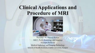
1. Clinical Applications and
Procedure of MRI
Presenter: Dr. Dheeraj Kumar
MRIT, Ph.D. (Radiology and Imaging)
Assistant Professor
Medical Radiology and Imaging Technology
School of Health Sciences, CSJM University, Kanpur
2. Clinical Applications of MRI
• Introduction: MRI, or Magnetic Resonance Imaging, is a versatile
medical imaging technique with a wide range of clinical applications.
• Soft Tissue Imaging: The unique ability of MRI to produce detailed
images of soft tissues, such as the brain, muscles, and organs.
• Non-Invasive Nature: MRI is a non-invasive and safe imaging
modality, making it invaluable for clinical diagnosis.
Friday, 27 October 2023 Clinical Application and Procedure By- Dheeraj Kumar 2
3. Neuroimaging
• Role in Neurology: The central role of MRI in
neuroimaging.
• Brain and Spinal Cord Imaging: MRI is used
to visualize the brain and spinal cord, allowing
for the diagnosis of conditions like tumors,
strokes, and multiple sclerosis.
• Clinical Significance: The critical clinical
applications of neuroimaging, such as guiding
surgical planning and monitoring disease
progression.
Friday, 27 October 2023 Clinical Application and Procedure By- Dheeraj Kumar 3
4. Friday, 27 October 2023 Clinical Application and Procedure By- Dheeraj Kumar 4
5. Cardiac Imaging
• Importance in Cardiology: The
significance of MRI in cardiology.
• Visualizing the Heart: MRI provides
detailed images of the heart's anatomy,
function, and blood flow.
• Clinical Applications: The use of
cardiac MRI in diagnosing heart
diseases, congenital anomalies, and
evaluating cardiac function.
Friday, 27 October 2023 Clinical Application and Procedure By- Dheeraj Kumar 5
6. Friday, 27 October 2023 Clinical Application and Procedure By- Dheeraj Kumar 6
7. Musculoskeletal Imaging
• Orthopedic Applications: The role of MRI in
orthopedics and musculoskeletal imaging.
• Joint and Soft Tissue Assessment: MRI is
used to assess joints, bones, and soft tissues,
making it ideal for diagnosing injuries,
fractures, and joint disorders.
• Clinical Significance: Its importance in the
evaluation of sports injuries and orthopedic
conditions.
Friday, 27 October 2023 Clinical Application and Procedure By- Dheeraj Kumar 7
8. Friday, 27 October 2023 Clinical Application and Procedure By- Dheeraj Kumar 8
9. Abdominal and Pelvic Imaging
• Gastroenterology and Urology: The
applications of MRI in abdominal and
pelvic imaging, particularly in
gastroenterology and urology.
• Diagnostic Capabilities: MRI aids in the
diagnosis of conditions such as tumors,
liver disease, and gynecological issues.
• Clinical Relevance: Its role in guiding
surgery and treatment planning.
Friday, 27 October 2023 Clinical Application and Procedure By- Dheeraj Kumar 9
10. Friday, 27 October 2023 Clinical Application and Procedure By- Dheeraj Kumar 10
11. Breast Imaging
• Breast Health: The importance of MRI
in breast health.
• High-Resolution Imaging: The use of
high-resolution breast MRI in the
detection and diagnosis of breast cancer.
• Applications: Its applications in
screening, staging, and treatment
planning for breast cancer patients.
Friday, 27 October 2023 Clinical Application and Procedure By- Dheeraj Kumar 11
12. Friday, 27 October 2023 Clinical Application and Procedure By- Dheeraj Kumar 12
13. Oncology
• Cancer Diagnosis and Treatment: The
significance of MRI in cancer diagnosis and
treatment.
• Visualizing Tumors: MRI allows for the
visualization of tumors, their size, location,
and characteristics.
• Monitoring Treatment Response: MRI is
used to monitor treatment response and
track disease progression in oncology.
Friday, 27 October 2023 Clinical Application and Procedure By- Dheeraj Kumar 13
14. Pediatric MRI
• Special Considerations for Pediatrics: The unique
considerations when performing MRI on pediatric
patients.
• Motion Control and Sedation: The importance of
motion control techniques and the use of sedation
for younger patients.
• Clinical Applications: The clinical applications of
pediatric MRI in neuroimaging and the diagnosis of
congenital anomalies.
Friday, 27 October 2023 Clinical Application and Procedure By- Dheeraj Kumar 14
15. MRI Procedure Overview
Introduction to the MRI Procedure:
• Start by explaining the purpose of this section, which is to provide a
comprehensive understanding of the MRI procedure, its components, and key
considerations.
• The widespread use of MRI in clinical diagnostics, making it essential for
radiology students to grasp the fundamental steps involved.
Friday, 27 October 2023 Clinical Application and Procedure By- Dheeraj Kumar 15
16. Purpose of the MRI Procedure
• The primary purpose of an MRI is to obtain images of the body's
internal structures, providing valuable information for diagnosis and
treatment planning.
• The versatility of MRI in imaging various anatomical regions,
including the brain, heart, musculoskeletal system, and more.
Friday, 27 October 2023 Clinical Application and Procedure By- Dheeraj Kumar 16
17. Components of an MRI Scanner
• The main components of an MRI scanner, including the strong
magnetic field, gradient coils, radiofrequency coils, and the computer
system.
• These components work together to create detailed images of the
body's internal structures.
Friday, 27 October 2023 Clinical Application and Procedure By- Dheeraj Kumar 17
18. • Patient Preparation:
• Transition to the second part of the MRI procedure overview, which focuses on
patient preparation.
• Stress the importance of patient preparation for a successful and comfortable MRI
experience.
• Informing Patients:
• The process begins with informing the patients about the MRI procedure. This
includes providing detailed information about what to expect during the scan.
• The importance of clear communication in alleviating anxiety and ensuring
cooperation from the patient.
Friday, 27 October 2023 Clinical Application and Procedure By- Dheeraj Kumar 18
19. Friday, 27 October 2023 Clinical Application and Procedure By- Dheeraj Kumar 19
20. • Screening for Contraindications:
• The critical step of screening patients for contraindications. This involves assessing the
patient's medical history to identify potential risks or conditions that may affect the MRI
procedure.
• Common contraindications such as the presence of metallic implants, pregnancy, or severe
claustrophobia.
• Informed Consent:
• Obtaining informed consent from the patient is a vital ethical and legal aspect of patient
preparation.
• Patients must acknowledge their understanding of the procedure and potential risks by signing
a consent form.
Friday, 27 October 2023 Clinical Application and Procedure By- Dheeraj Kumar 20
21. Patient Understanding
• Ensuring the patient fully understands the procedure and is
comfortable with it is paramount to a successful MRI.
• That patients can ask questions and seek clarification at any point
during the preparation process.
Friday, 27 October 2023 Clinical Application and Procedure By- Dheeraj Kumar 21
22. • Patient Comfort:
• Beyond the technical aspects of preparation, ensuring patient comfort is crucial.
• Strategies such as providing blankets, using headphones to listen to music, and offering
distractions to minimize anxiety.
• Preparing for the MRI Suite:
• The process of transitioning the patient to the MRI suite. Patients may be asked to change into
a hospital gown or specific attire, depending on the area being imaged.
• Stress the importance of complying with safety guidelines to avoid bringing metallic objects
into the MRI room.
Friday, 27 October 2023 Clinical Application and Procedure By- Dheeraj Kumar 22
23. • Positioning and Coils:
• The process of positioning the patient on the MRI table and using specialized coils
that help capture images of specific body areas.
• Immobilization devices may be used to reduce motion during the scan.
• Communication with the MRI Technologist:
• The role of the MRI technologist in ensuring patient comfort and safety.
• Encourage patients to communicate any concerns or discomfort with the
technologist, who will be able to address them during the procedure.
Friday, 27 October 2023 Clinical Application and Procedure By- Dheeraj Kumar 23
24. Safety Measures
• Additional safety measures taken, such as ear protection to dampen the
loud noise produced during the scan.
• Stress the importance of patient cooperation in remaining still during
the scan to avoid motion artifacts.
Friday, 27 October 2023 Clinical Application and Procedure By- Dheeraj Kumar 24
25. MRI Safety
• Safety Protocols: The safety protocols and considerations in MRI.
• Metallic Objects and Implants: The significance of checking for metallic
objects and implants that can interact with the strong magnetic field.
• Patient Monitoring: The continuous monitoring of patients during the scan
to ensure their safety and comfort.
Friday, 27 October 2023 Clinical Application and Procedure By- Dheeraj Kumar 25
26. Image Acquisition
Image acquisition is a critical step in the MRI process where raw data is collected and
will later be transformed into diagnostic images.
• Steps Involved:
• Spatial Encoding: MRI acquires spatial information by encoding the location of protons in the
body.
• Signal Reception: The role of specialized coils in detecting and receiving the signals emitted
by protons.
• Data Sampling: The signals are sampled over time to capture the complete image data.
Friday, 27 October 2023 Clinical Application and Procedure By- Dheeraj Kumar 26
27. Magnetic Fields
• Proton Alignment: The alignment of protons within the strong static
magnetic field.
• Precession: Protons precess (rotate) around the magnetic field lines
due to their magnetic moments.
• Frequency of Precession: The frequency of precession is determined
by the strength of the magnetic field, known as the Larmor frequency.
Friday, 27 October 2023 Clinical Application and Procedure By- Dheeraj Kumar 27
28. Radiofrequency Pulses
• Introduction: The role of radiofrequency (RF) pulses in the MRI process.
• Perturbing Proton Alignment: RF pulses are applied at the Larmor
frequency to perturb the alignment of protons.
• T1 and T2 Relaxation: During RF excitation, protons absorb energy and
move from their equilibrium positions, eventually relaxing back, emitting
signals with diagnostic information.
Friday, 27 October 2023 Clinical Application and Procedure By- Dheeraj Kumar 28
29. Gradient Coils
• Spatial Encoding: The role of gradient coils in
creating spatial variations in the magnetic field.
• Gradient Strength and Direction: Gradient coils
vary in strength and direction across the MRI
scanner, allowing precise spatial encoding.
• Spatial Localization: These gradients are essential
for pinpointing the exact location of protons in
different regions of the body.
Friday, 27 October 2023 Clinical Application and Procedure By- Dheeraj Kumar 29
30. Image Reconstruction
• Image Processing: Raw data acquired during the MRI scan is processed.
• Fourier Transform: The Fourier Transform, which is used to convert raw data
into images.
• Image Quality Factors: The factors that affect image quality, including
resolution, signal-to-noise ratio, and contrast.
• Adjustments: The adjustments made during image reconstruction to optimize
image quality.
Friday, 27 October 2023 Clinical Application and Procedure By- Dheeraj Kumar 30
31. Patient Comfort and Communication
• Patient Comfort: The importance of ensuring
patient comfort during the MRI procedure.
• Reducing Anxiety: Strategies to reduce patient
anxiety and claustrophobia, including
communication and the use of music or guided
imagery.
• Effective Communication: The need for effective
communication between the radiologic technologist
and the patient to ensure cooperation and minimal
movement during the scan.
Friday, 27 October 2023 Clinical Application and Procedure By- Dheeraj Kumar 31
32. Conclusion
• Patient-Centered Care: Stress the importance of patient comfort,
safety, and effective communication during the MRI procedure.
• Ongoing Learning: To continue their education and training in MRI
for the benefit of patient care and healthcare advancements.
Friday, 27 October 2023 Clinical Application and Procedure By- Dheeraj Kumar 32
33. References
1.Haacke, E. M., Brown, R. W., Thompson, M. R., & Venkatesan, R. (1999).
Magnetic resonance imaging: Physical principles and sequence design. Wiley-
Liss.
2.Stark, D. D., & Bradley, W. G. (1999). Magnetic resonance imaging. C.V. Mosby.
3.Edelman, R. R., & Hesse link, J. R. (2010). Clinical magnetic resonance imaging.
Saunders.
4.Rofsky, N. M., & De Corato, D. R. (2005). Magnetic resonance imaging in
clinical practice. Informa Healthcare.
Friday, 27 October 2023 Clinical Application and Procedure By- Dheeraj Kumar 33