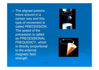
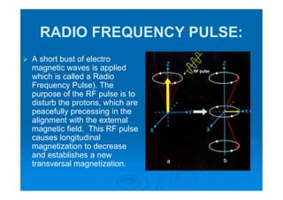
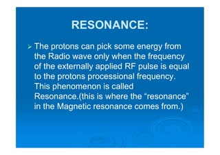
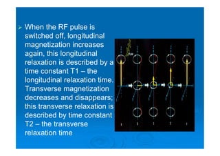
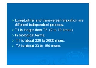
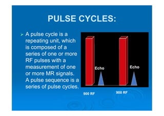
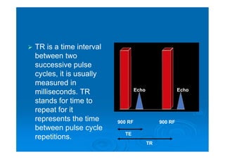
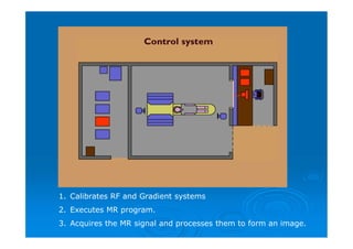
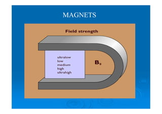
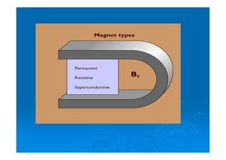
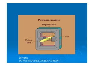
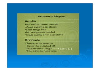
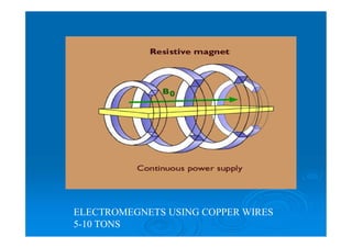
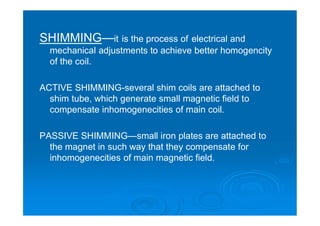
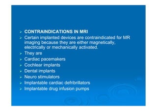
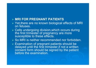
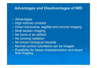
MRI BASICS
PRINCIPLES
 A proton has a spin and
thus the electrical charge
of the proton also moves.
A moving electrical
charge is an electrical
current, and this
accompanied by a
magnetic field. Thus the
proton had its own
magnetic field and it can
be seen as a little bar
magnet.
 When we put the
protons into external
magnetic field – the
protons being little
magnets align
themselves in the
external magnetic
field like a compass
needle in the
magnetic field of earth
 The aligned protons
move around in a
certain way and this
type of movement is
called PRECESSION.
The speed of the
precession is called
as PRECESSIONAL
FREQUENCY, which
is directly proportional
to the external
magnetic field
strength.
RADIO FREQUENCY PULSE:
 A short bust of electro
magnetic waves is applied
which is called a Radio
Frequency Pulse). The
purpose of the RF pulse is to
disturb the protons, which are
peacefully precessing in the
alignment with the external
magnetic field. This RF pulse
causes longitudinal
magnetization to decrease
and establishes a new
transversal magnetization.
RESONANCE:
 The protons can pick some energy from
the Radio wave only when the frequency
of the externally applied RF pulse is equal
to the protons processional frequency.
This phenomenon is called
Resonance.(this is where the “resonance”
in the Magnetic resonance comes from.)
RELAXATION:
 Is the process that occurs after terminating
the RF pulse in which the physical
changes that were caused by the RF pulse
to the state they were in prior to the
application to the pulse.
 When the RF pulse is
switched off, longitudinal
magnetization increases
again, this longitudinal
relaxation is described by a
time constant T1 – the
longitudinal relaxation time.
Transverse magnetization
decreases and disappears;
this transverse relaxation is
described by time constant
T2 – the transverse
relaxation time
 Longitudinal and transversal relaxation are
different independent process.
 T1 is longer than T2. (2 to 10 times).
 In biological terms,
 T1 is about 300 to 2000 msec.
 T2 is about 30 to 150 msec.
PULSE CYCLES:
 A pulse cycle is a
repeating unit, which
is composed of a
series of one or more
RF pulses with a
measurement of one
or more MR signals.
A pulse sequence is a
series of pulse cycles.
900 RF 900 RF
Echo Echo
 TR is a time interval
between two
successive pulse
cycles, it is usually
measured in
milliseconds. TR
stands for time to
repeat for it
represents the time
between pulse cycle
repetitions.
900 RF900 RF
EchoEcho
TE
TR
 TE is the time interval from one pulse (or a
series of pulses in a complicated pulse cycle) to
the measurements of MR signals. It is also
measured in milliseconds. TE stands for time to
echo. The application of RF pulse causes
energy to be input to the patient. The
measurement of the signal constitutes the
reflection or echo of this energy back to us.
 T1 and T2 refers to TISSUE PROPERTIES.
 TR and TE refers to EQUIPMENT
PARAMETERS
T1 & T2 WEIGHTING:
 T1 weighted image is the one in which the
intensity contrast between any two tissues in an
image is mainly due to the T1 relaxation
properties of the tissues.
 For a T1 Weighting: (short TR short TE)
 A T2 Weighted image is the one in which the
intensity contrast between any two tissues in an
image is mainly due to T2 relaxation properties
of the tissues.
 For a T2 weighting : Long TR Long TE
 A proton density image is a one which the
intensity contrast between any two tissues in an
image is mainly due to the density of the protons
of the tissues.
 For a proton density weighting:Long TR short TE
Pulse Sequences
 Pulse sequences enable us to control the
way in which the system applies pulses
and gradients. The different pulse
sequences are available and each is
designed for a specific purpose.
 Types
 Spin Echo
 Gradient Echo
 Inversion Recovery
 Spin Echo:
 It uses a 90degree
excitation pulse
followed by one or
more 180degree
rephasing pulses to
generate a spin echo.
 Uses
 Spin echo pulse sequences are the goal
standard for most imaging.
 T1 W images are useful for demonstrating
anatomical details.
 T2 W images are useful for pathological
details.
 Gradient Echo:
 It uses short flip angle therefore the scan
time is reduced, for rephrasing, gradient is
used instead of 1800 RF pulse.
Uses:
Very sensitive to flow study
Can produce angiographic type
images
 Inversion Recovery
 Begins with 1800 inverting
pulse followed by 900
excitation pulse to generate a
echo. The time gap between
1800 pulse and 900 pulse is
called time of inversion or TI .
Uses
 It produces heavily T1
weighted image to
demonstrate anatomical
details.
 Used for FAT suppression
MRI INTRUMENTATION
1. Calibrates RF and Gradient systems
2. Executes MR program.
3. Acquires the MR signal and processes them to form an image.
MAGNETS
DO NOT REQUIRE ELECTRIC CURRENT
20 TONS
0.05 TO 0.3 T
ELECTROMEGNETS USING COPPER WIRES
5-10 TONS
•NIOBIUM-TITANIUM (Nb-Ti) alloys.
•Powerful current is given initially and once high current
is flowing power supply is disconnected because the
resistance is virtually zero.
-267
degree
QUENCH—Loss of super conductivity of the
magnetic coil and resulting loss of magnetic
field is called quench.
Quenching is initiated by minute movements of
coil and this will release frictional energy to
helium.
To reduce quenching the supercon coil is
wrapped in copper for thermal insulation.
Field strength upto 15mt/ m
GRADIENT COILS
RF COILS
TYPES OF RF COILS
VOLUME COIL
SURFACE COILS
ACCORNDING TO THEIR PHYSICAL FORMS
SHIMMING—it is the process of electrical and
mechanical adjustments to achieve better homogencity
of the coil.
ACTIVE SHIMMING-several shim coils are attached to
shim tube, which generate small magnetic field to
compensate inhomogenecities of main coil.
PASSIVE SHIMMING—small iron plates are attached to
the magnet in such way that they compensate for
inhomogenecities of main magnetic field.
SAFETY ASPECTS IN MRI
 WHY SAFETY IN MRI?
 MRI has a very powerful magnet
 It is so strong that it will pull heavy items
such as oxygen cylinders, beds, stretchers
etc, into the scanner.
 TYPES OF SAFETY
 Magnetic safety
 RF safety
 MAGNETIC SAFETY
 Patients and personnel entering the scan room
must be screened.
 All metallic objects should be removed before
entering the scan room.
 Check objects such as wigs, hairpins, false teeth,
zips, buttons, wallets, make-up, Floppy disks,
Tapes , Credit cards etc. are all removed.
 Other magnetic objects such as
stretchers,wheelchairs,IV stands, fire
extinguisher,vacuum cleaner etc must not be
allowed to enter into the scan room.
 CONTRAINDICATIONS IN MRI
 Certain implanted devices are contraindicated for MR
imaging because they are either magnetically,
electrically or mechanically activated.
 They are
 Cardiac pacemakers
 Cochlear implants
 Dental implants
 Neuro stimulators
 Implantable cardiac defribrillators
 Implantable drug infusion pumps
 MRI FOR PREGNANT PATIENTS
 Yet,there are no known biological effects of MRI
on fetuses.
 Cells undergoing division,which occurs during
the first trimester of pregnancy are more
susceptible to these effects.
 So MRI is neither recommended nor forbidden.
 Examination of pregnant patients should be
delayed until the first trimester,if not a written
consent form should be signed by the patient
before the examination.
Advantages and Disadvantages of MRI
 Advantages
 High intrinsic contrast
 Direct transverse, sagittal and coronal imaging
 Multi section imaging
 No bone or air artifact
 No ionizing radiation
 No known biological hazards
 Normal control volunteers can be imaged
 Possibility for tissue characterization and blood
flow imaging
Disadvantages
 Long imaging time
 Many protocol options
 Correct choice of machine parameters essential
 Poor bone and calcium details
 Reduced facial resolution especially with body coil.
 Claustrophobia (2-5% cases studied)
 Difficulty to manage and monitor patients who critically
ill.
 ? Long term hazard
 High cost.
THANK YOU

Mri basics

  • 1.
  • 2.
    PRINCIPLES  A protonhas a spin and thus the electrical charge of the proton also moves. A moving electrical charge is an electrical current, and this accompanied by a magnetic field. Thus the proton had its own magnetic field and it can be seen as a little bar magnet.
  • 3.
     When weput the protons into external magnetic field – the protons being little magnets align themselves in the external magnetic field like a compass needle in the magnetic field of earth
  • 4.
     The alignedprotons move around in a certain way and this type of movement is called PRECESSION. The speed of the precession is called as PRECESSIONAL FREQUENCY, which is directly proportional to the external magnetic field strength.
  • 5.
    RADIO FREQUENCY PULSE: A short bust of electro magnetic waves is applied which is called a Radio Frequency Pulse). The purpose of the RF pulse is to disturb the protons, which are peacefully precessing in the alignment with the external magnetic field. This RF pulse causes longitudinal magnetization to decrease and establishes a new transversal magnetization.
  • 6.
    RESONANCE:  The protonscan pick some energy from the Radio wave only when the frequency of the externally applied RF pulse is equal to the protons processional frequency. This phenomenon is called Resonance.(this is where the “resonance” in the Magnetic resonance comes from.)
  • 7.
    RELAXATION:  Is theprocess that occurs after terminating the RF pulse in which the physical changes that were caused by the RF pulse to the state they were in prior to the application to the pulse.
  • 8.
     When theRF pulse is switched off, longitudinal magnetization increases again, this longitudinal relaxation is described by a time constant T1 – the longitudinal relaxation time. Transverse magnetization decreases and disappears; this transverse relaxation is described by time constant T2 – the transverse relaxation time
  • 9.
     Longitudinal andtransversal relaxation are different independent process.  T1 is longer than T2. (2 to 10 times).  In biological terms,  T1 is about 300 to 2000 msec.  T2 is about 30 to 150 msec.
  • 10.
    PULSE CYCLES:  Apulse cycle is a repeating unit, which is composed of a series of one or more RF pulses with a measurement of one or more MR signals. A pulse sequence is a series of pulse cycles. 900 RF 900 RF Echo Echo
  • 11.
     TR isa time interval between two successive pulse cycles, it is usually measured in milliseconds. TR stands for time to repeat for it represents the time between pulse cycle repetitions. 900 RF900 RF EchoEcho TE TR
  • 12.
     TE isthe time interval from one pulse (or a series of pulses in a complicated pulse cycle) to the measurements of MR signals. It is also measured in milliseconds. TE stands for time to echo. The application of RF pulse causes energy to be input to the patient. The measurement of the signal constitutes the reflection or echo of this energy back to us.  T1 and T2 refers to TISSUE PROPERTIES.  TR and TE refers to EQUIPMENT PARAMETERS
  • 13.
    T1 & T2WEIGHTING:  T1 weighted image is the one in which the intensity contrast between any two tissues in an image is mainly due to the T1 relaxation properties of the tissues.  For a T1 Weighting: (short TR short TE)  A T2 Weighted image is the one in which the intensity contrast between any two tissues in an image is mainly due to T2 relaxation properties of the tissues.  For a T2 weighting : Long TR Long TE  A proton density image is a one which the intensity contrast between any two tissues in an image is mainly due to the density of the protons of the tissues.  For a proton density weighting:Long TR short TE
  • 14.
    Pulse Sequences  Pulsesequences enable us to control the way in which the system applies pulses and gradients. The different pulse sequences are available and each is designed for a specific purpose.  Types  Spin Echo  Gradient Echo  Inversion Recovery
  • 15.
     Spin Echo: It uses a 90degree excitation pulse followed by one or more 180degree rephasing pulses to generate a spin echo.
  • 16.
     Uses  Spinecho pulse sequences are the goal standard for most imaging.  T1 W images are useful for demonstrating anatomical details.  T2 W images are useful for pathological details.
  • 17.
     Gradient Echo: It uses short flip angle therefore the scan time is reduced, for rephrasing, gradient is used instead of 1800 RF pulse. Uses: Very sensitive to flow study Can produce angiographic type images
  • 18.
     Inversion Recovery Begins with 1800 inverting pulse followed by 900 excitation pulse to generate a echo. The time gap between 1800 pulse and 900 pulse is called time of inversion or TI . Uses  It produces heavily T1 weighted image to demonstrate anatomical details.  Used for FAT suppression
  • 19.
  • 25.
    1. Calibrates RFand Gradient systems 2. Executes MR program. 3. Acquires the MR signal and processes them to form an image.
  • 29.
  • 31.
    DO NOT REQUIREELECTRIC CURRENT 20 TONS
  • 32.
  • 33.
  • 35.
    •NIOBIUM-TITANIUM (Nb-Ti) alloys. •Powerfulcurrent is given initially and once high current is flowing power supply is disconnected because the resistance is virtually zero. -267 degree
  • 37.
    QUENCH—Loss of superconductivity of the magnetic coil and resulting loss of magnetic field is called quench. Quenching is initiated by minute movements of coil and this will release frictional energy to helium. To reduce quenching the supercon coil is wrapped in copper for thermal insulation.
  • 38.
    Field strength upto15mt/ m GRADIENT COILS
  • 39.
  • 40.
  • 41.
  • 42.
    SURFACE COILS ACCORNDING TOTHEIR PHYSICAL FORMS
  • 43.
    SHIMMING—it is theprocess of electrical and mechanical adjustments to achieve better homogencity of the coil. ACTIVE SHIMMING-several shim coils are attached to shim tube, which generate small magnetic field to compensate inhomogenecities of main coil. PASSIVE SHIMMING—small iron plates are attached to the magnet in such way that they compensate for inhomogenecities of main magnetic field.
  • 44.
    SAFETY ASPECTS INMRI  WHY SAFETY IN MRI?  MRI has a very powerful magnet  It is so strong that it will pull heavy items such as oxygen cylinders, beds, stretchers etc, into the scanner.  TYPES OF SAFETY  Magnetic safety  RF safety
  • 45.
     MAGNETIC SAFETY Patients and personnel entering the scan room must be screened.  All metallic objects should be removed before entering the scan room.  Check objects such as wigs, hairpins, false teeth, zips, buttons, wallets, make-up, Floppy disks, Tapes , Credit cards etc. are all removed.  Other magnetic objects such as stretchers,wheelchairs,IV stands, fire extinguisher,vacuum cleaner etc must not be allowed to enter into the scan room.
  • 46.
     CONTRAINDICATIONS INMRI  Certain implanted devices are contraindicated for MR imaging because they are either magnetically, electrically or mechanically activated.  They are  Cardiac pacemakers  Cochlear implants  Dental implants  Neuro stimulators  Implantable cardiac defribrillators  Implantable drug infusion pumps
  • 47.
     MRI FORPREGNANT PATIENTS  Yet,there are no known biological effects of MRI on fetuses.  Cells undergoing division,which occurs during the first trimester of pregnancy are more susceptible to these effects.  So MRI is neither recommended nor forbidden.  Examination of pregnant patients should be delayed until the first trimester,if not a written consent form should be signed by the patient before the examination.
  • 48.
    Advantages and Disadvantagesof MRI  Advantages  High intrinsic contrast  Direct transverse, sagittal and coronal imaging  Multi section imaging  No bone or air artifact  No ionizing radiation  No known biological hazards  Normal control volunteers can be imaged  Possibility for tissue characterization and blood flow imaging
  • 49.
    Disadvantages  Long imagingtime  Many protocol options  Correct choice of machine parameters essential  Poor bone and calcium details  Reduced facial resolution especially with body coil.  Claustrophobia (2-5% cases studied)  Difficulty to manage and monitor patients who critically ill.  ? Long term hazard  High cost.
  • 50.