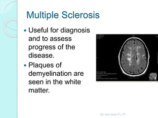
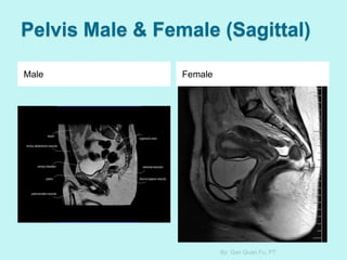
The document discusses the importance of physiotherapists understanding basic MRI interpretation. It notes that some patients come for treatment without their MRI interpretation sheet, leaving blank images for the physiotherapist to interpret. Having a basic understanding of MRI principles, machine components, image types and interpretation helps physiotherapists confirm assessments when the interpretation sheet is missing. The document aims to provide this foundational MRI knowledge for physiotherapists.


![Strength of Magnetic Field Used
in MRI
Measured in Tesla (T)
[Developed by Nikola
Tesla]
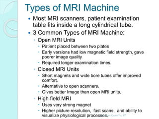
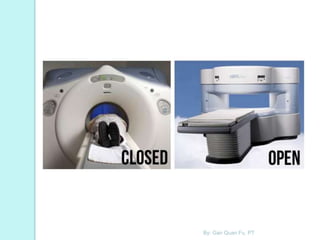
Open MRI units
◦ 0.1T to 0.3 T
Closed MRI unit
◦ 0.5 T to 0.6 T
High field MRI
◦ 1.0 T to 3.0 T or higher
By: Gan Quan Fu, PT](https://image.slidesharecdn.com/basicunderstandingonmagneticresonanceimagingmri-141231045409-conversion-gate01-150514070319-lva1-app6891/85/Basicunderstandingonmagneticresonanceimagingmri-141231045409-conversion-gate01-17-320.jpg)