Download to read offline


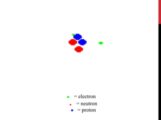
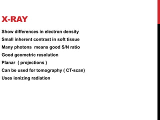
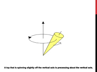
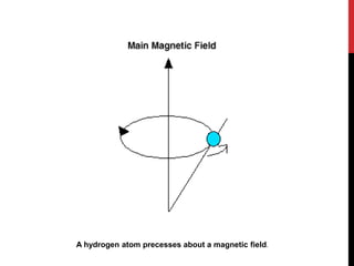

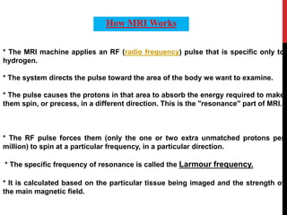
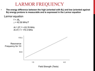

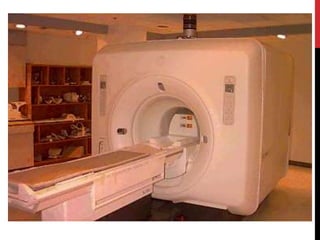
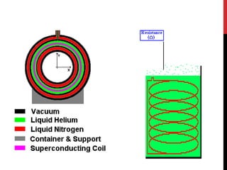


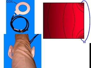
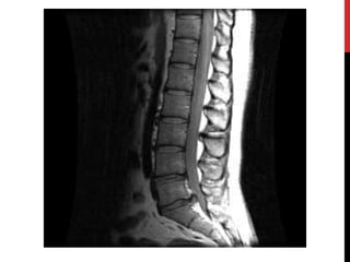

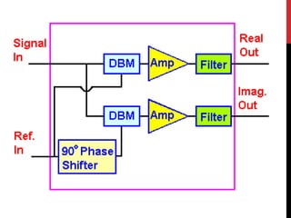


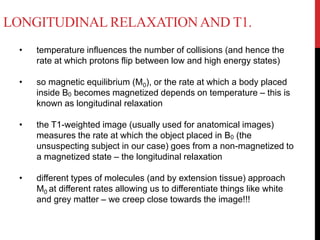
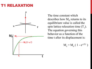

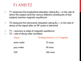
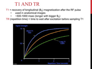


Magnetic resonance imaging (MRI) is a medical imaging technique that uses strong magnetic fields and radio waves to produce detailed images of the inside of the body. MRI is based on nuclear magnetic resonance, which uses magnetic fields to detect atomic nuclei within tissues from different angles in order to form cross-sectional images of internal structures. The document discusses the physics principles behind MRI, including how hydrogen protons are aligned by magnetic fields and how their signal can be localized to different regions of the body. It also covers differences between T1-weighted and T2-weighted MRI sequences, common artifacts that can appear on images, and advantages and risks of MRI compared to other imaging techniques like X-ray and CT scans.