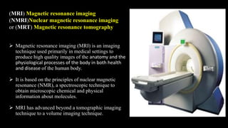
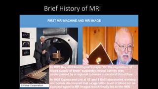
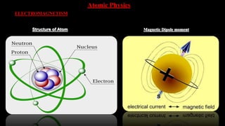
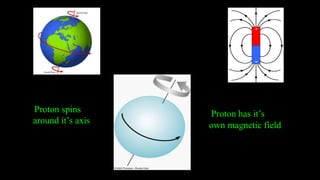
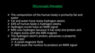
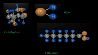
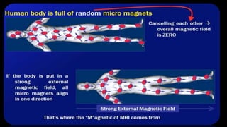
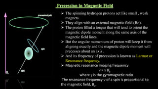
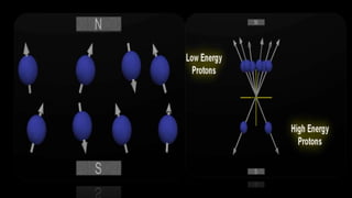
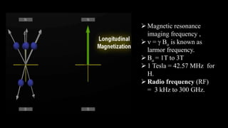
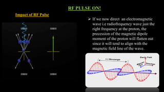
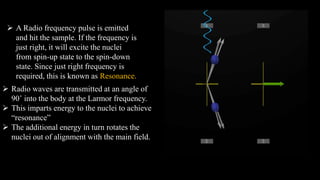
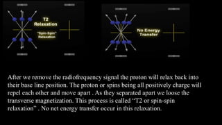
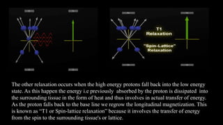
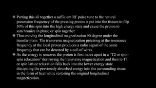
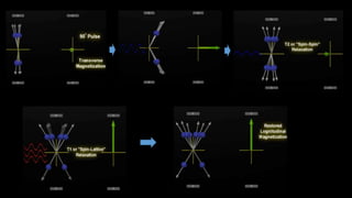
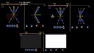
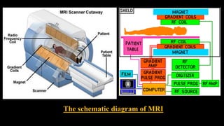
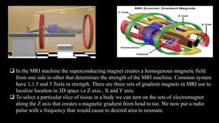
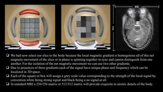
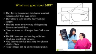
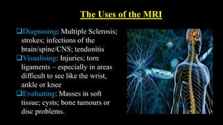
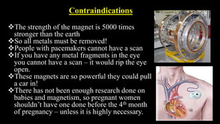
MRI uses strong magnetic fields and radio waves to produce detailed images of the inside of the body. It has advanced beyond a tomographic imaging technique to a volume imaging technique. The first MRI experiment was conducted in 1946. Important developments included Raymond Damadian constructing the first MRI scanner in 1977 and Peter Mansfield developing echo planar imaging. MRI works by aligning hydrogen protons in water and fat using magnetism and radio waves, and using magnetic field gradients to spatially encode the signal from tissues to form images. It is useful for diagnosing conditions, injuries and evaluating masses without using ionizing radiation.
this power-point slide presentation includes lots of information like how MRI coil works. what is shimming, magnet, fringe, and design of mri coil and also magnet. this will help a lot for radiologist and technician radiographers.. thanks.
Blood supply of face /certified fixed orthodontic courses by Indian dental a...Indian dental academy
Welcome to Indian Dental Academy
The Indian Dental Academy is the Leader in continuing dental education , training dentists in all aspects of dentistry and offering a wide range of dental certified courses in different formats.
Indian dental academy has a unique training program & curriculum that provides students with exceptional clinical skills and enabling them to return to their office with high level confidence and start treating patients
State of the art comprehensive training-Faculty of world wide repute &Very affordable.
MRI uses a strong magnetic field and radio waves to create detailed images of the organs and tissues within the body.
Developed by the Lauterbur in 1972 at Stony brook in New York.
MRI does not involve radiation
MRI contrasting agent is less likely to produce an allergic reaction that may occur when iodine-based substances are used for x-rays and CT scans
MRI gives extremely clear, detailed images of soft-tissue structures that other imaging techniques cannot achieve
The MRI machine cannot just simply “see the hydrogen nuclei which lie “hidden” in the water molecules distributed in the patient.
It needs to do ‘something’ to the hydrogen nuclei to detect their presence.
Here I discussed about the concept,types, types of images obtained, the advantages and disadvantages of MRI shortly...anyone who wants to know about MRI just go through it. I just prepared it in very simple language for the convenience of the readers all over the world. Thank you.
Nuclear magnetic resonance spectroscopy, most commonly known as NMR spectroscopy or magnetic resonance spectroscopy, is a spectroscopic technique to observe local magnetic fields around atomic nuclei.
CONCEPT
HISTORY OF XENOTRANSPLANTATION
IMPORTANCE OF XENOTRANSPLANTATION
CHOOSING OF DONOR SPECIES
XENOTRANSPLANTATION REJECTION
GUIDELINES ON XENO-TRANSPLANTATION BY ICMR
RECENT RESEARCH ON XENOTRANSPLANTATION
Title: Sense of Smell
Presenter: Dr. Faiza, Assistant Professor of Physiology
Qualifications:
MBBS (Best Graduate, AIMC Lahore)
FCPS Physiology
ICMT, CHPE, DHPE (STMU)
MPH (GC University, Faisalabad)
MBA (Virtual University of Pakistan)
Learning Objectives:
Describe the primary categories of smells and the concept of odor blindness.
Explain the structure and location of the olfactory membrane and mucosa, including the types and roles of cells involved in olfaction.
Describe the pathway and mechanisms of olfactory signal transmission from the olfactory receptors to the brain.
Illustrate the biochemical cascade triggered by odorant binding to olfactory receptors, including the role of G-proteins and second messengers in generating an action potential.
Identify different types of olfactory disorders such as anosmia, hyposmia, hyperosmia, and dysosmia, including their potential causes.
Key Topics:
Olfactory Genes:
3% of the human genome accounts for olfactory genes.
400 genes for odorant receptors.
Olfactory Membrane:
Located in the superior part of the nasal cavity.
Medially: Folds downward along the superior septum.
Laterally: Folds over the superior turbinate and upper surface of the middle turbinate.
Total surface area: 5-10 square centimeters.
Olfactory Mucosa:
Olfactory Cells: Bipolar nerve cells derived from the CNS (100 million), with 4-25 olfactory cilia per cell.
Sustentacular Cells: Produce mucus and maintain ionic and molecular environment.
Basal Cells: Replace worn-out olfactory cells with an average lifespan of 1-2 months.
Bowman’s Gland: Secretes mucus.
Stimulation of Olfactory Cells:
Odorant dissolves in mucus and attaches to receptors on olfactory cilia.
Involves a cascade effect through G-proteins and second messengers, leading to depolarization and action potential generation in the olfactory nerve.
Quality of a Good Odorant:
Small (3-20 Carbon atoms), volatile, water-soluble, and lipid-soluble.
Facilitated by odorant-binding proteins in mucus.
Membrane Potential and Action Potential:
Resting membrane potential: -55mV.
Action potential frequency in the olfactory nerve increases with odorant strength.
Adaptation Towards the Sense of Smell:
Rapid adaptation within the first second, with further slow adaptation.
Psychological adaptation greater than receptor adaptation, involving feedback inhibition from the central nervous system.
Primary Sensations of Smell:
Camphoraceous, Musky, Floral, Pepperminty, Ethereal, Pungent, Putrid.
Odor Detection Threshold:
Examples: Hydrogen sulfide (0.0005 ppm), Methyl-mercaptan (0.002 ppm).
Some toxic substances are odorless at lethal concentrations.
Characteristics of Smell:
Odor blindness for single substances due to lack of appropriate receptor protein.
Behavioral and emotional influences of smell.
Transmission of Olfactory Signals:
From olfactory cells to glomeruli in the olfactory bulb, involving lateral inhibition.
Primitive, less old, and new olfactory systems with different path
micro teaching on communication m.sc nursing.pdfAnurag Sharma
Microteaching is a unique model of practice teaching. It is a viable instrument for the. desired change in the teaching behavior or the behavior potential which, in specified types of real. classroom situations, tends to facilitate the achievement of specified types of objectives.
Knee anatomy and clinical tests 2024.pdfvimalpl1234
This includes all relevant anatomy and clinical tests compiled from standard textbooks, Campbell,netter etc..It is comprehensive and best suited for orthopaedicians and orthopaedic residents.
Title: Sense of Taste
Presenter: Dr. Faiza, Assistant Professor of Physiology
Qualifications:
MBBS (Best Graduate, AIMC Lahore)
FCPS Physiology
ICMT, CHPE, DHPE (STMU)
MPH (GC University, Faisalabad)
MBA (Virtual University of Pakistan)
Learning Objectives:
Describe the structure and function of taste buds.
Describe the relationship between the taste threshold and taste index of common substances.
Explain the chemical basis and signal transduction of taste perception for each type of primary taste sensation.
Recognize different abnormalities of taste perception and their causes.
Key Topics:
Significance of Taste Sensation:
Differentiation between pleasant and harmful food
Influence on behavior
Selection of food based on metabolic needs
Receptors of Taste:
Taste buds on the tongue
Influence of sense of smell, texture of food, and pain stimulation (e.g., by pepper)
Primary and Secondary Taste Sensations:
Primary taste sensations: Sweet, Sour, Salty, Bitter, Umami
Chemical basis and signal transduction mechanisms for each taste
Taste Threshold and Index:
Taste threshold values for Sweet (sucrose), Salty (NaCl), Sour (HCl), and Bitter (Quinine)
Taste index relationship: Inversely proportional to taste threshold
Taste Blindness:
Inability to taste certain substances, particularly thiourea compounds
Example: Phenylthiocarbamide
Structure and Function of Taste Buds:
Composition: Epithelial cells, Sustentacular/Supporting cells, Taste cells, Basal cells
Features: Taste pores, Taste hairs/microvilli, and Taste nerve fibers
Location of Taste Buds:
Found in papillae of the tongue (Fungiform, Circumvallate, Foliate)
Also present on the palate, tonsillar pillars, epiglottis, and proximal esophagus
Mechanism of Taste Stimulation:
Interaction of taste substances with receptors on microvilli
Signal transduction pathways for Umami, Sweet, Bitter, Sour, and Salty tastes
Taste Sensitivity and Adaptation:
Decrease in sensitivity with age
Rapid adaptation of taste sensation
Role of Saliva in Taste:
Dissolution of tastants to reach receptors
Washing away the stimulus
Taste Preferences and Aversions:
Mechanisms behind taste preference and aversion
Influence of receptors and neural pathways
Impact of Sensory Nerve Damage:
Degeneration of taste buds if the sensory nerve fiber is cut
Abnormalities of Taste Detection:
Conditions: Ageusia, Hypogeusia, Dysgeusia (parageusia)
Causes: Nerve damage, neurological disorders, infections, poor oral hygiene, adverse drug effects, deficiencies, aging, tobacco use, altered neurotransmitter levels
Neurotransmitters and Taste Threshold:
Effects of serotonin (5-HT) and norepinephrine (NE) on taste sensitivity
Supertasters:
25% of the population with heightened sensitivity to taste, especially bitterness
Increased number of fungiform papillae
Lung Cancer: Artificial Intelligence, Synergetics, Complex System Analysis, S...Oleg Kshivets
RESULTS: Overall life span (LS) was 2252.1±1742.5 days and cumulative 5-year survival (5YS) reached 73.2%, 10 years – 64.8%, 20 years – 42.5%. 513 LCP lived more than 5 years (LS=3124.6±1525.6 days), 148 LCP – more than 10 years (LS=5054.4±1504.1 days).199 LCP died because of LC (LS=562.7±374.5 days). 5YS of LCP after bi/lobectomies was significantly superior in comparison with LCP after pneumonectomies (78.1% vs.63.7%, P=0.00001 by log-rank test). AT significantly improved 5YS (66.3% vs. 34.8%) (P=0.00000 by log-rank test) only for LCP with N1-2. Cox modeling displayed that 5YS of LCP significantly depended on: phase transition (PT) early-invasive LC in terms of synergetics, PT N0—N12, cell ratio factors (ratio between cancer cells- CC and blood cells subpopulations), G1-3, histology, glucose, AT, blood cell circuit, prothrombin index, heparin tolerance, recalcification time (P=0.000-0.038). Neural networks, genetic algorithm selection and bootstrap simulation revealed relationships between 5YS and PT early-invasive LC (rank=1), PT N0—N12 (rank=2), thrombocytes/CC (3), erythrocytes/CC (4), eosinophils/CC (5), healthy cells/CC (6), lymphocytes/CC (7), segmented neutrophils/CC (8), stick neutrophils/CC (9), monocytes/CC (10); leucocytes/CC (11). Correct prediction of 5YS was 100% by neural networks computing (area under ROC curve=1.0; error=0.0).
CONCLUSIONS: 5YS of LCP after radical procedures significantly depended on: 1) PT early-invasive cancer; 2) PT N0--N12; 3) cell ratio factors; 4) blood cell circuit; 5) biochemical factors; 6) hemostasis system; 7) AT; 8) LC characteristics; 9) LC cell dynamics; 10) surgery type: lobectomy/pneumonectomy; 11) anthropometric data. Optimal diagnosis and treatment strategies for LC are: 1) screening and early detection of LC; 2) availability of experienced thoracic surgeons because of complexity of radical procedures; 3) aggressive en block surgery and adequate lymph node dissection for completeness; 4) precise prediction; 5) adjuvant chemoimmunoradiotherapy for LCP with unfavorable prognosis.
MANAGEMENT OF ATRIOVENTRICULAR CONDUCTION BLOCK.pdfJim Jacob Roy
Cardiac conduction defects can occur due to various causes.
Atrioventricular conduction blocks ( AV blocks ) are classified into 3 types.
This document describes the acute management of AV block.
Recomendações da OMS sobre cuidados maternos e neonatais para uma experiência pós-natal positiva.
Em consonância com os ODS – Objetivos do Desenvolvimento Sustentável e a Estratégia Global para a Saúde das Mulheres, Crianças e Adolescentes, e aplicando uma abordagem baseada nos direitos humanos, os esforços de cuidados pós-natais devem expandir-se para além da cobertura e da simples sobrevivência, de modo a incluir cuidados de qualidade.
Estas diretrizes visam melhorar a qualidade dos cuidados pós-natais essenciais e de rotina prestados às mulheres e aos recém-nascidos, com o objetivo final de melhorar a saúde e o bem-estar materno e neonatal.
Uma “experiência pós-natal positiva” é um resultado importante para todas as mulheres que dão à luz e para os seus recém-nascidos, estabelecendo as bases para a melhoria da saúde e do bem-estar a curto e longo prazo. Uma experiência pós-natal positiva é definida como aquela em que as mulheres, pessoas que gestam, os recém-nascidos, os casais, os pais, os cuidadores e as famílias recebem informação consistente, garantia e apoio de profissionais de saúde motivados; e onde um sistema de saúde flexível e com recursos reconheça as necessidades das mulheres e dos bebês e respeite o seu contexto cultural.
Estas diretrizes consolidadas apresentam algumas recomendações novas e já bem fundamentadas sobre cuidados pós-natais de rotina para mulheres e neonatos que recebem cuidados no pós-parto em unidades de saúde ou na comunidade, independentemente dos recursos disponíveis.
É fornecido um conjunto abrangente de recomendações para cuidados durante o período puerperal, com ênfase nos cuidados essenciais que todas as mulheres e recém-nascidos devem receber, e com a devida atenção à qualidade dos cuidados; isto é, a entrega e a experiência do cuidado recebido. Estas diretrizes atualizam e ampliam as recomendações da OMS de 2014 sobre cuidados pós-natais da mãe e do recém-nascido e complementam as atuais diretrizes da OMS sobre a gestão de complicações pós-natais.
O estabelecimento da amamentação e o manejo das principais intercorrências é contemplada.
Recomendamos muito.
Vamos discutir essas recomendações no nosso curso de pós-graduação em Aleitamento no Instituto Ciclos.
Esta publicação só está disponível em inglês até o momento.
Prof. Marcus Renato de Carvalho
www.agostodourado.com
TEST BANK for Operations Management, 14th Edition by William J. Stevenson, Ve...kevinkariuki227
TEST BANK for Operations Management, 14th Edition by William J. Stevenson, Verified Chapters 1 - 19, Complete Newest Version.pdf
These simplified slides by Dr. Sidra Arshad present an overview of the non-respiratory functions of the respiratory tract.
Learning objectives:
1. Enlist the non-respiratory functions of the respiratory tract
2. Briefly explain how these functions are carried out
3. Discuss the significance of dead space
4. Differentiate between minute ventilation and alveolar ventilation
5. Describe the cough and sneeze reflexes
Study Resources:
1. Chapter 39, Guyton and Hall Textbook of Medical Physiology, 14th edition
2. Chapter 34, Ganong’s Review of Medical Physiology, 26th edition
3. Chapter 17, Human Physiology by Lauralee Sherwood, 9th edition
4. Non-respiratory functions of the lungs https://academic.oup.com/bjaed/article/13/3/98/278874
Ozempic: Preoperative Management of Patients on GLP-1 Receptor Agonists Saeid Safari
Preoperative Management of Patients on GLP-1 Receptor Agonists like Ozempic and Semiglutide
ASA GUIDELINE
NYSORA Guideline
2 Case Reports of Gastric Ultrasound
Flu Vaccine Alert in Bangalore Karnatakaaddon Scans
As flu season approaches, health officials in Bangalore, Karnataka, are urging residents to get their flu vaccinations. The seasonal flu, while common, can lead to severe health complications, particularly for vulnerable populations such as young children, the elderly, and those with underlying health conditions.
Dr. Vidisha Kumari, a leading epidemiologist in Bangalore, emphasizes the importance of getting vaccinated. "The flu vaccine is our best defense against the influenza virus. It not only protects individuals but also helps prevent the spread of the virus in our communities," he says.
This year, the flu season is expected to coincide with a potential increase in other respiratory illnesses. The Karnataka Health Department has launched an awareness campaign highlighting the significance of flu vaccinations. They have set up multiple vaccination centers across Bangalore, making it convenient for residents to receive their shots.
To encourage widespread vaccination, the government is also collaborating with local schools, workplaces, and community centers to facilitate vaccination drives. Special attention is being given to ensuring that the vaccine is accessible to all, including marginalized communities who may have limited access to healthcare.
Residents are reminded that the flu vaccine is safe and effective. Common side effects are mild and may include soreness at the injection site, mild fever, or muscle aches. These side effects are generally short-lived and far less severe than the flu itself.
Healthcare providers are also stressing the importance of continuing COVID-19 precautions. Wearing masks, practicing good hand hygiene, and maintaining social distancing are still crucial, especially in crowded places.
Protect yourself and your loved ones by getting vaccinated. Together, we can help keep Bangalore healthy and safe this flu season. For more information on vaccination centers and schedules, residents can visit the Karnataka Health Department’s official website or follow their social media pages.
Stay informed, stay safe, and get your flu shot today!
How STIs Influence the Development of Pelvic Inflammatory Disease.pptx
Basic of mri
1. Presented by : ACHYUT BORA and NEELOTPAL SHARMA
GAUHATI UNIVERSITY, ASSAM, INDIA
2. (MRI) Magnetic resonance imaging
(NMRI)Nuclear magnetic resonance imaging
or (MRT) Magnetic resonance tomography
Magnetic resonance imaging (MRI) is an imaging
technique used primarily in medical settings to
produce high quality images of the anatomy and the
physiological processes of the body in both health
and disease of the human body.
It is based on the principles of nuclear magnetic
resonance (NMR), a spectroscopic technique to
obtain microscopic chemical and physical
information about molecules.
MRI has advanced beyond a tomographic imaging
technique to a volume imaging technique.
3. Brief History of MRI
Felix Bloch Edward PurcellJoseph Fourier Joseph Larmor
Raymond Damadian C Lauterbur Peter Mansfield Seiji OgawaRichard Ernst
The first Nuclear magnetic resonance
experiment was conducted
independently by two scientist in 1946.
Felix Bloch working at Stanford
University
Edward Purcell working at Harvard
University
Bloch and Purcell were awarded Nobel
Prize for Physics in1952
Joseph Fourier
1768- 1830
Joseph Larmor
1857-1942
Published his
collected papers on
electromagnetism
in 1900 in a famous
book entitled
“Aether and Matter”
In 1970 Raymond Damadian found
that it is possible to characterize
different body tissues using NMR
Technology
In 1977 he completed the
construction of the first MRI scanner
In 1978 he founded the FONAR
corporation which manufactured the
first commercial MRI
In 1973 Paul C Lauterbur discovered the
possibility to create a two dimensional
picture by introducing gradients in the
magnetic field. He used the back
projection technique to reconstruct the
image. He termed his new imaging
technique zeumatography. He shared
the 2003 Nobel prize for medicine with
Peter Mansfield
Peter Mansfield
developed the technique
of detecting the emitted
signals rapidly
mathematically analyzing
them and turning them
into an image.
He evolved a very fast
imaging technique known
as Echo Planar Imaging
In 1975 Richard Ernst
introduced 2D NMR
using phase and
frequency encoding
and the Fourier
transform instead of
Lauterbur’s back
projection technique
In 1992 Functional
MRI was developed
by Seiji Ogawa.
FIRST MRI MACHINE AND MRI IMAGE
In 1890 Roy and Sherrington’s paper ‘On the regulation of
blood supply of brain’ suggested neural activity was
accompanied by a regional increase in cerebral blood flow.
In 1992 Ogawa and Lee at AT and T Bell laboratories working
on rodents discovered that oxygenation level of blood act as
contrast agent in MR images which finally led to the fMRI
6. Microscopic Principles
The composition of the human body is primarily fat and
water
Fat and water have many hydrogen atoms
63% of human body is hydrogen atoms
Hydrogen nuclei have an NMR signal
MRI uses hydrogen because it has only one proton and
it aligns easily with the MRI magnet.
The hydrogen atom’s proton, possesses a property
called spin
A small magnetic field
Will cause the nucleus to produce an NMR signal
9. Precession in Magnetic Field
The spinning hydrogen protons act like small , weak
magnets.
They align with an external magnetic field (Bø).
The proton filled a torque that will tend to orient the
magnetic dipole moment along the same axis of the
magnetic field lines.
But the angular momentum of proton will keep it from
aligning exactly and the magnetic dipole moment will
precesses about an axis .
And its frequency of precession is known as Larmor or
Resonance frequency.
Magnetic resonance imaging frequency
n = g Bo
where g is the gyromagnetic ratio
The resonance frequency n of a spin is proportional to
the magnetic field, Bo.
10.
11. Magnetic resonance
imaging frequency ,
n = g Bo is known as
larmor frequency.
Bo = 1T to 3T
1 Tesla = 42.57 MHz for
H.
Radio frequency (RF)
= 3 kHz to 300 GHz.
12. RF PULSE ON!
Impact of RF Pulse
If we now direct an electromagnetic
wave i.e radiofrequency wave just the
right frequency at the proton, the
precession of the magnetic dipole
moment of the proton will flatten out
since it will tend to align with the
magnetic field line of the wave.
13. A Radio frequency pulse is emitted
and hit the sample. If the frequency is
just right, it will excite the nuclei
from spin-up state to the spin-down
state. Since just right frequency is
required, this is known as Resonance.
Radio waves are transmitted at an angle of
90˚ into the body at the Larmor frequency.
This imparts energy to the nuclei to achieve
“resonance”
The additional energy in turn rotates the
nuclei out of alignment with the main field.
14. The transverse magnetization rotates around as the proton precise and
generates a small but measurable current in a regional coil of wire. This is
the result we looking for the NMR.
15. After we remove the radiofrequency signal the proton will relax back into
their base line position. The proton or spins being all positively charge will
repel each other and move apart . As they separated apart we loose the
transverse magnetization. This process is called “T2 or spin-spin
relaxation” . No net energy transfer occur in this relaxation.
16. The other relaxation occurs when the high energy protons fall back into the low energy
state. As this happen the energy i.e previously absorbed by the proton is dissipated into
the surrounding tissue in the form of heat and thus involves in actual transfer of energy.
As the proton falls back to the base line we regrow the longitudinal magnetization. This
is known as “T1 or Spin-lattice relaxation” because it involves the transfer of energy
from the spin to the surrounding tissue's or lattice.
17. Putting this all together a sufficient RF pulse tune to the natural
precession frequency of the pressing proton is put into the tissues to flip
50% of this spin into the high energy state and cause the proton to
synchronize in phase or spin together.
Thus moving the longitudinal magnetization 90 degree under the
transfer plain. The transverse magnetization précising at the resonance
frequency at the local proton produces a radio signal of the same
frequency that can be detected by a coil of wires.
As the energy is removes the proton is first move apart in a “T2 or spin-
spin relaxation” destroying the transverse magnetization and then to T1
or spin lattice relaxation falls back into the lower energy state
dissipating the previously absorbed energy into the surrounding tissue
in the form of heat while restoring the original longitudinal
magnetization.
18.
19. As our body have different local environment like free flowing water molecules and others fixed
in position as on fats, they have characteristic difference in T1 AND T2 relaxation.
We use a sample of Fat and water to demonstrate this differences.
When we put a resonant frequency pulse all the proton absorbed that energy flip into the higher
energy state and spins together to produce a 90 degree pulse or transverse magnetization.
If we wait a sufficient amount of time the proton will move apart and the T2 or spin-spin
relaxation and a transverse magnetization will decay the proton associated with the free fatty acid
being relatively fixed in a position decay rapidly as the proton pushed away more rapidly as they
fall back to base line and T1 or spin lattice relaxation depositing heat energy into the surrounding
tissues and re-growing their longitudinal magnetization.
On the other hand protons in freely flowing water can hold on to their energy and continue to spin
together in phase thus maintaining the transverse magnetization. At this point when we turn on
our receiving coil and measure the signal coming back from the proton,
The relatively large transverse magnetization in water give strong signal while the smaller or
absent transverse magnetization in fat will give weak signal. By convention the strong water
signal will be assigned a grey scale color of white and the weak signal of fat will be dark grey or
black.
22. GRADIENT COILS
Gradient coils are used to produce deliberate variations in the main magnetic field
There are usually three sets of gradient coils, one for each direction.
The variation in the magnetic field permits localization of image slices as well as
phase encoding and frequency encoding.
The set of gradient coils for the z axis are helmholtz pairs, and for the x and y axis
paired saddle coils.
RADIOFREQUENCY COIL
RF coils act as transmitter and receiver
RF coils are the "antenna" of the MRI system
That transmit the RF signal and receives the return signal.
They are simply a loop of wire either circular or rectangular
Helmholtz pair coils consist of two circular coils parallel to each other.
They are used as the z gradient coils in MRI scanners
Paired saddle coils are also used for the x and y gradient coils.
23. In the MRI machine the superconducting magnet creates a homogenous magnetic field
from one side to other that determines the strength of the MRI machine. Common system
have 1,1.5 and 3 Tesla in strength. There are three sets of gradient magnets in MRI use to
localize location in 3D space i.e Z axis , X and Y axis.
To select a particular slice of tissue in a body we can turn on the sets of electromagnet
along the Z axis that creates a magnetic gradient from head to toe. We now put a radio
pulse with a frequency that would cause to desired area to resonate.
24. We had now select our slice to the body because the local magnetic gradient is homogenous all of this net
magnetic movement of the slice or in phase is spinning together in sync and cannot distinguish from one
another. For the isolation of the net magnetic movement we can use two other gradients.
Due to presences of three gradients each of the signal have unique phase and frequency which can be
localized in 3D space.
Each of the square or box will assign a grey scale value corresponding to the strength of the local signal by
convention white being strong signal and black being a no signal at all.
In standard MRI a 256/256 matrix or 512/512 matrix will provide exquisite in atomic details of the body.
25. What is so good about MRI?
They have given doctors the chance to detect
cancers earlier than ever before.
They allow a view into the body without
surgery.
They are a non invasive way of diagnosing
diseases and conditions.
Gives a clearer set of images than CAT scans
do.
The MRI does not use ionizing radiation,
which is a comfort to patients.
Also the contrast dye has a very low chance
of side effects.
‘Slice’ images can be taken on many planes.
26. The Uses of the MRI
Diagnosing: Multiple Sclerosis;
strokes; infections of the
brain/spine/CNS; tendonitis
Visualising: Injuries; torn
ligaments – especially in areas
difficult to see like the wrist,
ankle or knee
Evaluating: Masses in soft
tissue; cysts; bone tumours or
disc problems.
27. Contraindications
The strength of the magnet is 5000 times
stronger than the earth
So all metals must be removed!
People with pacemakers cannot have a scan
If you have any metal fragments in the eye
you cannot have a scan – it would rip the eye
open.
These magnets are so powerful they could pull
a car in!
There has not been enough research done on
babies and magnetism, so pregnant women
shouldn’t have one done before the 4th month
of pregnancy – unless it is highly necessary.
28. And the disadvantages?
Claustrophobia. Patients are in a very
enclosed space.
Weight and size. There are limitations to
how big a patient can be.
Noise. The scanner is very noisy.
Keeping still. Patients have to keep very
still for extended periods of time.
Cost. A scanner is very expensive,
therefore scanning is also costly.
Medical Contraindications. Pacemakers,
metal objects in body etc.
29. SHAPES OF MRI MACHINE
CLOSED MRI OPEN MRI UPRIGHT MRI
30. FUNCTIONAL MRI
Since the early 1990s, FMRI has come
FMRI is based on the same technology as MRI
FMRI looks at blood flow
It is a technique for measuring brain activity
It works by detecting the changes in blood
oxygenation and flow that occur in response to
neural activity
DIFFERENCE BETWEEN MRI AND FMRI
MRI
Views anatomical structure
Focuses on protons in
hydrogen nuclei
High spatial resolution
Utilized for experimental
purposes
FMRI
Views metabolic function
Calculates oxygen levels
Long-distance resolution
Utilized for diagnostic
purposes
31. The Future of MRI
MRI research is ever changing.
Smaller, lighter machines are always
been developed.
Work is on going to develop area
specific machines to scan small areas
like feet, arms, hands.
Ventilation dynamic research is being
tested with Helium to examine lung
function
Brain mapping is having and will
continue to grow and give us a better
image of how the brain works than
ever before