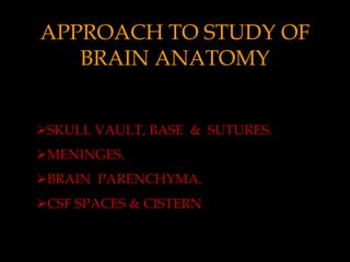
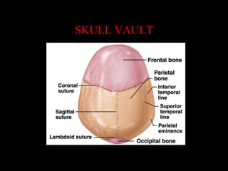
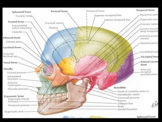
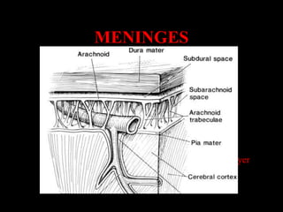
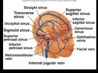
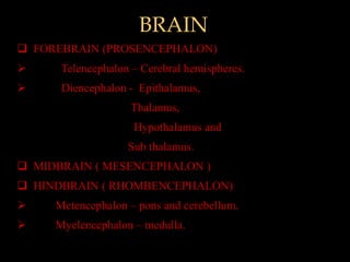
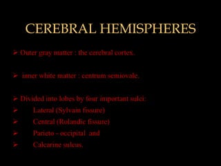
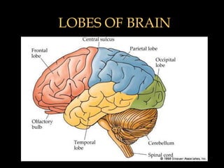
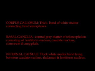
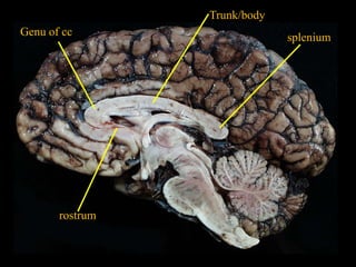
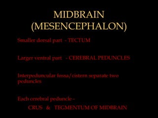
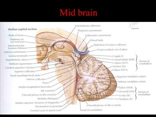
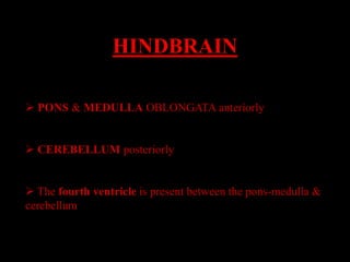
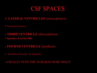
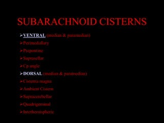
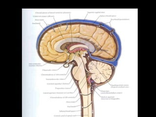
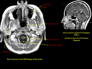
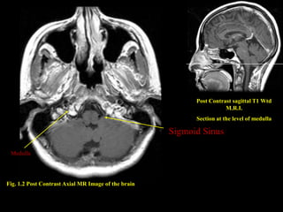
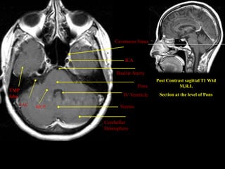
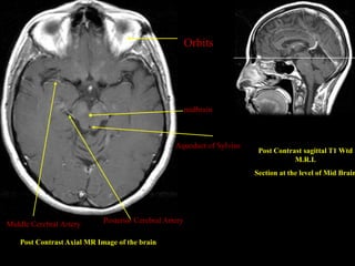
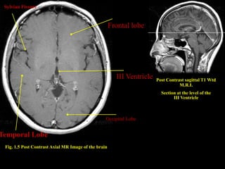
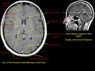
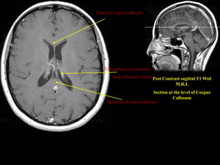
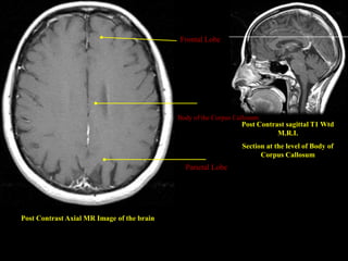
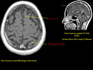
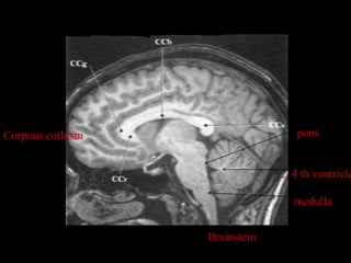
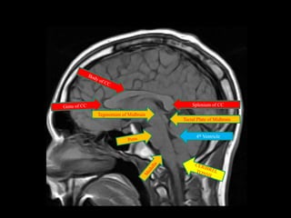
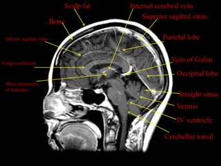
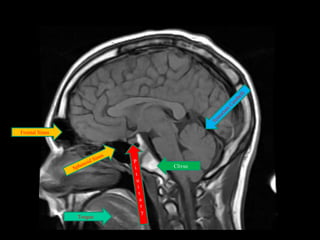
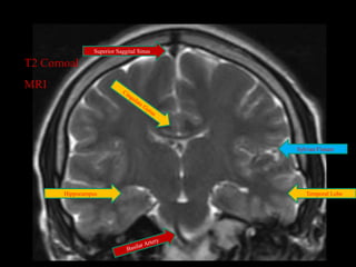
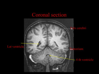
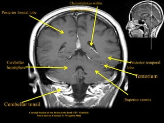
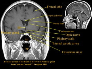
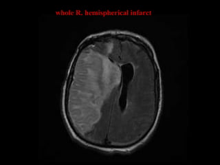
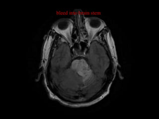
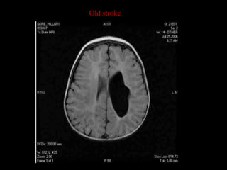
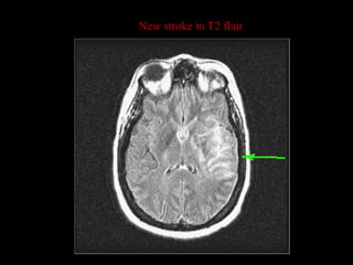
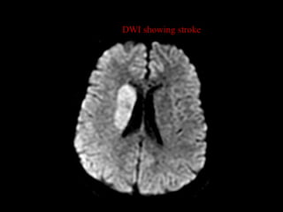
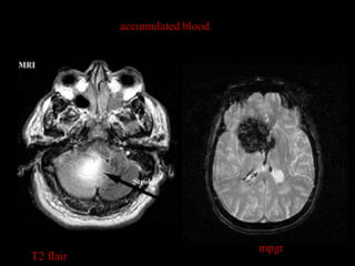
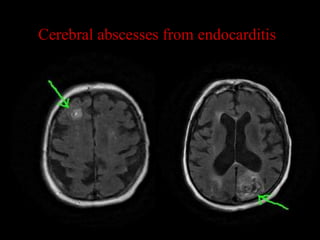
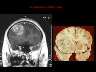
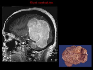
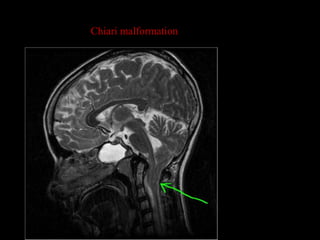
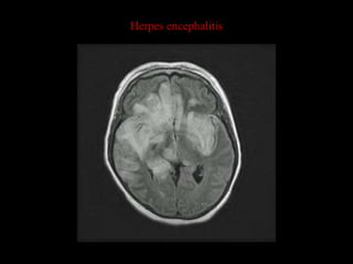
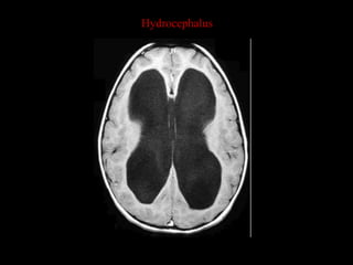
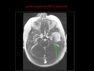
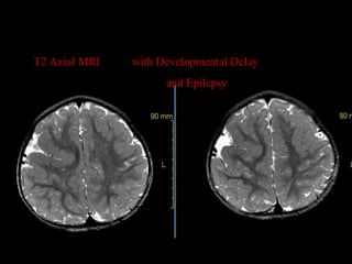
Sectional Anatomy of
       Brain


                Vipin kumar
                Msc medical imaging
APPROACH TO STUDY OF
   BRAIN ANATOMY


SKULL VAULT, BASE & SUTURES.
MENINGES.
BRAIN PARENCHYMA.
CSF SPACES & CISTERN
SKULL VAULT
MENINGES
 DURAMATER :


 ARACHNOID MATER


 PIAMATER


 outer periosteal layer & inner meningeal layer
MENINGEAL FOLDS
 DURAL REFLECTIONS

                        DURAL VENOUS SINUSES
 FALX CEREBRI
                          B/W TWO DURAMATERS


 TENTORIUM CEREBELLI


FALX CEREBELLI


DIAPHRAGMATIC SELLA
BRAIN
 FOREBRAIN (PROSENCEPHALON)
     Telencephalon – Cerebral hemispheres.
     Diencephalon - Epithalamus,
                   Thalamus,
                    Hypothalamus and
                   Sub thalamus.
 MIDBRAIN ( MESENCEPHALON )
 HINDBRAIN ( RHOMBENCEPHALON)
    Metencephalon – pons and cerebellum.
    Myelencephalon – medulla.
CEREBRAL HEMISPHERES
 Outer gray matter : the cerebral cortex.


 inner white matter : centrum semiovale.


 Divided into lobes by four important sulci:
      Lateral (Sylvain fissure)
      Central (Rolandic fissure)
      Parieto - occipital and
      Calcarine sulcus.
LOBES OF BRAIN
CORPUS CALLOSUM: Thick band of white matter
connecting two hemispheres.


BASAL GANGLIA : central gray matter of telencephalon
consisting of lentiform nucleus, caudate nucleus,
claustrum & amygdala.


INTERNAL CAPSULE: Thick white matter band lying
between caudate nucleus, thalamus & lentiform nucleus.
Trunk/body
Genu of cc                    splenium


       rostrum
MIDBRAIN
     (MESENCEPHALON)
Smaller dorsal part - TECTUM


Larger ventral part - CEREBRAL PEDUNCLES


Interpeduncular fossa/cistern separate two
peduncles


Each cerebral peduncle -
    CRUS & TEGMENTUM OF MIDBRAIN
Mid brain
HINDBRAIN

 PONS & MEDULLA OBLONGATA anteriorly


 CEREBELLUM posteriorly


 The fourth ventricle is present between the pons-medulla &
cerebellum
CSF SPACES
 LATERAL VENTRICLES (telencephalon)

 Foramina of monro


 THIRD     VENTRICLE (diencephalon)
 Aqueduct of sylvius(MB)


 FOURTH       VENTRICLE (hindbrain)

 Foramina of luschca & magendie


 FINALLY      INTO THE SUBARACNOID SPACE
SUBARACHNOID CISTERNS
VENTRAL (median & paramedian)
Perimedullary
Prepontine
Suprasellar
Cp angle
DORSAL (median & paramedian)
Cisterna magna
Ambient Cistern
Supracerebellar
Quadrigeminal
Interhemispheric
. Maxillary
                                              Sinus


                                                  . Nasopharynx

                                                                        Post Contrast sagittal T1 Weighted
                                                                                      M.R.I.
                                                                         Section at the level of Foramen
                                                   . Cervical Cord                   Magnum

                                                       Cisterna Magna


. Mandible

      Post Contrast Axial MR Image of the brain
Post Contrast sagittal T1 Wtd
                                                                      M.R.I.
                                                           Section at the level of medulla

                                                     Sigmoid Sinus

 Medulla


Fig. 1.2 Post Contrast Axial MR Image of the brain
Cavernous Sinus


                            ICA

                        Basilar Artery
                                          Post Contrast sagittal T1 Wtd
                                  Pons               M.R.I.
TMP
lobe                       IV Ventricle    Section at the level of Pons

       IAC   MCP         Vermis


                   Cerebellar
                   Hemisphere
Orbits


                                                         midbrain


                                                   Aqueduct of Sylvius
                                                                          Post Contrast sagittal T1 Wtd
                                                                                     M.R.I.
                                                                         Section at the level of Mid Brain


Middle Cerebral Artery       Posterior Cerebral Artery

    Post Contrast Axial MR Image of the brain
Sylvian Fissure


                                                      Frontal lobe


                                                        III Ventricle   Post Contrast sagittal T1 Wtd
                                                                                   M.R.I.
                                                                          Section at the level of the
                                                                                III Ventricle


                                                  Occipital Lobe


Temporal Lobe
 Fig. 1.5 Post Contrast Axial MR Image of the brain
fontal
  Lobe


                                                        Frontal Horn
 caudate Nucleus
. Putamen
                                                      . Internal Cerebral Vein
Internal Capsule


                                                          Choroid Plexus within the Post Contrast
                                                                                                sagittal T1 Wtd
                                                             occipital horn                   M.R.I.
                                                                               Section at the level of Thalamus

                                                       Occipital Lobe


Temp Lobe

              Thalamus          Superior Sagittal Sinus
 Fig. 1.6 Post Contrast Axial MR Image of the brain
Genu of corpus callosum


    Choroid plexus within the
        body of lateral ventricle
                                Post Contrast sagittal T1 Wtd
                                           M.R.I.
                                Section at the level of Corpus
                                          Callosum
     Splenium of corpus callosum
Frontal Lobe


                                            Body of the Corpus Callosum
                                                                 Post Contrast sagittal T1 Wtd
                                                                            M.R.I.
                                                                 Section at the level of Body of
                                                                       Corpus Callosum
                                              Parietal Lobe


Post Contrast Axial MR Image of the brain
Frontal Lobe


                                                             Post Contrast sagittal T1 Wtd
                                                                        M.R.I.
                                                           Section above the Corpus Callosum

                                            Parietal Lobe


Post Contrast Axial MR Image of the brain
Corpous collusm                pons


                              4 th ventricle

                              medulla


                  Brainstem
Splenium of CC
Tegmentum of Midbrain
                        Tectal Plate of Midbrain


                              4th Ventricle
Scalp fat   Internal cerebral vein
                                           Superior sagittal sinus
                      . Bone

 Inferior sagittal sinus                          Parietal lobe


Corpus callosum
                                                     Vein of Galen
                                                       Occipital lobe
 Mass intermedia
 of thalamus
                                                       Straight sinus
                                                      . Vermis
                                                      . IV ventricle

                                                   Cerebellar tonsil
Frontal Sinus


                         Clivus


                Tongue
Superior Saggital Sinus

T2 Cornoal
MRI


                                           Sylvian Fissure


       Hippocampus                            Temporal Lobe
Coronal section

                                  Falx cerebri


Lat ventricle
                                  Tentorium


                                      4 th ventricle
Choroid plexus within
                                                        lateral ventricle
Posterior frontal lobe


   Cerebellar                                                                   . Posterior temporal
   hemisphere                                                                   lobe

                                                                                       Tentorium


                                                                               Superior vermis
 Cerebellar tonsil
                 Coronal Section of the Brain at the level of IV Ventricle
                       Post Contrast Coronal T1 Weighted MRI
Frontal lobe

                                                    Corpus callosum


                                                           Frontal horn
                 III
                                                                  Caudate nucleus
Pituitary gland
                                                                        Optic nerve
                           sp                                    Pituitary stalk
                                                           Internal carotid artery
                           np
                                                                    Cavernous sinus


  Coronal Section of the Brain at the level of Pituitary gland
         Post Contrast Coronal T1 Weighted MRI
PATHOLOGIS
whole R. hemispherical infarct
bleed into brain stem
Old stroke
New stroke in T2 flair
DWI showing stroke
accumulated blood


                               mpgr
T2 flair
Cerebral abscesses from endocarditis
Glioblastoma Multiforme
Giant meningioma
Chiari malformation
Herpes encephalitis
Hydrocephalus
giant unruptured MCA aneurysm
T2 Axial MRI   with Developmental Delay
                     and Epilepsy
MRI SECTIONAL ANATOMY OF BRAIN

MRI SECTIONAL ANATOMY OF BRAIN

  • 1.
    Sectional Anatomy of Brain Vipin kumar Msc medical imaging
  • 2.
    APPROACH TO STUDYOF BRAIN ANATOMY SKULL VAULT, BASE & SUTURES. MENINGES. BRAIN PARENCHYMA. CSF SPACES & CISTERN
  • 3.
  • 5.
    MENINGES  DURAMATER : ARACHNOID MATER  PIAMATER  outer periosteal layer & inner meningeal layer
  • 6.
    MENINGEAL FOLDS  DURALREFLECTIONS DURAL VENOUS SINUSES  FALX CEREBRI B/W TWO DURAMATERS  TENTORIUM CEREBELLI FALX CEREBELLI DIAPHRAGMATIC SELLA
  • 8.
    BRAIN  FOREBRAIN (PROSENCEPHALON)  Telencephalon – Cerebral hemispheres.  Diencephalon - Epithalamus, Thalamus, Hypothalamus and Sub thalamus.  MIDBRAIN ( MESENCEPHALON )  HINDBRAIN ( RHOMBENCEPHALON)  Metencephalon – pons and cerebellum.  Myelencephalon – medulla.
  • 9.
    CEREBRAL HEMISPHERES  Outergray matter : the cerebral cortex.  inner white matter : centrum semiovale.  Divided into lobes by four important sulci:  Lateral (Sylvain fissure)  Central (Rolandic fissure)  Parieto - occipital and  Calcarine sulcus.
  • 10.
  • 11.
    CORPUS CALLOSUM: Thickband of white matter connecting two hemispheres. BASAL GANGLIA : central gray matter of telencephalon consisting of lentiform nucleus, caudate nucleus, claustrum & amygdala. INTERNAL CAPSULE: Thick white matter band lying between caudate nucleus, thalamus & lentiform nucleus.
  • 12.
    Trunk/body Genu of cc splenium rostrum
  • 13.
    MIDBRAIN (MESENCEPHALON) Smaller dorsal part - TECTUM Larger ventral part - CEREBRAL PEDUNCLES Interpeduncular fossa/cistern separate two peduncles Each cerebral peduncle - CRUS & TEGMENTUM OF MIDBRAIN
  • 14.
  • 15.
    HINDBRAIN  PONS &MEDULLA OBLONGATA anteriorly  CEREBELLUM posteriorly  The fourth ventricle is present between the pons-medulla & cerebellum
  • 16.
    CSF SPACES  LATERALVENTRICLES (telencephalon)  Foramina of monro  THIRD VENTRICLE (diencephalon)  Aqueduct of sylvius(MB)  FOURTH VENTRICLE (hindbrain)  Foramina of luschca & magendie  FINALLY INTO THE SUBARACNOID SPACE
  • 17.
    SUBARACHNOID CISTERNS VENTRAL (median& paramedian) Perimedullary Prepontine Suprasellar Cp angle DORSAL (median & paramedian) Cisterna magna Ambient Cistern Supracerebellar Quadrigeminal Interhemispheric
  • 19.
    . Maxillary Sinus . Nasopharynx Post Contrast sagittal T1 Weighted M.R.I. Section at the level of Foramen . Cervical Cord Magnum Cisterna Magna . Mandible Post Contrast Axial MR Image of the brain
  • 20.
    Post Contrast sagittalT1 Wtd M.R.I. Section at the level of medulla Sigmoid Sinus Medulla Fig. 1.2 Post Contrast Axial MR Image of the brain
  • 21.
    Cavernous Sinus ICA Basilar Artery Post Contrast sagittal T1 Wtd Pons M.R.I. TMP lobe IV Ventricle Section at the level of Pons IAC MCP Vermis Cerebellar Hemisphere
  • 22.
    Orbits midbrain Aqueduct of Sylvius Post Contrast sagittal T1 Wtd M.R.I. Section at the level of Mid Brain Middle Cerebral Artery Posterior Cerebral Artery Post Contrast Axial MR Image of the brain
  • 23.
    Sylvian Fissure Frontal lobe III Ventricle Post Contrast sagittal T1 Wtd M.R.I. Section at the level of the III Ventricle Occipital Lobe Temporal Lobe Fig. 1.5 Post Contrast Axial MR Image of the brain
  • 24.
    fontal Lobe Frontal Horn caudate Nucleus . Putamen . Internal Cerebral Vein Internal Capsule Choroid Plexus within the Post Contrast sagittal T1 Wtd occipital horn M.R.I. Section at the level of Thalamus Occipital Lobe Temp Lobe Thalamus Superior Sagittal Sinus Fig. 1.6 Post Contrast Axial MR Image of the brain
  • 25.
    Genu of corpuscallosum Choroid plexus within the body of lateral ventricle Post Contrast sagittal T1 Wtd M.R.I. Section at the level of Corpus Callosum Splenium of corpus callosum
  • 26.
    Frontal Lobe Body of the Corpus Callosum Post Contrast sagittal T1 Wtd M.R.I. Section at the level of Body of Corpus Callosum Parietal Lobe Post Contrast Axial MR Image of the brain
  • 27.
    Frontal Lobe Post Contrast sagittal T1 Wtd M.R.I. Section above the Corpus Callosum Parietal Lobe Post Contrast Axial MR Image of the brain
  • 28.
    Corpous collusm pons 4 th ventricle medulla Brainstem
  • 29.
    Splenium of CC Tegmentumof Midbrain Tectal Plate of Midbrain 4th Ventricle
  • 30.
    Scalp fat Internal cerebral vein Superior sagittal sinus . Bone Inferior sagittal sinus Parietal lobe Corpus callosum Vein of Galen Occipital lobe Mass intermedia of thalamus Straight sinus . Vermis . IV ventricle Cerebellar tonsil
  • 31.
    Frontal Sinus Clivus Tongue
  • 32.
    Superior Saggital Sinus T2Cornoal MRI Sylvian Fissure Hippocampus Temporal Lobe
  • 33.
    Coronal section Falx cerebri Lat ventricle Tentorium 4 th ventricle
  • 34.
    Choroid plexus within lateral ventricle Posterior frontal lobe Cerebellar . Posterior temporal hemisphere lobe Tentorium Superior vermis Cerebellar tonsil Coronal Section of the Brain at the level of IV Ventricle Post Contrast Coronal T1 Weighted MRI
  • 35.
    Frontal lobe Corpus callosum Frontal horn III Caudate nucleus Pituitary gland Optic nerve sp Pituitary stalk Internal carotid artery np Cavernous sinus Coronal Section of the Brain at the level of Pituitary gland Post Contrast Coronal T1 Weighted MRI
  • 36.
  • 37.
  • 38.
  • 39.
  • 40.
    New stroke inT2 flair
  • 41.
  • 42.
    accumulated blood mpgr T2 flair
  • 43.
  • 44.
  • 45.
  • 46.
  • 47.
  • 48.
  • 49.
  • 50.
    T2 Axial MRI with Developmental Delay and Epilepsy

Editor's Notes

  • #22 . Internal Carotid ArteryInternal Auditory Canal
  • #40 Usually cystify and develop firm borders
  • #41 New strokes usually show up as white on T2.
  • #43 Usually cystify and develop firm borders
  • #45 Glios are rapid growing and cause death by brain compression. They do not usually metastasize, but occasionally can following debulking surgery.
  • #47 Some believe cranium too small for brain, Others believe the foramen magnum is malformed. Symptoms of headache, ataxia and nystagmus with progressive pressure on brain stem
  • #48 Bi-temporal distribution is typical. Thought to occur by re-activation of herpes virus much like “cold sores” except through different nerve distribution