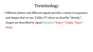
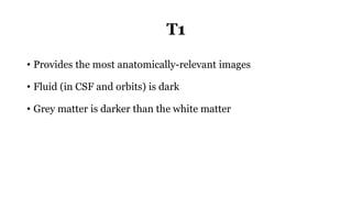
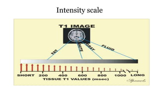
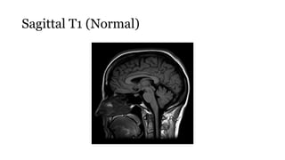
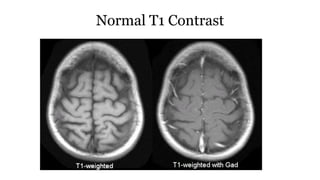
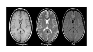
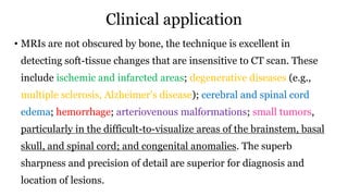
Magnetic Resonance
imaging
Evaluator: Mr L Anand Presenter: Shruti Shirke
[Asso professor, CON AIIMS BBSR] M.Sc Neuroscience Nursing
Introduction
• MRI (an abbreviation of magnetic resonance imaging) is
an imaging modality that uses non-ionizing radiation to create useful
diagnostic images.
• In simple terms, an MRI scanner consists of a large, powerful magnet in
which the patient lies. A radio wave antenna is used to send signals to
the body and then a radiofrequency receiver detects the emitted signals.
These returning signals are converted into images by a computer
attached to the scanner. Imaging of any part of the body can be obtained
in any plane.
Advantages
1. Ability to image without the use of ionizing x-rays, Eliminate the radio exposure risk.
2. Images may be acquired in multiple planes (axial, sagittal, coronal, or oblique) without repositioning the
patient.
3. MRI images demonstrate superior soft-tissue contrast as compared to CT scans and plain radiographs
making it the ideal examination of the brain, spine, joints, and other soft tissue body parts
4. Some angiographic images can be obtained without the use of contrast material, unlike CT or
conventional angiography
5. Advanced techniques such: as diffusion, spectroscopy, and perfusion allow for precise tissue
characterization rather than merely 'macroscopic' imaging
6. Functional MRI allows visualization of active parts of the brain during certain activities and also
understanding of the underlying networks
Advantages cont..
• Tissue resolution is far superior with MRI with the exception of bone,
which cannot be seen with MRI except for bone marrow
• Provides better differentiation than CT for water, iron, fat, and blood
using physical and biochemical characteristics of tissues imaged
• Can detect soft-tissue changes not seen on radiographs such as small
tumors along cranial nerves and other tumors as small as 0.3 mm
• Provides higher-resolution detail of the posterior fossa, skull base, and
orbits compared with CT scan
Disadvantages
1. MRI scans are more expensive than CT scans
2. MRI scans take significantly longer to acquire than CT and patient comfort
can be an issue, maybe exacerbated by:
• MR image acquisition is noisy compared to CT
• MRI scanner bores tend to be more enclosed than CT with associated claustrophobia
3. MR images are subject to unique artifacts that must be recognized.
4. MRI scanning is not safe for patients with some metal implants and foreign
bodies. Careful attention to safety measures is necessary to avoid serious
injury to patients and staff, and this requires special MRI compatible
equipment and stringent adherence to safety protocols.
Terminology
• Different plaines and different signals provide a variety of sequences
and images that we use. Unlike CT where we describe "density",
images are described by signal intensity ("hyper-" bright, "hypo-"
dark).
T1
• Provides the most anatomically-relevant images
• Fluid (in CSF and orbits) is dark
• Grey matter is darker than the white matter
Intensity scale
Sagittal T1 (Normal)
Chiari Malformation (Sagittal T1)
Chiari Malformation (Axial T1)
Normal T1 Contrast
T2
• Standard sequence
• Fluid is bright (hyper-intense)
• White matter is darker than grey
Axial T2
Axial T2
A coronal T2 weighted image shows a low T2 signal adenoma in
the left side of the gland (arrow) which represented a densely
granulated GH secreting adenoma.
FLAIR (fluid attenuation inversion
recovery)
• Commonly used sequence
• Similar to T2, but the fluid is darker or "suppressed"
• Useful for areas of edema or inflammation
• Used to identify plaques in multiple sclerosis (especially
periventricular)
Metastatic melanoma
Multiple sclerosis with incidental cerebellar
gangliocytoma (Sagittal FLAIR)
DWI and ADC (diffusion-weighted
imaging and apparent diffusion coefficient)
• These "blocky" images show how easily water moves around
• Restricted diffusion occurs in stroke, abscesses and cellular
tumors
DWI and ADC
• is a commonly performed MRI sequence for evaluation of
acute ischemic stroke, and is sensitive in the detection of small and
early infarcts. Conventional MRI sequences (T1WI, T2WI) may not
demonstrate an infarct for 6 hours, and small infarcts may be hard to
appreciate on CT for days, especially without the benefit of prior
imaging.
• Increased DWI signal in ischemic brain tissue is observed within a few
minutes after arterial occlusion and progresses through a stereotypic
sequence of apparent diffusion coefficient (ADC) reduction, followed
by subsequent increase, pseudo-normalization and, finally, permanent
elevation. Reported sensitivity ranges from 88-100% and specificity
ranges from 86-100%.
DWI
ADC
MCA Stroke (Axial DWI)
Tractography
Contraindications
1. Patients with ferromagnetic devices, such as artificial pacemakers,
defibrillators, implanted stimulators of the brain or spinal cord, cochlear
implants, prosthetic devices(e.g., metallic hip replacements, orthopedic
pins), artificial limbs, respirators, and other metallic equipment
2. Patients with older ferromagnetic intracranial aneurysm clips or metal
bullet or other metal fragments in the orbits or brain
3. Women in the first trimester of pregnancy (gadolinium contraindicated
during entire pregnancy)
4. Placement of a vascular stent, coil, or filter within the past 6 weeks
Relative contraindications:
1. Agitated, uncooperative patients or patients with uncontrolled
movement disorders
2. Grossly obese patients who cannot fit into the MRI tube
3. Claustrophobic patients who cannot tolerate the closeness of the
tube to their faces
4. Critically ill patients requiring a ventilator and intravenous (IV)
pump (some ventilators are specially designed nonmagnetic; IV
pumps are generally magnetic and cannot be used)
Clinical application
• MRIs are not obscured by bone, the technique is excellent in
detecting soft-tissue changes that are insensitive to CT scan. These
include ischemic and infarcted areas; degenerative diseases (e.g.,
multiple sclerosis, Alzheimer’s disease); cerebral and spinal cord
edema; hemorrhage; arteriovenous malformations; small tumors,
particularly in the difficult-to-visualize areas of the brainstem, basal
skull, and spinal cord; and congenital anomalies. The superb
sharpness and precision of detail are superior for diagnosis and
location of lesions.
Procedure
• The patient is screened for contraindications. After removing all metal objects
and credit cards (magnet can desensitize the black strip on the back of a credit
card).
• The patient lies on a padded stretcher that slides into a tunnel-like chamber.
• The head is placed in a plastic helmet-like structure; the arms are at the side of
the body and are held in place with Velcro straps.
• The head of the table is then rolled several feet into the scanner. The scanner tube
has a restricted opening; it has been compared to the opening of a barrel or large
pipe.
Procedure cont..
• During the scan, noise, caused by the pulsating RF waves, is heard.
• The patient must be prepared for this and for the requirement to remain
absolutely motionless for the scan. Although the patient is alone in the
room, voice contact is maintained with the technician through an
intercom.
• If contrast enhancement is desired, gadolinium is administered
intravenously. The MRI takes approximately 5 to 50 minutes to complete.
Patient Preparation and Post-procedural Care
• Carefully question and screen for the presence of any metal implants or other
contraindications.
• For the patient who is vague about possible contraindications, radiographic
screening may be necessary.
• The patient should be briefed about the procedure and the necessity to remain
still for several minutes in very close quarters.
• Antianxiety medication may be ordered for some patients.
• All jewelry, eyeglasses, and hair clips or pins and any other metal objects should
be removed.
Patient Preparation and Post-procedural Care
• Credit cards should not be in the room because the magnetic field will erase the strip on the card.
• Unless a contrast medium will be administered, there are no dietary restrictions before the
procedure.
• Several points need attention if contrast medium is to be used. Adequate kidney function is
verified through adequate blood urea nitrogen and creatinine levels.
• Many protocols require nothing by mouth for 4 to 6 hours before the MRI.
• If contrast media are to be used, skin testing may be done to check for an allergic reaction.
• There are no special aftercare requirements. When contrast medium is used, adequate hydration
is needed.
Summary
Introduction
Advantages
Disadvantages
Contraindications
Sequences
Patient preparation
Post procedure care
•Thankyou
MRI BRAIN
MRI BRAIN
MRI BRAIN
MRI BRAIN
MRI BRAIN

MRI BRAIN

  • 1.
    Magnetic Resonance imaging Evaluator: MrL Anand Presenter: Shruti Shirke [Asso professor, CON AIIMS BBSR] M.Sc Neuroscience Nursing
  • 2.
    Introduction • MRI (anabbreviation of magnetic resonance imaging) is an imaging modality that uses non-ionizing radiation to create useful diagnostic images. • In simple terms, an MRI scanner consists of a large, powerful magnet in which the patient lies. A radio wave antenna is used to send signals to the body and then a radiofrequency receiver detects the emitted signals. These returning signals are converted into images by a computer attached to the scanner. Imaging of any part of the body can be obtained in any plane.
  • 3.
    Advantages 1. Ability toimage without the use of ionizing x-rays, Eliminate the radio exposure risk. 2. Images may be acquired in multiple planes (axial, sagittal, coronal, or oblique) without repositioning the patient. 3. MRI images demonstrate superior soft-tissue contrast as compared to CT scans and plain radiographs making it the ideal examination of the brain, spine, joints, and other soft tissue body parts 4. Some angiographic images can be obtained without the use of contrast material, unlike CT or conventional angiography 5. Advanced techniques such: as diffusion, spectroscopy, and perfusion allow for precise tissue characterization rather than merely 'macroscopic' imaging 6. Functional MRI allows visualization of active parts of the brain during certain activities and also understanding of the underlying networks
  • 4.
    Advantages cont.. • Tissueresolution is far superior with MRI with the exception of bone, which cannot be seen with MRI except for bone marrow • Provides better differentiation than CT for water, iron, fat, and blood using physical and biochemical characteristics of tissues imaged • Can detect soft-tissue changes not seen on radiographs such as small tumors along cranial nerves and other tumors as small as 0.3 mm • Provides higher-resolution detail of the posterior fossa, skull base, and orbits compared with CT scan
  • 5.
    Disadvantages 1. MRI scansare more expensive than CT scans 2. MRI scans take significantly longer to acquire than CT and patient comfort can be an issue, maybe exacerbated by: • MR image acquisition is noisy compared to CT • MRI scanner bores tend to be more enclosed than CT with associated claustrophobia 3. MR images are subject to unique artifacts that must be recognized. 4. MRI scanning is not safe for patients with some metal implants and foreign bodies. Careful attention to safety measures is necessary to avoid serious injury to patients and staff, and this requires special MRI compatible equipment and stringent adherence to safety protocols.
  • 6.
    Terminology • Different plainesand different signals provide a variety of sequences and images that we use. Unlike CT where we describe "density", images are described by signal intensity ("hyper-" bright, "hypo-" dark).
  • 7.
    T1 • Provides themost anatomically-relevant images • Fluid (in CSF and orbits) is dark • Grey matter is darker than the white matter
  • 8.
  • 9.
  • 10.
  • 11.
  • 12.
  • 13.
    T2 • Standard sequence •Fluid is bright (hyper-intense) • White matter is darker than grey
  • 14.
  • 15.
  • 17.
    A coronal T2weighted image shows a low T2 signal adenoma in the left side of the gland (arrow) which represented a densely granulated GH secreting adenoma.
  • 18.
    FLAIR (fluid attenuationinversion recovery) • Commonly used sequence • Similar to T2, but the fluid is darker or "suppressed" • Useful for areas of edema or inflammation • Used to identify plaques in multiple sclerosis (especially periventricular)
  • 20.
  • 21.
    Multiple sclerosis withincidental cerebellar gangliocytoma (Sagittal FLAIR)
  • 22.
    DWI and ADC(diffusion-weighted imaging and apparent diffusion coefficient) • These "blocky" images show how easily water moves around • Restricted diffusion occurs in stroke, abscesses and cellular tumors
  • 23.
    DWI and ADC •is a commonly performed MRI sequence for evaluation of acute ischemic stroke, and is sensitive in the detection of small and early infarcts. Conventional MRI sequences (T1WI, T2WI) may not demonstrate an infarct for 6 hours, and small infarcts may be hard to appreciate on CT for days, especially without the benefit of prior imaging. • Increased DWI signal in ischemic brain tissue is observed within a few minutes after arterial occlusion and progresses through a stereotypic sequence of apparent diffusion coefficient (ADC) reduction, followed by subsequent increase, pseudo-normalization and, finally, permanent elevation. Reported sensitivity ranges from 88-100% and specificity ranges from 86-100%.
  • 24.
  • 26.
  • 27.
  • 30.
    Contraindications 1. Patients withferromagnetic devices, such as artificial pacemakers, defibrillators, implanted stimulators of the brain or spinal cord, cochlear implants, prosthetic devices(e.g., metallic hip replacements, orthopedic pins), artificial limbs, respirators, and other metallic equipment 2. Patients with older ferromagnetic intracranial aneurysm clips or metal bullet or other metal fragments in the orbits or brain 3. Women in the first trimester of pregnancy (gadolinium contraindicated during entire pregnancy) 4. Placement of a vascular stent, coil, or filter within the past 6 weeks
  • 31.
    Relative contraindications: 1. Agitated,uncooperative patients or patients with uncontrolled movement disorders 2. Grossly obese patients who cannot fit into the MRI tube 3. Claustrophobic patients who cannot tolerate the closeness of the tube to their faces 4. Critically ill patients requiring a ventilator and intravenous (IV) pump (some ventilators are specially designed nonmagnetic; IV pumps are generally magnetic and cannot be used)
  • 32.
    Clinical application • MRIsare not obscured by bone, the technique is excellent in detecting soft-tissue changes that are insensitive to CT scan. These include ischemic and infarcted areas; degenerative diseases (e.g., multiple sclerosis, Alzheimer’s disease); cerebral and spinal cord edema; hemorrhage; arteriovenous malformations; small tumors, particularly in the difficult-to-visualize areas of the brainstem, basal skull, and spinal cord; and congenital anomalies. The superb sharpness and precision of detail are superior for diagnosis and location of lesions.
  • 33.
    Procedure • The patientis screened for contraindications. After removing all metal objects and credit cards (magnet can desensitize the black strip on the back of a credit card). • The patient lies on a padded stretcher that slides into a tunnel-like chamber. • The head is placed in a plastic helmet-like structure; the arms are at the side of the body and are held in place with Velcro straps. • The head of the table is then rolled several feet into the scanner. The scanner tube has a restricted opening; it has been compared to the opening of a barrel or large pipe.
  • 34.
    Procedure cont.. • Duringthe scan, noise, caused by the pulsating RF waves, is heard. • The patient must be prepared for this and for the requirement to remain absolutely motionless for the scan. Although the patient is alone in the room, voice contact is maintained with the technician through an intercom. • If contrast enhancement is desired, gadolinium is administered intravenously. The MRI takes approximately 5 to 50 minutes to complete.
  • 35.
    Patient Preparation andPost-procedural Care • Carefully question and screen for the presence of any metal implants or other contraindications. • For the patient who is vague about possible contraindications, radiographic screening may be necessary. • The patient should be briefed about the procedure and the necessity to remain still for several minutes in very close quarters. • Antianxiety medication may be ordered for some patients. • All jewelry, eyeglasses, and hair clips or pins and any other metal objects should be removed.
  • 36.
    Patient Preparation andPost-procedural Care • Credit cards should not be in the room because the magnetic field will erase the strip on the card. • Unless a contrast medium will be administered, there are no dietary restrictions before the procedure. • Several points need attention if contrast medium is to be used. Adequate kidney function is verified through adequate blood urea nitrogen and creatinine levels. • Many protocols require nothing by mouth for 4 to 6 hours before the MRI. • If contrast media are to be used, skin testing may be done to check for an allergic reaction. • There are no special aftercare requirements. When contrast medium is used, adequate hydration is needed.
  • 37.
  • 38.