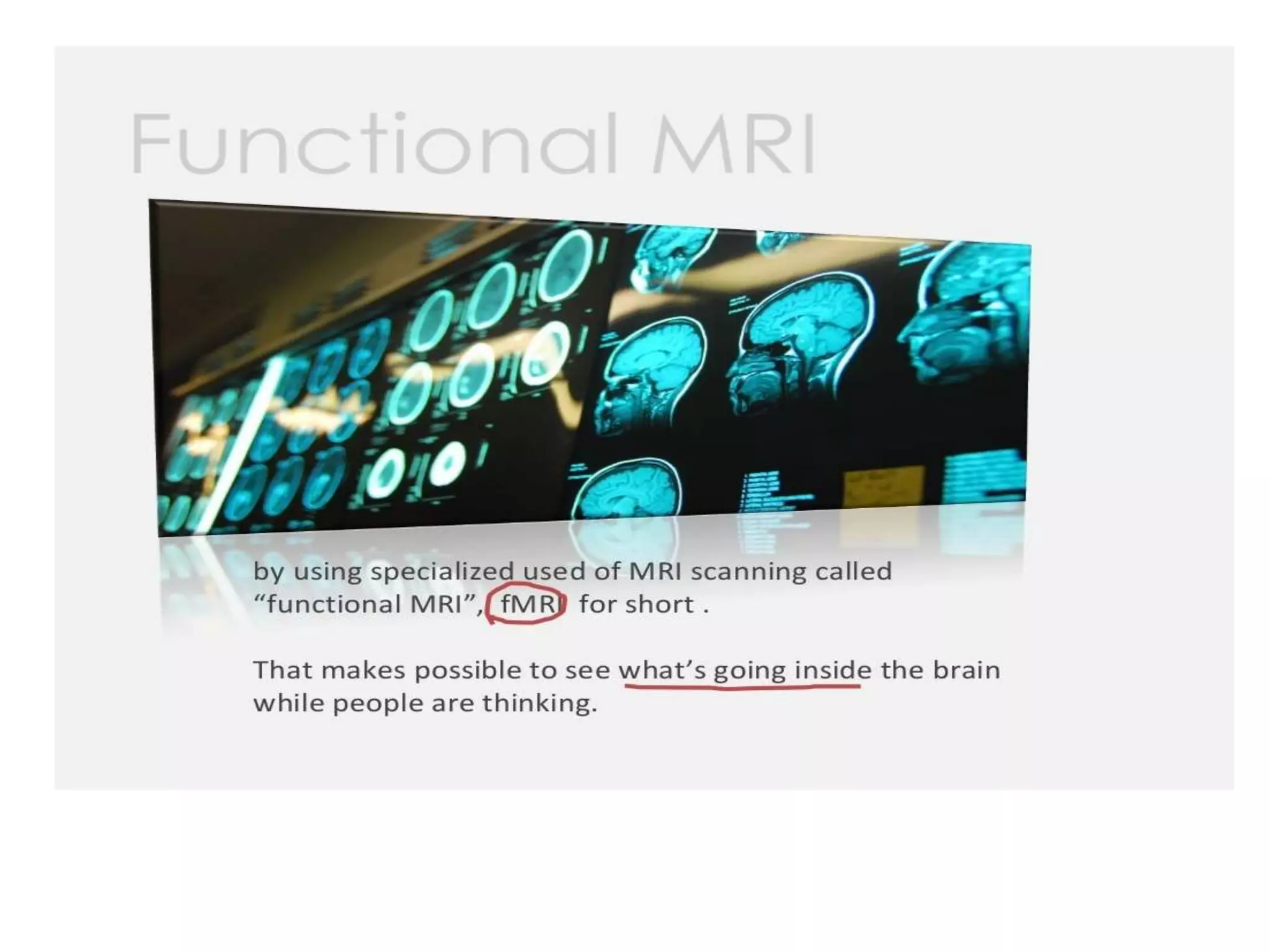
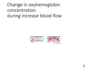
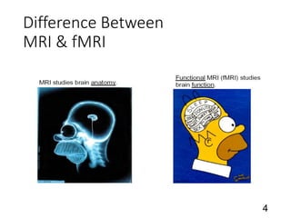
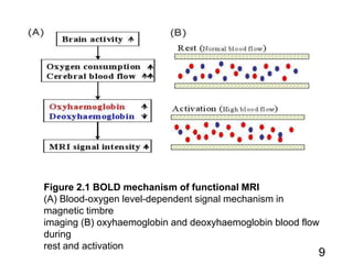
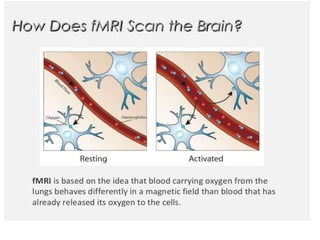
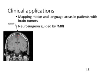
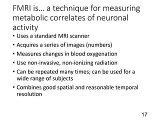
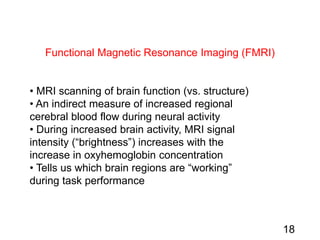
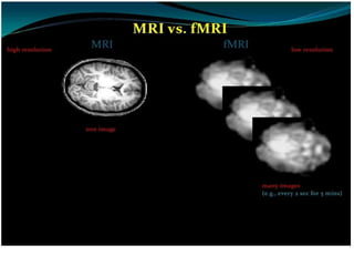
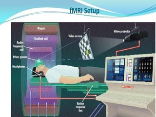
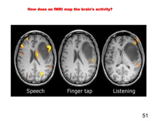
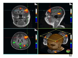
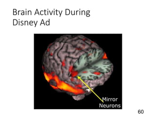
VIP Call Girls Pune Vrinda 9907093804 Short 1500 Night 6000 Best call girls S...
FMRI.ppt
1.
2. 2
An fMRI scan is a functional magnetic resonance imaging scan
that measures and maps the brain’s activity. An fMRI scan uses
the same technology as an MRI scan. An MRI is a noninvasive test
that uses a strong magnetic field and radio waves to create an
image of the brain. The image an MRI scan produces is just of
organs/tissue, but an fMRI will produce an image showing the
blood flow in the brain. By showing the blood flow it will display
which parts of the brain are being stimulated.
What is an fMRI scan and how does it work?
5. 5
fMRI Vs. MRI
The MRI and fMRI differ from each other in a way
that the MRI views the anatomical structure while
the fMRI views the metabolic function.
An MRI studies the water molecule’s hydrogen
nuclei whereas an fMRI calculates the levels of
oxygen.
An MRI’s structural imaging views at a high
resolution the differences between tissue types
with respect to space. On the other hand, an fMRI’s
functional imaging views the tissue differences with
respect to time.
6. Early Uses of NMR
• Most early NMR was used for chemical analysis
• No medical applications
• 1971 – Damadian publishes and patents idea for using
NMR to distinguish healthy and malignant tissues
• “Tumor detection by nuclear magnetic resonance”, Science
• Proposes using differences in relaxation times
• No image formation method proposed
• 1973 – Lauterbur describes projection method for
creating NMR images
• Mansfield (1973) independently describes similar approach
6
7. Terms Used for MRI
•
NMR (Nuclear Magnetic Resonance)
MR (Magnetic Resonance)
MRI (Magnetic Resonance Imaging)
7
8. fMRIacquisition technique
•BOLD(BloodOxygenLevel Dependent), the most
common, provides a mixed signal dependant on:–
bloodFLOW–bloodVOLUME–bloodOXYGENATION.
•Variations on the technique can be used to emphasize
or de-emphasize one or another of these components:-
Pefusion-based fMRI * blood flow-Injections of various
compounds * blood volume-Diffusion-based fMRI cell
swelling (after excitation)
8
9. 9
Figure 2.1 BOLD mechanism of functional MRI
(A) Blood-oxygen level-dependent signal mechanism in
magnetic timbre
imaging (B) oxyhaemoglobin and deoxyhaemoglobin blood flow
during
rest and activation
10.
11. •fMRI-BOLD is best used for studying processes that can be rapidly turned on and off
like language, vision, movement, hearing, and memory.
•The study of emotion is hampered by its slow and variable onsetand its inability to be
quickly reversed. Some have, however, succeed in using this technique to study fear
(Whalen et al.)fMRI-BOLD paradigms generally have several periods of rest alternating with
several periods of activation. •Images are then compared over the entire activation to the rest
periods. Images over the first 3 to 6 seconds of each period are generally discarded due to the
delay in hemodynamicresponse. •Alternating paradimsare used because the signal intensity
generated by the MRI scanner drifts with time.Design of fMRIExperiments
11
13. Clinical applications
• Mapping motor and language areas in patients with
brain tumors
• Neurosurgeon guided by fMRI
13
tumor
14. fMRI Experiment Stages: Prep
14
1) Prepare subject
• Consent form
• Safety screening
• Instructions and practice trials if appropriate
2) Shimming
• putting body in magnetic field makes it non-uniform
• adjust 3 orthogonal weak magnets to make magnetic field as homogenous as
possible
3) Sagittals
Take images along the midline to use to plan slices
In this example, these are the functional
slices we want: 12 slices x 6 mm
15. fMRI Experiment Stages:
Anatomicals
15
4) Take anatomical (T1) images
• high-resolution images (e.g., 0.75 x 0.75 x 3.0 mm)
• 3D data: 3 spatial dimensions, sampled at one point in time
• 64 anatomical slices takes ~4 minutes
64 slices
x 3 mm
16. Slice Thickness
e.g., 6 mm
Number of Slices
e.g., 10
SAGITTAL SLICE IN-PLANE SLICE
Field of View (FOV)
e.g., 19.2 cm
VOXEL
(Volumetric Pixel)
3 mm
3 mm
6 mm
Slice Terminology
16
Matrix Size
e.g., 64 x 64
In-plane resolution
e.g., 192 mm / 64
= 3 mm
17. FMRI is… a technique for measuring
metabolic correlates of neuronal
activity
• Uses a standard MRI scanner
• Acquires a series of images (numbers)
• Measures changes in blood oxygenation
• Use non-invasive, non-ionizing radiation
• Can be repeated many times; can be used for a
wide range of subjects
• Combines good spatial and reasonable temporal
resolution
17
18. Functional Magnetic Resonance Imaging (FMRI)
• MRI scanning of brain function (vs. structure)
• An indirect measure of increased regional
cerebral blood flow during neural activity
• During increased brain activity, MRI signal
intensity (“brightness”) increases with the
increase in oxyhemoglobin concentration
• Tells us which brain regions are “working”
during task performance
18
19.
20.
21.
22. Main Components of a Scanner
• Magnetic: Static Magnetic Field Coils
• Resonance: Radiofrequency Coil
• Imaging: Gradient Field Coils
• Shimming Coils
• Data transfer and storage computers
• Physiological monitoring, stimulus display, and
behavioral recording hardware
22
23. 1. Magnetic: Static Field Coils
The scanner contains large
parallel coilings of wires.
These generate the main
magnetic field (B0), which gives
the scanner its field strength
(e.g., 3T).
23
24. Surface Coil Volume Coil
2. Resonance: Radiofrequency Coils
Electronic coils
tuned to radio
signals send
energy into the
brain and record
an emitted “echo”.
24
26. 3. Imaging: Gradient Coils
Three gradient coils
are used, one in
each of the cardinal
directions.
These allow spatial
encoding of the MR
signal.
26
28. RF Exposure Standards
• The FDA limits RF exposure to less than a 1 degree
C rise in core body temperature
28
29. RF Exposure Standards
• 4W/Kg whole body for 15 min
• 3W/Kg averaged over head for 10 min
• 8W/Kg in any gram of tissue in the head or
torso for 15 min
• 12W/Kg in any gram of tissue in the extremities
for 15 min
29
49. Tools Necessary for fMRI
• High-field MRI (1.5T or greater) scanner
• BOLD effect (fMRI signal) increases with field strength
• Fast imaging sequence
• Echo Planar Imaging (EPI)
• Stimulus presentation equipment
• Projector to show visual stimuli
• Response devices such as button box to record subject’s
response
• Headphones for auditory stimuli (and hearing
protection)
49
53. The big advantage of fMRI is that it doesn't use
radiation like X-rays, computed tomography (CT) and
positron emission tomography (PET) scans. If done
correctly, fMRI has virtually no risks. It can evaluate
brain function safely, noninvasively and effectively.
fMRI is easy to use, and the images it produces are
very high resolution (as detailed as 1 millimeter). Also,
compared to the traditional questionnaire methods of
psychological evaluation, fMRI is far more objective.
Yet fMRI also has its disadvantages. First, it's
expensive. Second, it can only capture a clear image if
the person being scanned stays completely still. And
third, researchers still don't completely understand how
it works. 53
What Are the Advantages and Disadvantages of fMRI?
54. MRI Safety
• Static B0 Field
• Projectiles
• Implants/other materials in the body
• RF Field
• tissue heating
• Gradient fields
• peripheral nerve stimulation
• acoustic noise
54
56. Projectile Effects: External
56
“Large ferromagnetic objects that were reported as having been drawn
into the MR equipment include a defibrillator, a wheelchair, a respirator,
ankle weights, an IV pole, a tool box, sand bags containing metal filings,
a vacuum cleaner, and mop buckets.”
-Chaljub et al., (2001) AJR
Chaljub (2001)
Chaljub (2001)
Schenck (1996)
The Scanner is Never Off!
58. Functional Brain Mapping with
MRI
• Basic concept - changes in neuronal activity
produces a measurable change in MR signal
• Collect 100-500 MRI scans continuously (1 every 2-
3s each typically cover 30-50 slices)
• Experimenter induces changes in activity at known
points in time by having subject perform some
cognitive or motoric task
• Analyses statistically tests for MR signal changes
that corresponding to experimental task
58
59. Super Bowl Ads
• Marco Iacoboni at UCLA used fMRI to examine the
brain’s response to different super bowl ads
• Ranked ads based on brain responses
• Found differences in the ads that stimulated the
brain most and those people reported as liking the
most
59