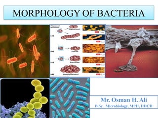
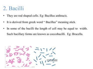
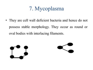
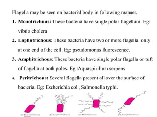
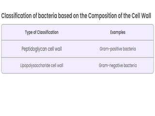
SlideShare a Scribd company logo
Mr. Osman H. Ali
B.Sc. Microbiology, MPH, HDCH
MORPHOLOGY OF BACTERIA
Contents
• Introduction
• Size of Bacteria
• Shape of Bacteria
• Arrangements of Bacterial Cells
• Structure of Bacterial Cell
Introduction
• A prokaryotic and single cell microorganism.
• Small in size.
• Don’t have nuclear membranes and also lack membrane
bound organelles.
• Can be able to see by using Microscope.
Conti……
• They occur in water, soil, air, food, and all
natural environment. They can survive extremes of temperature,
pH, oxygen, and atmospheric pressure.
Size of Bacteria
• Bacteria are very small microorganisms which are visible
under the microscope.
• They are having the size range in microns.
• Bacteria are stained by staining reagents and
then visualised under
(1000X)
high power of
of compound
magnification
microscope.
• An electron microscope is used for clear
visualization of internal structure of bacteria.
SHAPE OF BACTERIA
On the basis of shape bacteria are classified as
1. Cocci
2. Bacilli
3. Vibrios
4. Spirilla
5. Spirochetes
6. Actinomycetes
7. Mycoplasma
1. Cocci
• Cocci are small, spherical or oval cells. In greek
‘Kokkos’ means berry. Eg: micrococcus
2. Bacilli
• They are rod shaped cells. Eg: Bacillus anthracis.
• It is derived from greek word “ Bacillus” meaning stick.
• In some of the bacilli the length of cell may be equal to width.
Such bacillary forms are known as coccobacilli. Eg: Bracella.
3. Vibrios
• They are comma shaped curved rods. Eg: Vibrio
comma.
4. Spirilla
• They are longer rigid rods with several curves or
coils.
• They have a helical shape and rigid body.
• Eg: Spirillum ruprem.
5. Spirochetes
• They are slender and flexuous spiral forms.
6. Actinomycetes
• The characteristic shape is due to the presence of
rigid cell wall. Eg: Streptomyces.
• They are branching filamentous bacteria.
• Eg: Streptomyces species.
7. Mycoplasma
• They are cell wall deficient bacteria and hence do not
possess stable morphology. They occur as round or
oval bodies with interlacing filaments.
Arrangement of Bacterial Cells
Cocci appears as several
arrangement or grouping.
1.Diplococci
2.Streptococci
3.Tetracocci
4.Staphylococci
5.Sarcinae
characteristics
1. Diplococci
• They split in one plane and remains in pair. Eg:
diplococcus pneumoniae.
2. Streptococci
These cells divide in one planes and remain attached ,
to form chains. Eg: streptococcus lactis.
3. Tetracocci
• They divide in two planes and live in groups of four.
Eg: Gaffyka tetragena.
4. Staphylococci
• Cocci cells divide in three planes in an irregular pattern.
These cells produce bunches of cocci as in grapes. Eg:
staphylococcus aureus, staphylococcus albus.
5. Sarcinae
• Sarcinae cells divide in three planes in a regular
pattern.
• These cells produces a cuboidal arrangement of
group of a eight cells.
• Eg: Micrococcus tetragena.
Arrangement of grouping formed by bacilli
species
1. Diplobacilli
2. Streptobacilli
3. Trichomes
Trichomes
Diplobacilli
streptobacilli
Bacterial Structures
• Flagella
• Pili
• Capsule
• Plasma Membrane
• Cytoplasm
• Cell Wall
• Ribosomes
• mesosomes
• Inclusions
• Spores
Flagella
• Flagella are
appendages,
bacteria.
long, slender, thin hair-like cytoplasmic
which are responsible for the motility of
• These are the organs of locomotion.
• They are 0.01 to 0.02 µm in diameter, 3 to 20 µm in length.
• Flagella are made up of a protein- flagellin.
• The flagellum has three basic parts ,
1. Filament
2. Hook
3. Basal body
• Filament is the thin, cylindrical, long outermost region with a
constant diameter.
• The filament is attached to a slightly wider hook.
• The basal body is composed of a small central rod inserted
into a series of rings.
• Gram negative bacteria contain four rings as L-ring, P-ring, S-
ring, M-ring whereas gram positive bacteria have only S and M
rings in basal body.
Flagella may be seen on bacterial body in following manner.
1. Monotrichous: These bacteria have single polar flagellum. Eg:
vibrio cholera
2. Lophotrichous: These bacteria have two or more flagella only
at one end of the cell. Eg: pseudomonas fluorescence.
3. Amphitrichous: These bacteria have single polar flagella or tuft
of flagella at both poles. Eg :Aquaspirillum serpens.
4. Peritrichous: Several flagella present all over the surface of
bacteria. Eg: Escherichia coli, Salmonella typhi.
Pili or fimbriae
• Pili are hair-like microfibrils, 0.5 to 2 µm in length and 5 to 7
nm in diameter.
• They are thinner, shorter and more numerous than flagella.
• They are present only on gram negative cells.
• They are composed of protein known as pillin.
• They are unrelated to motility and are found on motile and non-
motile cells.
Conti……
• Fimbriae and pili, these two terms are used interchangeably but
they can be distinguished.
• Fimbriae can be evenly distributed over the entire surface of the
cell or they occurs at the poles of the bacterial cell. Each
bacteria possess 100 to 200 fimbriae.
• Pili are usually longer than fimbriae and number only one or two
per cell.
Function:
• Pili play an important role in attachment to surfaces. Hence pili
is also called organ of adhesion.
• The cytoplasmic (plasma) membrane is a thin ( 5 to 10 nm).
• It separates the cell wall and cytoplasm.
• It composed of phospholipids (20 to 30 %) and proteins ( 60 to
70 %).
• Prokaryotic plasma membranes are less rigid than eukaryotic
membrane due to lack of sterols.
Plasma membrane
Conti…….
Functions:
1. It acts as a semipermeable membrane controlling the inflow
and outflow of metabolites to and from the protoplasm.
2. It provides the mechanical strength to the bacterial cell.
3. It helps in DNA replication.
4. It contains enzyme, permease, which plays an important role
in the passage of selective nutrients through the membranes.
Cytoplasm
• The bacterial cytoplasm is a suspension of organic,
inorganic solutes in a viscous water solution.
• The cytoplasm of bacteria differs from that of
higher eukaryotic microorganisms in not
containing endoplasmic reticulum, Golgi
Conti…..
apparatus, mitochondria and lysosomes.
• It contains the ribosomes, proteins and other
water soluble components and reserve material.
• In most bacterial, extrachromosal DNA ( plasmid
DNA ) is also present.
Mesosomes
• Mesosomes are respiratory sites of bacteria.
• The mesosomes are attached to the bacterial
chromosomes and is involved in DNA segregation
during cell division.
• They are predominant in Gram positive bacteria.
Nucleoid
• The bacterial chromosomes is not surrounded by nuclear
membrane so it is called nucleoid.
• The bacterial chromosomes are made up of double strand
circular DNA.
Intra Cytoplasmic inclusion
Many species of bacteria produce cytoplasmic inclusion
bodies which appears as round granules.
• They are made up of either glycogen or starch.
• They appear reddish when stained with polychrome methylene
Capsule
• Bacteria synthesize loose amorphous organic exopolymer
which is deposited outside and tightly to cell wall called
capsules.
• Capsules may be composed of complex polypeptides or
of
polysaccharides. Water (98%) is the main component
bacterial capsule.
Conti……..
• Some times the capsular material is loosely associated with the
bacterium, it can be easily washed away. The loose layer is called
slime layer.
• Capsulated bacteria produces smooth colonies and non capsulated
bacteria produces rough colonies on the surface of agar media.
Functions
1. They protect the cell from drying.
2. They protects the bacterial cell against anti-bacterial agents and
phages.
Cell wall
• Cell wall is rigid structure which gives definite shape to cell,
situated between the capsule and cytoplasmic membrane.
• It is about 10 – 20 nm in thickness and constitutes 20-30 % of
dry weight of cell.
• The cell wall cannot be seen by direct light microscopy and does
not stain easily by different staining reagents.
Conti…….
• The cell wall of bacteria contains diaminopimelic acid (DAP),
muramic acid and teichoic acid. These substances are joined
together to give rise to a complex polymeric structure known as
peptidoglycan or murein or mucopeptide.
• Peptidoglycan is the major constituent of the cell wall of gram
positive bacteria (50 to 90 %) where as in gram negative
bacterial cell wall its presence is only 5 -10 %.
Chemical structure of a bacterial cell wall
A comparison of cell walls of gram positive and gram negative bacteria
Functions of cell wall
1. Cell wall is involved in growth and cell division of bacteria.
2. It gives shape to the cell.
3. It gives protection to internal structure and acts as
supporting layer.
4. To prevent rupture of bacteria caused by osmotic pressure
differences between intra cellular and extra cellular
environment.
5. To provide support for flagella.
6. To regulate a certain degree of passage of molecules into
and out of the cell.
7. It serve as the sites of attachment for most bacterial viruses.
Ribosomes
• Ribosomes are the center of protein synthesis.
• They are slightly smaller than eukaryotic ribosomes.
• The sedimentation constant is 70s.
• This 70s ribosomes are made up of two subunits namely a
large subunits 50s and a small subunit 30s.
30s
50s
• During active protein synthesis the ribosomes are associated
with mRNA and such associations are called polysomes.
mRNA
Polysomes
Spore
• The process of endospore formation is known as sporulation and it
may take 4 to 8 hours in a vegetative cell.
• Endospores are thick-walled, highly refractile bodies that are
produced one per cell.
• Each bacterial spore on germination forms a single vegetative cell.
Therefore, sporulation in bacteria is a method of preservation and not
reproduction.
Conti…….
• Spores are extremely resistant to dessication, staining,
disinfecting chemicals, radiation and heat.
• They remain viable for centuries and help bacteria to survive
for long period under unfavorable environment. Endospore
can remain dormant for thousand of years.
• Spores of all medically important bacteria are destroyed by
moist heat sterilization ( autoclave ) at 121 °C for 20 minutes.
BACTERIAL SPORULATION
Morphologyof Bacteria.pptx

More Related Content

What's hot

Aspergillus
AspergillusAspergillus
Aspergillus
vaishalidandge3
 
Embedding
EmbeddingEmbedding
COLLECTION AND TRANSPORTATION OF CLINICAL SAMPLES
COLLECTION AND TRANSPORTATION OF CLINICAL SAMPLESCOLLECTION AND TRANSPORTATION OF CLINICAL SAMPLES
COLLECTION AND TRANSPORTATION OF CLINICAL SAMPLES
NCRIMS, Meerut
 
Chromoblastomycosis and phaeohyphomycosis
Chromoblastomycosis and phaeohyphomycosisChromoblastomycosis and phaeohyphomycosis
Chromoblastomycosis and phaeohyphomycosis
adisutesfaye21
 
Klebsiella spp
Klebsiella sppKlebsiella spp
Klebsiella spp
Dr. Samira Fattah
 
Penicillium
PenicilliumPenicillium
Penicillium
Babar205761
 
Myxobacteria pptx
Myxobacteria pptxMyxobacteria pptx
Myxobacteria pptx
Asif nawaz khan (AUST)
 
STRUCTURE OF FUNGI.pptx
STRUCTURE OF FUNGI.pptxSTRUCTURE OF FUNGI.pptx
STRUCTURE OF FUNGI.pptx
NithyaNandapal
 
Colony characteristics of bacteria
Colony characteristics of bacteriaColony characteristics of bacteria
Colony characteristics of bacteria
Tahir Ali,Punjab University Lahore
 
Histokinetic by dr narmada
Histokinetic by  dr narmadaHistokinetic by  dr narmada
Histokinetic by dr narmadaNarmada Tiwari
 
Blood concentration Methods- PBF
Blood concentration Methods- PBFBlood concentration Methods- PBF
Blood concentration Methods- PBF
Shiksha Choytoo
 
Staphylococcus aureus bacteria ppt
Staphylococcus aureus  bacteria pptStaphylococcus aureus  bacteria ppt
Staphylococcus aureus bacteria ppt
Tusha Varshney
 
Grams staining
Grams stainingGrams staining
Grams staining
Dr.Dinesh Jain
 
Pseudomonas
PseudomonasPseudomonas
Pseudomonas
NCRIMS, Meerut
 
Actinomycetes
ActinomycetesActinomycetes
Actinomycetes
DR. ANKUR KUMAR
 
Capsule staining
Capsule stainingCapsule staining
Capsule staining
Mohit Hinsu
 
Parasitology (intestinal protozoa)
Parasitology (intestinal protozoa)Parasitology (intestinal protozoa)
Parasitology (intestinal protozoa)
Osama Al-Zahrani
 

What's hot (20)

Aspergillus
AspergillusAspergillus
Aspergillus
 
Embedding
EmbeddingEmbedding
Embedding
 
COLLECTION AND TRANSPORTATION OF CLINICAL SAMPLES
COLLECTION AND TRANSPORTATION OF CLINICAL SAMPLESCOLLECTION AND TRANSPORTATION OF CLINICAL SAMPLES
COLLECTION AND TRANSPORTATION OF CLINICAL SAMPLES
 
Chromoblastomycosis and phaeohyphomycosis
Chromoblastomycosis and phaeohyphomycosisChromoblastomycosis and phaeohyphomycosis
Chromoblastomycosis and phaeohyphomycosis
 
Klebsiella spp
Klebsiella sppKlebsiella spp
Klebsiella spp
 
Mycoplasma
MycoplasmaMycoplasma
Mycoplasma
 
Penicillium
PenicilliumPenicillium
Penicillium
 
Myxobacteria pptx
Myxobacteria pptxMyxobacteria pptx
Myxobacteria pptx
 
STRUCTURE OF FUNGI.pptx
STRUCTURE OF FUNGI.pptxSTRUCTURE OF FUNGI.pptx
STRUCTURE OF FUNGI.pptx
 
Colony characteristics of bacteria
Colony characteristics of bacteriaColony characteristics of bacteria
Colony characteristics of bacteria
 
stains
stainsstains
stains
 
Histokinetic by dr narmada
Histokinetic by  dr narmadaHistokinetic by  dr narmada
Histokinetic by dr narmada
 
Blood concentration Methods- PBF
Blood concentration Methods- PBFBlood concentration Methods- PBF
Blood concentration Methods- PBF
 
Staphylococcus aureus bacteria ppt
Staphylococcus aureus  bacteria pptStaphylococcus aureus  bacteria ppt
Staphylococcus aureus bacteria ppt
 
Grams staining
Grams stainingGrams staining
Grams staining
 
Pseudomonas
PseudomonasPseudomonas
Pseudomonas
 
Actinomycetes
ActinomycetesActinomycetes
Actinomycetes
 
Capsule staining
Capsule stainingCapsule staining
Capsule staining
 
Fungal cultures
Fungal culturesFungal cultures
Fungal cultures
 
Parasitology (intestinal protozoa)
Parasitology (intestinal protozoa)Parasitology (intestinal protozoa)
Parasitology (intestinal protozoa)
 

Similar to Morphologyof Bacteria.pptx

Morphology of bacteria
Morphology of bacteriaMorphology of bacteria
Morphology of bacteria
Snehal Patel
 
bcterialultrastructureall-201118081937.pdf
bcterialultrastructureall-201118081937.pdfbcterialultrastructureall-201118081937.pdf
bcterialultrastructureall-201118081937.pdf
dawitg2
 
Bcterial ultrastructure all
Bcterial ultrastructure allBcterial ultrastructure all
Bcterial ultrastructure all
Vasundhara Kakade Pisal
 
What is bacteria?(Structures Present in Bacteria And their Functions | Prokar...
What is bacteria?(Structures Present in Bacteria And their Functions | Prokar...What is bacteria?(Structures Present in Bacteria And their Functions | Prokar...
What is bacteria?(Structures Present in Bacteria And their Functions | Prokar...
sehriqayyum
 
Bacteria.pdf
Bacteria.pdfBacteria.pdf
Bacteria.pdf
Anup Bajracharya
 
Bacterial morphology
Bacterial morphologyBacterial morphology
Bacterial morphology
Ashfaq Ahmad
 
Bacterial cell
Bacterial cellBacterial cell
Bacterial cell
PalashGhosal1
 
Bacterialmorphologyanatomy 131001004402-phpapp02
Bacterialmorphologyanatomy 131001004402-phpapp02Bacterialmorphologyanatomy 131001004402-phpapp02
Bacterialmorphologyanatomy 131001004402-phpapp02bajuar
 
Bacterial morphology & anatomy
Bacterial morphology & anatomyBacterial morphology & anatomy
Bacterial morphology & anatomy
Kalpesh Zunjarrao
 
Bacterial Morphology and Atomy.pptx microbiology
Bacterial Morphology and Atomy.pptx microbiologyBacterial Morphology and Atomy.pptx microbiology
Bacterial Morphology and Atomy.pptx microbiology
osmanolow
 
Structureofbacterialcell 180806181503
Structureofbacterialcell 180806181503Structureofbacterialcell 180806181503
Structureofbacterialcell 180806181503
MICROBIOLOGYDEPARTME7
 
structureofbacterialcell-180806181503 (1).pdf
structureofbacterialcell-180806181503 (1).pdfstructureofbacterialcell-180806181503 (1).pdf
structureofbacterialcell-180806181503 (1).pdf
NdayisengaJeanBercky
 
Structure of bacterial cell
Structure of bacterial cellStructure of bacterial cell
Structure of bacterial cell
gohil sanjay bhagvanji
 
Morophology of bacteria
Morophology of bacteriaMorophology of bacteria
Lecture 2:- Bacterial morphology , bacterial cell structure
Lecture 2:- Bacterial morphology , bacterial cell structureLecture 2:- Bacterial morphology , bacterial cell structure
Lecture 2:- Bacterial morphology , bacterial cell structure
Raghda alomari
 
Bacteria
BacteriaBacteria
Bacteria
Dhanashri Mali
 
bacteria-200824080643.pdf
bacteria-200824080643.pdfbacteria-200824080643.pdf
bacteria-200824080643.pdf
dawitg2
 
Bacterial Morphologyand Atomy.pdf microbiology
Bacterial Morphologyand Atomy.pdf microbiologyBacterial Morphologyand Atomy.pdf microbiology
Bacterial Morphologyand Atomy.pdf microbiology
osmanolow
 
Presentation1.pptx
Presentation1.pptxPresentation1.pptx
Presentation1.pptx
SURYAPRAKASHDrKNModi
 
Ultrastructure and characterstic features of bacteria.
Ultrastructure and characterstic features of bacteria.Ultrastructure and characterstic features of bacteria.
Ultrastructure and characterstic features of bacteria.
Archana Shaw
 

Similar to Morphologyof Bacteria.pptx (20)

Morphology of bacteria
Morphology of bacteriaMorphology of bacteria
Morphology of bacteria
 
bcterialultrastructureall-201118081937.pdf
bcterialultrastructureall-201118081937.pdfbcterialultrastructureall-201118081937.pdf
bcterialultrastructureall-201118081937.pdf
 
Bcterial ultrastructure all
Bcterial ultrastructure allBcterial ultrastructure all
Bcterial ultrastructure all
 
What is bacteria?(Structures Present in Bacteria And their Functions | Prokar...
What is bacteria?(Structures Present in Bacteria And their Functions | Prokar...What is bacteria?(Structures Present in Bacteria And their Functions | Prokar...
What is bacteria?(Structures Present in Bacteria And their Functions | Prokar...
 
Bacteria.pdf
Bacteria.pdfBacteria.pdf
Bacteria.pdf
 
Bacterial morphology
Bacterial morphologyBacterial morphology
Bacterial morphology
 
Bacterial cell
Bacterial cellBacterial cell
Bacterial cell
 
Bacterialmorphologyanatomy 131001004402-phpapp02
Bacterialmorphologyanatomy 131001004402-phpapp02Bacterialmorphologyanatomy 131001004402-phpapp02
Bacterialmorphologyanatomy 131001004402-phpapp02
 
Bacterial morphology & anatomy
Bacterial morphology & anatomyBacterial morphology & anatomy
Bacterial morphology & anatomy
 
Bacterial Morphology and Atomy.pptx microbiology
Bacterial Morphology and Atomy.pptx microbiologyBacterial Morphology and Atomy.pptx microbiology
Bacterial Morphology and Atomy.pptx microbiology
 
Structureofbacterialcell 180806181503
Structureofbacterialcell 180806181503Structureofbacterialcell 180806181503
Structureofbacterialcell 180806181503
 
structureofbacterialcell-180806181503 (1).pdf
structureofbacterialcell-180806181503 (1).pdfstructureofbacterialcell-180806181503 (1).pdf
structureofbacterialcell-180806181503 (1).pdf
 
Structure of bacterial cell
Structure of bacterial cellStructure of bacterial cell
Structure of bacterial cell
 
Morophology of bacteria
Morophology of bacteriaMorophology of bacteria
Morophology of bacteria
 
Lecture 2:- Bacterial morphology , bacterial cell structure
Lecture 2:- Bacterial morphology , bacterial cell structureLecture 2:- Bacterial morphology , bacterial cell structure
Lecture 2:- Bacterial morphology , bacterial cell structure
 
Bacteria
BacteriaBacteria
Bacteria
 
bacteria-200824080643.pdf
bacteria-200824080643.pdfbacteria-200824080643.pdf
bacteria-200824080643.pdf
 
Bacterial Morphologyand Atomy.pdf microbiology
Bacterial Morphologyand Atomy.pdf microbiologyBacterial Morphologyand Atomy.pdf microbiology
Bacterial Morphologyand Atomy.pdf microbiology
 
Presentation1.pptx
Presentation1.pptxPresentation1.pptx
Presentation1.pptx
 
Ultrastructure and characterstic features of bacteria.
Ultrastructure and characterstic features of bacteria.Ultrastructure and characterstic features of bacteria.
Ultrastructure and characterstic features of bacteria.
 

More from OsmanHassan35

Antimicrobial Chemotherapy.pptx
Antimicrobial Chemotherapy.pptxAntimicrobial Chemotherapy.pptx
Antimicrobial Chemotherapy.pptx
OsmanHassan35
 
sterilization disinfection.pdf
sterilization disinfection.pdfsterilization disinfection.pdf
sterilization disinfection.pdf
OsmanHassan35
 
sterilization & disinfectiono- lecture 5.pptx
sterilization & disinfectiono- lecture 5.pptxsterilization & disinfectiono- lecture 5.pptx
sterilization & disinfectiono- lecture 5.pptx
OsmanHassan35
 
Bacterial taxonomy.pptx
Bacterial taxonomy.pptxBacterial taxonomy.pptx
Bacterial taxonomy.pptx
OsmanHassan35
 
sterilization disinfection.pptx
sterilization disinfection.pptxsterilization disinfection.pptx
sterilization disinfection.pptx
OsmanHassan35
 
Gram Staining PDF.pdf
Gram Staining PDF.pdfGram Staining PDF.pdf
Gram Staining PDF.pdf
OsmanHassan35
 
Culture Media.pdf
Culture Media.pdfCulture Media.pdf
Culture Media.pdf
OsmanHassan35
 
Foodquality ManagementSystems.pptx
Foodquality ManagementSystems.pptxFoodquality ManagementSystems.pptx
Foodquality ManagementSystems.pptx
OsmanHassan35
 
Microorganisms Associated with Food. chapter 2.pptx
Microorganisms Associated with Food. chapter 2.pptxMicroorganisms Associated with Food. chapter 2.pptx
Microorganisms Associated with Food. chapter 2.pptx
OsmanHassan35
 
bacterialphysiology- lecture 4.pptx
bacterialphysiology- lecture 4.pptxbacterialphysiology- lecture 4.pptx
bacterialphysiology- lecture 4.pptx
OsmanHassan35
 
Food FQC lecture 1.pdf
Food FQC lecture 1.pdfFood FQC lecture 1.pdf
Food FQC lecture 1.pdf
OsmanHassan35
 
Balantidium coli.pdf
Balantidium coli.pdfBalantidium coli.pdf
Balantidium coli.pdf
OsmanHassan35
 
Entamoeba histolytica.pptx
Entamoeba histolytica.pptxEntamoeba histolytica.pptx
Entamoeba histolytica.pptx
OsmanHassan35
 
trichomases.pdf
trichomases.pdftrichomases.pdf
trichomases.pdf
OsmanHassan35
 
necator-americanus.pptx
necator-americanus.pptxnecator-americanus.pptx
necator-americanus.pptx
OsmanHassan35
 
trichuristrichiura.pptx
trichuristrichiura.pptxtrichuristrichiura.pptx
trichuristrichiura.pptx
OsmanHassan35
 
enterobiusvermicularis.pptx
enterobiusvermicularis.pptxenterobiusvermicularis.pptx
enterobiusvermicularis.pptx
OsmanHassan35
 
onchocerca-160125115128 (2).pdf
onchocerca-160125115128 (2).pdfonchocerca-160125115128 (2).pdf
onchocerca-160125115128 (2).pdf
OsmanHassan35
 
trichinella spiralis.pptx
trichinella spiralis.pptxtrichinella spiralis.pptx
trichinella spiralis.pptx
OsmanHassan35
 
wuchereriabancroftibycarmel-160125115235.pdf
wuchereriabancroftibycarmel-160125115235.pdfwuchereriabancroftibycarmel-160125115235.pdf
wuchereriabancroftibycarmel-160125115235.pdf
OsmanHassan35
 

More from OsmanHassan35 (20)

Antimicrobial Chemotherapy.pptx
Antimicrobial Chemotherapy.pptxAntimicrobial Chemotherapy.pptx
Antimicrobial Chemotherapy.pptx
 
sterilization disinfection.pdf
sterilization disinfection.pdfsterilization disinfection.pdf
sterilization disinfection.pdf
 
sterilization & disinfectiono- lecture 5.pptx
sterilization & disinfectiono- lecture 5.pptxsterilization & disinfectiono- lecture 5.pptx
sterilization & disinfectiono- lecture 5.pptx
 
Bacterial taxonomy.pptx
Bacterial taxonomy.pptxBacterial taxonomy.pptx
Bacterial taxonomy.pptx
 
sterilization disinfection.pptx
sterilization disinfection.pptxsterilization disinfection.pptx
sterilization disinfection.pptx
 
Gram Staining PDF.pdf
Gram Staining PDF.pdfGram Staining PDF.pdf
Gram Staining PDF.pdf
 
Culture Media.pdf
Culture Media.pdfCulture Media.pdf
Culture Media.pdf
 
Foodquality ManagementSystems.pptx
Foodquality ManagementSystems.pptxFoodquality ManagementSystems.pptx
Foodquality ManagementSystems.pptx
 
Microorganisms Associated with Food. chapter 2.pptx
Microorganisms Associated with Food. chapter 2.pptxMicroorganisms Associated with Food. chapter 2.pptx
Microorganisms Associated with Food. chapter 2.pptx
 
bacterialphysiology- lecture 4.pptx
bacterialphysiology- lecture 4.pptxbacterialphysiology- lecture 4.pptx
bacterialphysiology- lecture 4.pptx
 
Food FQC lecture 1.pdf
Food FQC lecture 1.pdfFood FQC lecture 1.pdf
Food FQC lecture 1.pdf
 
Balantidium coli.pdf
Balantidium coli.pdfBalantidium coli.pdf
Balantidium coli.pdf
 
Entamoeba histolytica.pptx
Entamoeba histolytica.pptxEntamoeba histolytica.pptx
Entamoeba histolytica.pptx
 
trichomases.pdf
trichomases.pdftrichomases.pdf
trichomases.pdf
 
necator-americanus.pptx
necator-americanus.pptxnecator-americanus.pptx
necator-americanus.pptx
 
trichuristrichiura.pptx
trichuristrichiura.pptxtrichuristrichiura.pptx
trichuristrichiura.pptx
 
enterobiusvermicularis.pptx
enterobiusvermicularis.pptxenterobiusvermicularis.pptx
enterobiusvermicularis.pptx
 
onchocerca-160125115128 (2).pdf
onchocerca-160125115128 (2).pdfonchocerca-160125115128 (2).pdf
onchocerca-160125115128 (2).pdf
 
trichinella spiralis.pptx
trichinella spiralis.pptxtrichinella spiralis.pptx
trichinella spiralis.pptx
 
wuchereriabancroftibycarmel-160125115235.pdf
wuchereriabancroftibycarmel-160125115235.pdfwuchereriabancroftibycarmel-160125115235.pdf
wuchereriabancroftibycarmel-160125115235.pdf
 

Recently uploaded

Lung Cancer: Artificial Intelligence, Synergetics, Complex System Analysis, S...
Lung Cancer: Artificial Intelligence, Synergetics, Complex System Analysis, S...Lung Cancer: Artificial Intelligence, Synergetics, Complex System Analysis, S...
Lung Cancer: Artificial Intelligence, Synergetics, Complex System Analysis, S...
Oleg Kshivets
 
Are There Any Natural Remedies To Treat Syphilis.pdf
Are There Any Natural Remedies To Treat Syphilis.pdfAre There Any Natural Remedies To Treat Syphilis.pdf
Are There Any Natural Remedies To Treat Syphilis.pdf
Little Cross Family Clinic
 
ACUTE SCROTUM.....pdf. ACUTE SCROTAL CONDITIOND
ACUTE SCROTUM.....pdf. ACUTE SCROTAL CONDITIONDACUTE SCROTUM.....pdf. ACUTE SCROTAL CONDITIOND
ACUTE SCROTUM.....pdf. ACUTE SCROTAL CONDITIOND
DR SETH JOTHAM
 
Triangles of Neck and Clinical Correlation by Dr. RIG.pptx
Triangles of Neck and Clinical Correlation by Dr. RIG.pptxTriangles of Neck and Clinical Correlation by Dr. RIG.pptx
Triangles of Neck and Clinical Correlation by Dr. RIG.pptx
Dr. Rabia Inam Gandapore
 
micro teaching on communication m.sc nursing.pdf
micro teaching on communication m.sc nursing.pdfmicro teaching on communication m.sc nursing.pdf
micro teaching on communication m.sc nursing.pdf
Anurag Sharma
 
Prix Galien International 2024 Forum Program
Prix Galien International 2024 Forum ProgramPrix Galien International 2024 Forum Program
Prix Galien International 2024 Forum Program
Levi Shapiro
 
NVBDCP.pptx Nation vector borne disease control program
NVBDCP.pptx Nation vector borne disease control programNVBDCP.pptx Nation vector borne disease control program
NVBDCP.pptx Nation vector borne disease control program
Sapna Thakur
 
Maxilla, Mandible & Hyoid Bone & Clinical Correlations by Dr. RIG.pptx
Maxilla, Mandible & Hyoid Bone & Clinical Correlations by Dr. RIG.pptxMaxilla, Mandible & Hyoid Bone & Clinical Correlations by Dr. RIG.pptx
Maxilla, Mandible & Hyoid Bone & Clinical Correlations by Dr. RIG.pptx
Dr. Rabia Inam Gandapore
 
Superficial & Deep Fascia of the NECK.pptx
Superficial & Deep Fascia of the NECK.pptxSuperficial & Deep Fascia of the NECK.pptx
Superficial & Deep Fascia of the NECK.pptx
Dr. Rabia Inam Gandapore
 
How STIs Influence the Development of Pelvic Inflammatory Disease.pptx
How STIs Influence the Development of Pelvic Inflammatory Disease.pptxHow STIs Influence the Development of Pelvic Inflammatory Disease.pptx
How STIs Influence the Development of Pelvic Inflammatory Disease.pptx
FFragrant
 
Surat @ℂall @Girls ꧁❤8527049040❤꧂@ℂall @Girls Service Vip Top Model Safe
Surat @ℂall @Girls ꧁❤8527049040❤꧂@ℂall @Girls Service Vip Top Model SafeSurat @ℂall @Girls ꧁❤8527049040❤꧂@ℂall @Girls Service Vip Top Model Safe
Surat @ℂall @Girls ꧁❤8527049040❤꧂@ℂall @Girls Service Vip Top Model Safe
Savita Shen $i11
 
HOT NEW PRODUCT! BIG SALES FAST SHIPPING NOW FROM CHINA!! EU KU DB BK substit...
HOT NEW PRODUCT! BIG SALES FAST SHIPPING NOW FROM CHINA!! EU KU DB BK substit...HOT NEW PRODUCT! BIG SALES FAST SHIPPING NOW FROM CHINA!! EU KU DB BK substit...
HOT NEW PRODUCT! BIG SALES FAST SHIPPING NOW FROM CHINA!! EU KU DB BK substit...
GL Anaacs
 
Phone Us ❤85270-49040❤ #ℂall #gIRLS In Surat By Surat @ℂall @Girls Hotel With...
Phone Us ❤85270-49040❤ #ℂall #gIRLS In Surat By Surat @ℂall @Girls Hotel With...Phone Us ❤85270-49040❤ #ℂall #gIRLS In Surat By Surat @ℂall @Girls Hotel With...
Phone Us ❤85270-49040❤ #ℂall #gIRLS In Surat By Surat @ℂall @Girls Hotel With...
Savita Shen $i11
 
Flu Vaccine Alert in Bangalore Karnataka
Flu Vaccine Alert in Bangalore KarnatakaFlu Vaccine Alert in Bangalore Karnataka
Flu Vaccine Alert in Bangalore Karnataka
addon Scans
 
Alcohol_Dr. Jeenal Mistry MD Pharmacology.pdf
Alcohol_Dr. Jeenal Mistry MD Pharmacology.pdfAlcohol_Dr. Jeenal Mistry MD Pharmacology.pdf
Alcohol_Dr. Jeenal Mistry MD Pharmacology.pdf
Dr Jeenal Mistry
 
Charaka Samhita Sutra sthana Chapter 15 Upakalpaniyaadhyaya
Charaka Samhita Sutra sthana Chapter 15 UpakalpaniyaadhyayaCharaka Samhita Sutra sthana Chapter 15 Upakalpaniyaadhyaya
Charaka Samhita Sutra sthana Chapter 15 Upakalpaniyaadhyaya
Dr KHALID B.M
 
heat stroke and heat exhaustion in children
heat stroke and heat exhaustion in childrenheat stroke and heat exhaustion in children
heat stroke and heat exhaustion in children
SumeraAhmad5
 
Ocular injury ppt Upendra pal optometrist upums saifai etawah
Ocular injury  ppt  Upendra pal  optometrist upums saifai etawahOcular injury  ppt  Upendra pal  optometrist upums saifai etawah
Ocular injury ppt Upendra pal optometrist upums saifai etawah
pal078100
 
Report Back from SGO 2024: What’s the Latest in Cervical Cancer?
Report Back from SGO 2024: What’s the Latest in Cervical Cancer?Report Back from SGO 2024: What’s the Latest in Cervical Cancer?
Report Back from SGO 2024: What’s the Latest in Cervical Cancer?
bkling
 
POST OPERATIVE OLIGURIA and its management
POST OPERATIVE OLIGURIA and its managementPOST OPERATIVE OLIGURIA and its management
POST OPERATIVE OLIGURIA and its management
touseefaziz1
 

Recently uploaded (20)

Lung Cancer: Artificial Intelligence, Synergetics, Complex System Analysis, S...
Lung Cancer: Artificial Intelligence, Synergetics, Complex System Analysis, S...Lung Cancer: Artificial Intelligence, Synergetics, Complex System Analysis, S...
Lung Cancer: Artificial Intelligence, Synergetics, Complex System Analysis, S...
 
Are There Any Natural Remedies To Treat Syphilis.pdf
Are There Any Natural Remedies To Treat Syphilis.pdfAre There Any Natural Remedies To Treat Syphilis.pdf
Are There Any Natural Remedies To Treat Syphilis.pdf
 
ACUTE SCROTUM.....pdf. ACUTE SCROTAL CONDITIOND
ACUTE SCROTUM.....pdf. ACUTE SCROTAL CONDITIONDACUTE SCROTUM.....pdf. ACUTE SCROTAL CONDITIOND
ACUTE SCROTUM.....pdf. ACUTE SCROTAL CONDITIOND
 
Triangles of Neck and Clinical Correlation by Dr. RIG.pptx
Triangles of Neck and Clinical Correlation by Dr. RIG.pptxTriangles of Neck and Clinical Correlation by Dr. RIG.pptx
Triangles of Neck and Clinical Correlation by Dr. RIG.pptx
 
micro teaching on communication m.sc nursing.pdf
micro teaching on communication m.sc nursing.pdfmicro teaching on communication m.sc nursing.pdf
micro teaching on communication m.sc nursing.pdf
 
Prix Galien International 2024 Forum Program
Prix Galien International 2024 Forum ProgramPrix Galien International 2024 Forum Program
Prix Galien International 2024 Forum Program
 
NVBDCP.pptx Nation vector borne disease control program
NVBDCP.pptx Nation vector borne disease control programNVBDCP.pptx Nation vector borne disease control program
NVBDCP.pptx Nation vector borne disease control program
 
Maxilla, Mandible & Hyoid Bone & Clinical Correlations by Dr. RIG.pptx
Maxilla, Mandible & Hyoid Bone & Clinical Correlations by Dr. RIG.pptxMaxilla, Mandible & Hyoid Bone & Clinical Correlations by Dr. RIG.pptx
Maxilla, Mandible & Hyoid Bone & Clinical Correlations by Dr. RIG.pptx
 
Superficial & Deep Fascia of the NECK.pptx
Superficial & Deep Fascia of the NECK.pptxSuperficial & Deep Fascia of the NECK.pptx
Superficial & Deep Fascia of the NECK.pptx
 
How STIs Influence the Development of Pelvic Inflammatory Disease.pptx
How STIs Influence the Development of Pelvic Inflammatory Disease.pptxHow STIs Influence the Development of Pelvic Inflammatory Disease.pptx
How STIs Influence the Development of Pelvic Inflammatory Disease.pptx
 
Surat @ℂall @Girls ꧁❤8527049040❤꧂@ℂall @Girls Service Vip Top Model Safe
Surat @ℂall @Girls ꧁❤8527049040❤꧂@ℂall @Girls Service Vip Top Model SafeSurat @ℂall @Girls ꧁❤8527049040❤꧂@ℂall @Girls Service Vip Top Model Safe
Surat @ℂall @Girls ꧁❤8527049040❤꧂@ℂall @Girls Service Vip Top Model Safe
 
HOT NEW PRODUCT! BIG SALES FAST SHIPPING NOW FROM CHINA!! EU KU DB BK substit...
HOT NEW PRODUCT! BIG SALES FAST SHIPPING NOW FROM CHINA!! EU KU DB BK substit...HOT NEW PRODUCT! BIG SALES FAST SHIPPING NOW FROM CHINA!! EU KU DB BK substit...
HOT NEW PRODUCT! BIG SALES FAST SHIPPING NOW FROM CHINA!! EU KU DB BK substit...
 
Phone Us ❤85270-49040❤ #ℂall #gIRLS In Surat By Surat @ℂall @Girls Hotel With...
Phone Us ❤85270-49040❤ #ℂall #gIRLS In Surat By Surat @ℂall @Girls Hotel With...Phone Us ❤85270-49040❤ #ℂall #gIRLS In Surat By Surat @ℂall @Girls Hotel With...
Phone Us ❤85270-49040❤ #ℂall #gIRLS In Surat By Surat @ℂall @Girls Hotel With...
 
Flu Vaccine Alert in Bangalore Karnataka
Flu Vaccine Alert in Bangalore KarnatakaFlu Vaccine Alert in Bangalore Karnataka
Flu Vaccine Alert in Bangalore Karnataka
 
Alcohol_Dr. Jeenal Mistry MD Pharmacology.pdf
Alcohol_Dr. Jeenal Mistry MD Pharmacology.pdfAlcohol_Dr. Jeenal Mistry MD Pharmacology.pdf
Alcohol_Dr. Jeenal Mistry MD Pharmacology.pdf
 
Charaka Samhita Sutra sthana Chapter 15 Upakalpaniyaadhyaya
Charaka Samhita Sutra sthana Chapter 15 UpakalpaniyaadhyayaCharaka Samhita Sutra sthana Chapter 15 Upakalpaniyaadhyaya
Charaka Samhita Sutra sthana Chapter 15 Upakalpaniyaadhyaya
 
heat stroke and heat exhaustion in children
heat stroke and heat exhaustion in childrenheat stroke and heat exhaustion in children
heat stroke and heat exhaustion in children
 
Ocular injury ppt Upendra pal optometrist upums saifai etawah
Ocular injury  ppt  Upendra pal  optometrist upums saifai etawahOcular injury  ppt  Upendra pal  optometrist upums saifai etawah
Ocular injury ppt Upendra pal optometrist upums saifai etawah
 
Report Back from SGO 2024: What’s the Latest in Cervical Cancer?
Report Back from SGO 2024: What’s the Latest in Cervical Cancer?Report Back from SGO 2024: What’s the Latest in Cervical Cancer?
Report Back from SGO 2024: What’s the Latest in Cervical Cancer?
 
POST OPERATIVE OLIGURIA and its management
POST OPERATIVE OLIGURIA and its managementPOST OPERATIVE OLIGURIA and its management
POST OPERATIVE OLIGURIA and its management
 

Morphologyof Bacteria.pptx

  • 1. Mr. Osman H. Ali B.Sc. Microbiology, MPH, HDCH MORPHOLOGY OF BACTERIA
  • 2. Contents • Introduction • Size of Bacteria • Shape of Bacteria • Arrangements of Bacterial Cells • Structure of Bacterial Cell
  • 3. Introduction • A prokaryotic and single cell microorganism. • Small in size. • Don’t have nuclear membranes and also lack membrane bound organelles. • Can be able to see by using Microscope.
  • 4. Conti…… • They occur in water, soil, air, food, and all natural environment. They can survive extremes of temperature, pH, oxygen, and atmospheric pressure.
  • 5. Size of Bacteria • Bacteria are very small microorganisms which are visible under the microscope. • They are having the size range in microns. • Bacteria are stained by staining reagents and then visualised under (1000X) high power of of compound magnification microscope. • An electron microscope is used for clear visualization of internal structure of bacteria.
  • 6. SHAPE OF BACTERIA On the basis of shape bacteria are classified as 1. Cocci 2. Bacilli 3. Vibrios 4. Spirilla 5. Spirochetes 6. Actinomycetes 7. Mycoplasma
  • 7.
  • 8. 1. Cocci • Cocci are small, spherical or oval cells. In greek ‘Kokkos’ means berry. Eg: micrococcus
  • 9. 2. Bacilli • They are rod shaped cells. Eg: Bacillus anthracis. • It is derived from greek word “ Bacillus” meaning stick. • In some of the bacilli the length of cell may be equal to width. Such bacillary forms are known as coccobacilli. Eg: Bracella.
  • 10. 3. Vibrios • They are comma shaped curved rods. Eg: Vibrio comma.
  • 11. 4. Spirilla • They are longer rigid rods with several curves or coils. • They have a helical shape and rigid body. • Eg: Spirillum ruprem.
  • 12. 5. Spirochetes • They are slender and flexuous spiral forms.
  • 13. 6. Actinomycetes • The characteristic shape is due to the presence of rigid cell wall. Eg: Streptomyces. • They are branching filamentous bacteria. • Eg: Streptomyces species.
  • 14. 7. Mycoplasma • They are cell wall deficient bacteria and hence do not possess stable morphology. They occur as round or oval bodies with interlacing filaments.
  • 15. Arrangement of Bacterial Cells Cocci appears as several arrangement or grouping. 1.Diplococci 2.Streptococci 3.Tetracocci 4.Staphylococci 5.Sarcinae characteristics
  • 16. 1. Diplococci • They split in one plane and remains in pair. Eg: diplococcus pneumoniae.
  • 17. 2. Streptococci These cells divide in one planes and remain attached , to form chains. Eg: streptococcus lactis.
  • 18. 3. Tetracocci • They divide in two planes and live in groups of four. Eg: Gaffyka tetragena.
  • 19. 4. Staphylococci • Cocci cells divide in three planes in an irregular pattern. These cells produce bunches of cocci as in grapes. Eg: staphylococcus aureus, staphylococcus albus.
  • 20. 5. Sarcinae • Sarcinae cells divide in three planes in a regular pattern. • These cells produces a cuboidal arrangement of group of a eight cells. • Eg: Micrococcus tetragena.
  • 21. Arrangement of grouping formed by bacilli species 1. Diplobacilli 2. Streptobacilli 3. Trichomes Trichomes Diplobacilli streptobacilli
  • 22. Bacterial Structures • Flagella • Pili • Capsule • Plasma Membrane • Cytoplasm • Cell Wall • Ribosomes • mesosomes • Inclusions • Spores
  • 23. Flagella • Flagella are appendages, bacteria. long, slender, thin hair-like cytoplasmic which are responsible for the motility of • These are the organs of locomotion. • They are 0.01 to 0.02 µm in diameter, 3 to 20 µm in length. • Flagella are made up of a protein- flagellin. • The flagellum has three basic parts , 1. Filament 2. Hook 3. Basal body
  • 24. • Filament is the thin, cylindrical, long outermost region with a constant diameter. • The filament is attached to a slightly wider hook. • The basal body is composed of a small central rod inserted into a series of rings. • Gram negative bacteria contain four rings as L-ring, P-ring, S- ring, M-ring whereas gram positive bacteria have only S and M rings in basal body.
  • 25. Flagella may be seen on bacterial body in following manner. 1. Monotrichous: These bacteria have single polar flagellum. Eg: vibrio cholera 2. Lophotrichous: These bacteria have two or more flagella only at one end of the cell. Eg: pseudomonas fluorescence. 3. Amphitrichous: These bacteria have single polar flagella or tuft of flagella at both poles. Eg :Aquaspirillum serpens. 4. Peritrichous: Several flagella present all over the surface of bacteria. Eg: Escherichia coli, Salmonella typhi.
  • 26. Pili or fimbriae • Pili are hair-like microfibrils, 0.5 to 2 µm in length and 5 to 7 nm in diameter. • They are thinner, shorter and more numerous than flagella. • They are present only on gram negative cells. • They are composed of protein known as pillin. • They are unrelated to motility and are found on motile and non- motile cells.
  • 27. Conti…… • Fimbriae and pili, these two terms are used interchangeably but they can be distinguished. • Fimbriae can be evenly distributed over the entire surface of the cell or they occurs at the poles of the bacterial cell. Each bacteria possess 100 to 200 fimbriae. • Pili are usually longer than fimbriae and number only one or two per cell. Function: • Pili play an important role in attachment to surfaces. Hence pili is also called organ of adhesion.
  • 28. • The cytoplasmic (plasma) membrane is a thin ( 5 to 10 nm). • It separates the cell wall and cytoplasm. • It composed of phospholipids (20 to 30 %) and proteins ( 60 to 70 %). • Prokaryotic plasma membranes are less rigid than eukaryotic membrane due to lack of sterols. Plasma membrane
  • 29. Conti……. Functions: 1. It acts as a semipermeable membrane controlling the inflow and outflow of metabolites to and from the protoplasm. 2. It provides the mechanical strength to the bacterial cell. 3. It helps in DNA replication. 4. It contains enzyme, permease, which plays an important role in the passage of selective nutrients through the membranes.
  • 30. Cytoplasm • The bacterial cytoplasm is a suspension of organic, inorganic solutes in a viscous water solution. • The cytoplasm of bacteria differs from that of higher eukaryotic microorganisms in not containing endoplasmic reticulum, Golgi
  • 31. Conti….. apparatus, mitochondria and lysosomes. • It contains the ribosomes, proteins and other water soluble components and reserve material. • In most bacterial, extrachromosal DNA ( plasmid DNA ) is also present.
  • 32. Mesosomes • Mesosomes are respiratory sites of bacteria. • The mesosomes are attached to the bacterial chromosomes and is involved in DNA segregation during cell division. • They are predominant in Gram positive bacteria.
  • 33. Nucleoid • The bacterial chromosomes is not surrounded by nuclear membrane so it is called nucleoid. • The bacterial chromosomes are made up of double strand circular DNA. Intra Cytoplasmic inclusion Many species of bacteria produce cytoplasmic inclusion bodies which appears as round granules. • They are made up of either glycogen or starch. • They appear reddish when stained with polychrome methylene
  • 34. Capsule • Bacteria synthesize loose amorphous organic exopolymer which is deposited outside and tightly to cell wall called capsules. • Capsules may be composed of complex polypeptides or of polysaccharides. Water (98%) is the main component bacterial capsule.
  • 35. Conti…….. • Some times the capsular material is loosely associated with the bacterium, it can be easily washed away. The loose layer is called slime layer. • Capsulated bacteria produces smooth colonies and non capsulated bacteria produces rough colonies on the surface of agar media. Functions 1. They protect the cell from drying. 2. They protects the bacterial cell against anti-bacterial agents and phages.
  • 36. Cell wall • Cell wall is rigid structure which gives definite shape to cell, situated between the capsule and cytoplasmic membrane. • It is about 10 – 20 nm in thickness and constitutes 20-30 % of dry weight of cell. • The cell wall cannot be seen by direct light microscopy and does not stain easily by different staining reagents.
  • 37. Conti……. • The cell wall of bacteria contains diaminopimelic acid (DAP), muramic acid and teichoic acid. These substances are joined together to give rise to a complex polymeric structure known as peptidoglycan or murein or mucopeptide. • Peptidoglycan is the major constituent of the cell wall of gram positive bacteria (50 to 90 %) where as in gram negative bacterial cell wall its presence is only 5 -10 %.
  • 38. Chemical structure of a bacterial cell wall
  • 39. A comparison of cell walls of gram positive and gram negative bacteria
  • 40. Functions of cell wall 1. Cell wall is involved in growth and cell division of bacteria. 2. It gives shape to the cell. 3. It gives protection to internal structure and acts as supporting layer. 4. To prevent rupture of bacteria caused by osmotic pressure differences between intra cellular and extra cellular environment. 5. To provide support for flagella. 6. To regulate a certain degree of passage of molecules into and out of the cell. 7. It serve as the sites of attachment for most bacterial viruses.
  • 41.
  • 42. Ribosomes • Ribosomes are the center of protein synthesis. • They are slightly smaller than eukaryotic ribosomes. • The sedimentation constant is 70s. • This 70s ribosomes are made up of two subunits namely a large subunits 50s and a small subunit 30s. 30s 50s • During active protein synthesis the ribosomes are associated with mRNA and such associations are called polysomes. mRNA Polysomes
  • 43. Spore • The process of endospore formation is known as sporulation and it may take 4 to 8 hours in a vegetative cell. • Endospores are thick-walled, highly refractile bodies that are produced one per cell. • Each bacterial spore on germination forms a single vegetative cell. Therefore, sporulation in bacteria is a method of preservation and not reproduction.
  • 44. Conti……. • Spores are extremely resistant to dessication, staining, disinfecting chemicals, radiation and heat. • They remain viable for centuries and help bacteria to survive for long period under unfavorable environment. Endospore can remain dormant for thousand of years. • Spores of all medically important bacteria are destroyed by moist heat sterilization ( autoclave ) at 121 °C for 20 minutes.
  • 45.
  • 46.