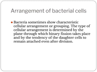
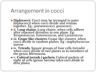
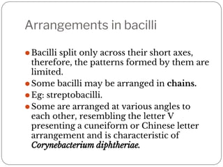
SlideShare a Scribd company logo
BACTERIAL MORPHOLOGY
Introduction
⚫Bacteria are unicellular free living
organisms without chlorophyll having
both DNA and RNA.
⚫They are prokaryotic organism i.e they
does not have a true nucleus.
⚫Size of bacteria are usually measured in
terms of microns or micrometers.
⚫Bacteria of medical importance generally
measures 0.2 – 1.5 micrometer.
⚫The smallest body that can be resolved
clearly by human eye is 200micron. Hence
to see bacteria , a light microscope must be
used.
Classification of bacteria based on
morphology
This classification is based on their shape.
⚫ Cocci : Spherical or near spherical shaped. Eg: streptococcus,
staphylococcus
⚫ Bacilli : relatively straight or rod shaped. Eg : Salmonella
i. Coccobacilli : Bacilli whose length and width are almost
equal. Eg: brucella
ii. Vibrio : curved or comma-shaped bacilli. Eg: Vibrio cholera
iii. Spirilla : rigid spiral or helical shaped. They are non-
flexible.
iv. Spirochete : these are also spiral shaped bacteria but they are
flexible.
v. Mycoplasma : these are cell wall deficient bacteria and hence
do not possess a stable morphology.
vi. Actinomycetes : these bacteria are long and show branching
similar to fungi.
Arrangement of bacterial cells
⚫ Bacteria sometimes show characteristic
cellular arrangement or grouping. The type of
cellular arrangement is determined by the
plane through which binary fission takes place
and by the tendency of the daughter cells to
remain attached even after division.
Arrangement in cocci
⚫ Diplococci: Cocci may be arranged in pairs
(diplococci) when cocci divide and remain
together. Eg ; pneumococci, gonococci
⚫ ii. Long chains: Long chains ,when cells adhere
after repeated divisions in one plane. Eg:
Streptococcus, Enterococcus, and Lactococcus
⚫ iii. Grape like clusters: Grape like clusters ,when
cocci divide in random planes. Eg : staphylococcus
aureus
⚫ iv. Tetrads: Square groups of four cells (tetrads)
when cocci divide in two planes as in members of
the genus Micrococcus.
⚫ v. Cubical (octads ) packets: Cubical packets of
eight of cells (genus Sarcina) when cocci divide in
three planes.
Arrangements in bacilli
⚫Bacilli split only across their short axes,
therefore, the patterns formed by them are
limited.
⚫Some bacilli may be arranged in chains.
⚫Eg: streptobacilli.
⚫Some are arranged at various angles to
each other, resembling the letter V
presenting a cuneiform or Chinese letter
arrangement and is characteristic of
Corynebacterium diphtheriae.
Anatomy of Bacterial Cell
⚫The bacterial cell has an outer layer or cell
envelop consisting of two components – a
rigid cell wall and beneath it a cytoplasmic
or plasma membrane.
⚫The cell envelop encloses the protoplasm,
comprising the cytoplasm, ribosomes,
vacuoles, and the nuclear body.
⚫Some bacteria may possess additional
structures such as capsule, fimbriae and
flagella
Cell wall
⚫The cell wall accounts for the shape of the
bacteria.
⚫Chemically the cell wall is composed of
peptidoglycan or murein scaffoldings
(formed by N Acetyl glucosamine and N
acetyl muramic acid alternating in chain
crosslinked by peptide chains.)
⚫In general, the walls of the Gram positive
bacteria have simpler chemical nature than
those of Gram negative bacteria.
⚫The bacterial cell wall contains antigens
that are important in virulence and
immunity.
Gram positive bacteria cell wall
⚫ It is about 80nm thick.
⚫ It is composed mostly of peptidoglycan ( 60-
90% of the cell wall is peptidoglycan) and are
responsible for retaining the crystal violet
during gram staining.
⚫ In addition, gram positive bacteria contain
theichoic acid (which consist primarily of an
alcohol and phosphate ). Thecoic acid acts as a
surface antigen, it binds with magnesium ion
and protect bacteria from thermal injuries.
⚫ Some bacterias like the Mycobacterium have
large amount of lipid in their cell wall and
hence they exhibit acid fast staining due to the
presence of mycolic acid.
Gram negative bacteria cell wall
⚫ Cell wall of gram negative bacteria is a complex structure.
⚫ Peptidoglycan layer in cell wall of gram negative bacteria is
only single-unit thick i.e it contribute only 5-10% of cell wall.
⚫ The outer portion of cell wall consist of an outer membrane
which is attached to the peptidoglycan layer by lipoproteins.
⚫ Outer membrane
⚫ The outer membrane is a bilayered structure. The inner layer is
formed by phospholipid while the outer layer is formed by
Lipopolysaccharide (LPS).
⚫ The outer membrane provides resistance to the bacterial cell
from various chemical including antibiotics, dyes, disinfectants
and lysosymes.
⚫ The outer membrane has unique proteins called porins which
permit passage of molecules into periplasm. ( Periplasm is a gel
like fluid between the outermembrane and plasma membrane.
Lipopolysaccharide (LPS)
⚫it is a complex molecule that contains lipid
and carbohydrate and consist of 3
components.
1. Lipid A : it is present in the top layer of
outer membrane. It acts as an endotoxin
and is responsible for the endotoxic
activities like fever, tissue necrosis etc
2. Core polysaccharide : these are sugars and
their role is to provide stability.
3. Polysaccharide O : is a sugar molecule
extending outwards from the core
polysaccharide. It is also called as O
Antigen. It is useful for distinguishing
species of gram negative bacteria.
Functions of cell wall
1. Provide shape to bacteria.
2. Prevent bacterial cell from bursting.
3. Role in division of bacteria
4. Resistance to harmful effects of
environment
Cytoplasmic (Plasma) Membrane
⚫ It is a semipermeable membrane which lies beneath the cell wall.
⚫ Chemically it is composed of phospholipids and proteins (integral
proteins and peripheral proteins).
⚫ Lipid molecules are arranged in a double layer with their
hydrophilic polar regions externally aligned and in contact with a
layer of protein at each surface.
⚫ Functions of Cytoplasmic Membrane
i. Semipermeable membrane—controlling the inflow and outflow
of metabolites to and from the protoplasm i.e transport of
nutrients into celland elimination of waste material.
ii. Housing enzymes involved in outer membrane synthesis, cell
wall synthesis, and manufacturing of extracellular structures.
iii. Housing many sensory and chemotaxis proteins that monitor
chemical and physical changes in the environment.
iv. Generation of chemical energy (i.e, ATP).
v. It helps in DNA replication.
Cytoplasm
⚫ Bacterial cytoplasm is a viscous watery
solution containing organic and inorganic
solutes like ribosomes, vacuoles, inclusion
bodies etc.
⚫ They lack mitochondria and endoplasmic
reticulum.
Ribosomes
⚫ These are the centre of protein synthesis.
⚫ They are composed of ribosomal RNA and ribosomal protein.
⚫ They are 10-20 nm size, with a sedimentation constant of 70 S
(S for Svedberg units).
⚫ Each 70 S unit consists of a 30 Sand a 50 S subunits.
Intracytoplasmic inclusions
⚫ These are source of stored energy.
⚫ These are usually seen in bacteria growing in conditions of
nutritional deficiencies and disappear when deficit nutrients
are supplied.
⚫ These inclusions may contain polysaccharides, lipids, sulphur
granules, volutin granules ( phosphate granules seen in
diphtheria bacilli.)
Mesosomes
⚫ Mesosomes are invaginations of the plasma
membrane in the shape of vesicles, tubules, or
lamellae.
⚫ They are generally more prominent in gram-
positive bacteria.
⚫ There are 2 types of mesosomes:- lateral and septal
Function
⚫ They have respiratory enzymes and are believed to
be involved in bacterial respiration ( analogous too
mitochondria of eukaryotes)
⚫ They (septal mesosomes) may be involved in cell
wall formation during cell division.
⚫ They also probably play a role in chromosome
replication and distribution to daughter cells.
Nucleoid
⚫ Bacteria lacks true nucleus, but the genetic
material is located in an irregular shaped
region called nucleoide.
⚫ They does not possess a nuclear membrane
(separating them from the cytoplasm)or
nucleolus.
⚫ The genetic material of a bacterial cell is
contained in a single, long molecule of
double-stranded deoxyribonucleic acid
(DNA) which can be extracted in the form
of a closed circular thread. It measures
about 1mm.
⚫ The bacterial chromosome is haploid and
replicates by simple fission instead of by
mitosis as in higher cells
Plasmid
⚫ These are extranuclear genetic elements
in the cytoplasm and it consists of DNA.
⚫ Also called as episomes.
⚫ Plasmids are not essential for host growth
and reproduction, but may confer on it
certain properties such as drug resistance,
resistance to toxic metal ,ability to
degrade complex organic molecules etc
which may constitute a survival
advantage.
⚫ These are transmitted from one bacteria
to another by conjugation (sexual
reproduction) or binary fission.
Cellular Appendages
Capsule and slime layer
⚫ Some bacteria possesses a layer of amorphous viscous
material lying outside cell wall called glycocalyx.
⚫ When it is well organised and not easily washed off, it
is called capsule. Eg: streptococcus pneumonia
⚫ If it is diffuse, unorganised and easily washed off it is
referred as a slime layer as in Leuconostoc.
⚫ Capsules too thin to be seen under the light
microscope are called microcapsules. Eg :Neisseria
meningitidis
⚫ Some bacteria may have both a capsule and a slime
layer (for example, Streptococcus salivarius).
⚫ Capsules and slime layers usually are composed of
polysaccharide (for example pneumococcus) or of
polypeptide in some bacteria (for example, Bacillus
anthracis and Yersinia pestis).
Capsule and slime layer
Functions of Capsule
i. Virulence factor: Capsules often act as a
virulence factor by protecting the bacterium
from ingestion by phagocytosis, and
noncapsulate mutant of these bacteria are
nonvirulent. Repeated subcultures in vitro
lead to the loss of capsule and also of
virulence.
ii. Protection of the cell: In protecting the
cell from attack by various kinds of
antibacterial agents, e.g. bacteriophages,
colicines, complement, lysozyme and other
lytic enzymes.
iii. Can act as a source of nutrients and
energy. Eg : Streptococcus mutans, which
colonizes teeth, ferments the sugar in the
capsule and so formed acid by-products
contribute to the tooth decay.
iv. Identification and typing of bacteria:
Capsular antigen is specific for bacteria and
can be used for identification and typing of
bacteria.
Flagella
⚫ These are cytoplasmic Appendages protruding through cell wall. They are the
organ of locomotion.
⚫ It has three parts :
i. Filament: The filament is the longest and most obvious portion which
extends from the cell surface to the tip. It is a hollow cylinder made of
single protein called flagellin.
ii. Hook: The hook is a short, curved segment which links the filament to
its basal body.
iii. Basal body: The basal body is embedded in the cytoplasmic
membrane.
In the gram-negative bacteria, the basal body has four rings connected to
a central rod (L, P, S and M). The outer L and P rings associated with the
lipopolysaccharide and peptidoglycan layers respectively. S ring is
located just above the cytoplasmic membrane and the inner M ring
contacts the cytoplasmic membrane.
Gram-positive bacteria have only two basal body rings, an inner ring
connected to the cytoplasmic membrane and an outer one probably
attached to peptidoglycan.
Arrangement of flagella
The number and location of flagella are
distinctive for each genus. There are four types
of flagella arrangement:
• Monotrichous —Single polar flagellum (e.g.
Cholera vibrio).
• Amphitrichous—Single flagellum at both ends
(e.g. Alcaligenes faecalis).
• Lophotrichous—Tuft of flagella at one or both
ends (e.g. spirilla).
• Peritrichous—Flagella surrounding the cell
(e.g. Typhoid bacilli).
Fimbria or Pili
⚫ These are hair like appendages projecting
from cell surface.
⚫ They are made of proteins called pilin.
⚫ They are generally seen in gram negative
bacteria.
⚫ They are antigenic.
⚫ They are not involved in motility and
hence can be seen in both motile and
non- motile bacteria.
⚫ Fimbria are organ of adhesion. This
enhances virulence of bacteria.
Types:
1. Common: They function as organs of adhesions
that allow attachment of a bacterial cell to other
cells or surfaces.
Based on morphology, adhesive property and
antigenic nature they are of 6 types.
1. Sex pili : These are longer and fewer in number
than other fimbriae. They are found on ‘male’
bacteria and help in the attachment of those cells
to ‘female’ bacteria, forming hollow conjugation
tubes through which, it is assumed, genetic
material is transferred from the donor to the
recipient cell.
2. Col 1 : These are associated with colicin factor 1.
Types of common pili
Type I Thick, haemagglutination positive, mannose sensitive
Type II Thick, haemagglutination positive, mannose sensitive
Type III Thin, haemagglutination positive, mannose resistant
Type IV Thinner, haemagglutination positive
Type V Monopolar fimbriae seen in pseudomonas
Type VI Fimbriae are very long and seen in klebsiella.
BACTERIAL SPORES
⚫ Spores are highly resistant resting (or dormat) stage of
the bacteria formed in unfavorable environmemal
conditions as a result of depletion of exogenous
nutrients.
⚫ Bacterial spores formed within the parent cell, are
called endospores and the remaining part of the
bacteria is called the sporangium.
⚫ Structure: Bacterial spore comprises of several layers.
From innermost towards the outermost, the layers are:
core- inner membrane - spore coat- exosporium
⚫ The core is the inner most part containing the DNA
material and is walled off from the cortex by an inner
membrane and 1he germ cell wall.
⚫ Cortex and the coat layers lies external to the core,
and are separated from each other by an outer
membrane.
⚫ The outermost layer is called as the exosporium.
⚫ Example of spore forming bacteria are bacillus and
clostridium.
Sporulation
It refers tonthe process of formation of spore from vegetative
stage of bacteria. It is not a method of reproduction because the
bacteria do not divide during sporulation.
Sporulation commences when growth of bacteria ceases due to
lack of nutrients.
1. The bacterial DNA replicates and divides into two DNA
molecules.
2. A transverse septum grows across the cell from the cell
membrane forming forespore and sporangium.
3. The forespore is subsequently completely encircled by
dividing septum as a dou ble layered membrane.The inner
layer becomes the inner membrane. Between the two layers
is laid spore cortex and outer layer is transformed into spore
coat which consists of several layers
4. In some species from outer layer also develops exosporium
which bears ridges and folds
5. Maturation of spore occurs and it acquires properties of heat
resistance and refractility.
6. Finally lytic enzymes destroys the sporangium releasing the
spore.
Germination
⚫ It is the process of conversion of a spore into
vegetative cell under suitable condition.
⚫ It occurs in 3 stages:
a. Activation : This process requires agents like
heat, low pH etc which damage the coat of
spore and help in germination.
b. Initiation : Once activated, the spore loses its
refractility and swells up if the conditions are
favourable.
c. Outgrowth : The spore wall is shed and the
germ cell appears by rupturing the remaining
spore coat.The germ cell elongates to form
the vegetative bacterium.
Thank you

More Related Content

Similar to BACTERIA MORPHOLOGY-1.pptx

EUBACTERIA :OVERVIEW
EUBACTERIA :OVERVIEWEUBACTERIA :OVERVIEW
EUBACTERIA :OVERVIEW
Himanshi Chauhan
 
structure of mo.pptx
structure of mo.pptxstructure of mo.pptx
structure of mo.pptx
DiptiPriya6
 
basic introdution on bacteria.pdf
basic introdution on bacteria.pdfbasic introdution on bacteria.pdf
basic introdution on bacteria.pdf
ShubhamkumarMaurya7
 
Ultrastructure and characterstic features of bacteria.
Ultrastructure and characterstic features of bacteria.Ultrastructure and characterstic features of bacteria.
Ultrastructure and characterstic features of bacteria.
Archana Shaw
 
Introduction of Microbiology
Introduction of MicrobiologyIntroduction of Microbiology
Introduction of Microbiology
ProfNori
 
Morphology of bacteria
Morphology of bacteriaMorphology of bacteria
Morphology of bacteria
Snehal Patel
 
1. Introduction Microbiology.pdf
1. Introduction Microbiology.pdf1. Introduction Microbiology.pdf
1. Introduction Microbiology.pdf
GOALLWORLD
 
Chapter1 cell structure of bacteria
Chapter1 cell structure of bacteriaChapter1 cell structure of bacteria
Chapter1 cell structure of bacteria
shobejee
 
Morphology of bacterial cell presentation new 1.pptx
Morphology of bacterial cell  presentation new 1.pptxMorphology of bacterial cell  presentation new 1.pptx
Morphology of bacterial cell presentation new 1.pptx
ArnabSamanta26
 
Bacteriology3.pdf
Bacteriology3.pdfBacteriology3.pdf
Bacteriology3.pdf
FkFo
 
Bacterial morphology
Bacterial morphologyBacterial morphology
Bacterial morphology
Ashfaq Ahmad
 
Morophology of bacteria
Morophology of bacteriaMorophology of bacteria
Prokaryotic cell (Anjali Bisen )
Prokaryotic cell (Anjali Bisen )Prokaryotic cell (Anjali Bisen )
Prokaryotic cell (Anjali Bisen )
AnjaliBisen
 
Study of bacteria
Study of bacteriaStudy of bacteria
Study of bacteria
Ravikumar Patil
 
bacteria- lecture 3.pptx microbiology and Immunology
bacteria- lecture 3.pptx microbiology and Immunologybacteria- lecture 3.pptx microbiology and Immunology
bacteria- lecture 3.pptx microbiology and Immunology
osmanolow
 
bacteria- lecture 3.pptx
bacteria- lecture 3.pptxbacteria- lecture 3.pptx
bacteria- lecture 3.pptx
OsmanAli92
 
Prokaryotes vs Eukaryotes
Prokaryotes vs EukaryotesProkaryotes vs Eukaryotes
Prokaryotes vs Eukaryotes
mgsonline
 
Prokaryotes _Eukaryotes
Prokaryotes _EukaryotesProkaryotes _Eukaryotes
Prokaryotes _Eukaryotes
musselburghgrammar
 
structure & classification of microbes
structure & classification of microbesstructure & classification of microbes
structure & classification of microbes
KHyati CHaudhari
 
sturcture of bacteria lecture 3[1].pptx
sturcture of bacteria  lecture 3[1].pptxsturcture of bacteria  lecture 3[1].pptx
sturcture of bacteria lecture 3[1].pptx
tehseenmalik450
 

Similar to BACTERIA MORPHOLOGY-1.pptx (20)

EUBACTERIA :OVERVIEW
EUBACTERIA :OVERVIEWEUBACTERIA :OVERVIEW
EUBACTERIA :OVERVIEW
 
structure of mo.pptx
structure of mo.pptxstructure of mo.pptx
structure of mo.pptx
 
basic introdution on bacteria.pdf
basic introdution on bacteria.pdfbasic introdution on bacteria.pdf
basic introdution on bacteria.pdf
 
Ultrastructure and characterstic features of bacteria.
Ultrastructure and characterstic features of bacteria.Ultrastructure and characterstic features of bacteria.
Ultrastructure and characterstic features of bacteria.
 
Introduction of Microbiology
Introduction of MicrobiologyIntroduction of Microbiology
Introduction of Microbiology
 
Morphology of bacteria
Morphology of bacteriaMorphology of bacteria
Morphology of bacteria
 
1. Introduction Microbiology.pdf
1. Introduction Microbiology.pdf1. Introduction Microbiology.pdf
1. Introduction Microbiology.pdf
 
Chapter1 cell structure of bacteria
Chapter1 cell structure of bacteriaChapter1 cell structure of bacteria
Chapter1 cell structure of bacteria
 
Morphology of bacterial cell presentation new 1.pptx
Morphology of bacterial cell  presentation new 1.pptxMorphology of bacterial cell  presentation new 1.pptx
Morphology of bacterial cell presentation new 1.pptx
 
Bacteriology3.pdf
Bacteriology3.pdfBacteriology3.pdf
Bacteriology3.pdf
 
Bacterial morphology
Bacterial morphologyBacterial morphology
Bacterial morphology
 
Morophology of bacteria
Morophology of bacteriaMorophology of bacteria
Morophology of bacteria
 
Prokaryotic cell (Anjali Bisen )
Prokaryotic cell (Anjali Bisen )Prokaryotic cell (Anjali Bisen )
Prokaryotic cell (Anjali Bisen )
 
Study of bacteria
Study of bacteriaStudy of bacteria
Study of bacteria
 
bacteria- lecture 3.pptx microbiology and Immunology
bacteria- lecture 3.pptx microbiology and Immunologybacteria- lecture 3.pptx microbiology and Immunology
bacteria- lecture 3.pptx microbiology and Immunology
 
bacteria- lecture 3.pptx
bacteria- lecture 3.pptxbacteria- lecture 3.pptx
bacteria- lecture 3.pptx
 
Prokaryotes vs Eukaryotes
Prokaryotes vs EukaryotesProkaryotes vs Eukaryotes
Prokaryotes vs Eukaryotes
 
Prokaryotes _Eukaryotes
Prokaryotes _EukaryotesProkaryotes _Eukaryotes
Prokaryotes _Eukaryotes
 
structure & classification of microbes
structure & classification of microbesstructure & classification of microbes
structure & classification of microbes
 
sturcture of bacteria lecture 3[1].pptx
sturcture of bacteria  lecture 3[1].pptxsturcture of bacteria  lecture 3[1].pptx
sturcture of bacteria lecture 3[1].pptx
 

Recently uploaded

NVBDCP.pptx Nation vector borne disease control program
NVBDCP.pptx Nation vector borne disease control programNVBDCP.pptx Nation vector borne disease control program
NVBDCP.pptx Nation vector borne disease control program
Sapna Thakur
 
Muscles of Mastication by Dr. Rabia Inam Gandapore.pptx
Muscles of Mastication by Dr. Rabia Inam Gandapore.pptxMuscles of Mastication by Dr. Rabia Inam Gandapore.pptx
Muscles of Mastication by Dr. Rabia Inam Gandapore.pptx
Dr. Rabia Inam Gandapore
 
Journal Article Review on Rasamanikya
Journal Article Review on RasamanikyaJournal Article Review on Rasamanikya
Journal Article Review on Rasamanikya
Dr. Jyothirmai Paindla
 
Vestibulocochlear Nerve by Dr. Rabia Inam Gandapore.pptx
Vestibulocochlear Nerve by Dr. Rabia Inam Gandapore.pptxVestibulocochlear Nerve by Dr. Rabia Inam Gandapore.pptx
Vestibulocochlear Nerve by Dr. Rabia Inam Gandapore.pptx
Dr. Rabia Inam Gandapore
 
Top-Vitamin-Supplement-Brands-in-India List
Top-Vitamin-Supplement-Brands-in-India ListTop-Vitamin-Supplement-Brands-in-India List
Top-Vitamin-Supplement-Brands-in-India List
SwisschemDerma
 
The Best Ayurvedic Antacid Tablets in India
The Best Ayurvedic Antacid Tablets in IndiaThe Best Ayurvedic Antacid Tablets in India
The Best Ayurvedic Antacid Tablets in India
Swastik Ayurveda
 
CHEMOTHERAPY_RDP_CHAPTER 6_Anti Malarial Drugs.pdf
CHEMOTHERAPY_RDP_CHAPTER 6_Anti Malarial Drugs.pdfCHEMOTHERAPY_RDP_CHAPTER 6_Anti Malarial Drugs.pdf
CHEMOTHERAPY_RDP_CHAPTER 6_Anti Malarial Drugs.pdf
rishi2789
 
Ear and its clinical correlations By Dr. Rabia Inam Gandapore.pptx
Ear and its clinical correlations By Dr. Rabia Inam Gandapore.pptxEar and its clinical correlations By Dr. Rabia Inam Gandapore.pptx
Ear and its clinical correlations By Dr. Rabia Inam Gandapore.pptx
Dr. Rabia Inam Gandapore
 
Novas diretrizes da OMS para os cuidados perinatais de mais qualidade
Novas diretrizes da OMS para os cuidados perinatais de mais qualidadeNovas diretrizes da OMS para os cuidados perinatais de mais qualidade
Novas diretrizes da OMS para os cuidados perinatais de mais qualidade
Prof. Marcus Renato de Carvalho
 
A Classical Text Review on Basavarajeeyam
A Classical Text Review on BasavarajeeyamA Classical Text Review on Basavarajeeyam
A Classical Text Review on Basavarajeeyam
Dr. Jyothirmai Paindla
 
Hemodialysis: Chapter 4, Dialysate Circuit - Dr.Gawad
Hemodialysis: Chapter 4, Dialysate Circuit - Dr.GawadHemodialysis: Chapter 4, Dialysate Circuit - Dr.Gawad
Hemodialysis: Chapter 4, Dialysate Circuit - Dr.Gawad
NephroTube - Dr.Gawad
 
THERAPEUTIC ANTISENSE MOLECULES .pptx
THERAPEUTIC ANTISENSE MOLECULES    .pptxTHERAPEUTIC ANTISENSE MOLECULES    .pptx
THERAPEUTIC ANTISENSE MOLECULES .pptx
70KRISHPATEL
 
Efficacy of Avartana Sneha in Ayurveda
Efficacy of Avartana Sneha in AyurvedaEfficacy of Avartana Sneha in Ayurveda
Efficacy of Avartana Sneha in Ayurveda
Dr. Jyothirmai Paindla
 
8 Surprising Reasons To Meditate 40 Minutes A Day That Can Change Your Life.pptx
8 Surprising Reasons To Meditate 40 Minutes A Day That Can Change Your Life.pptx8 Surprising Reasons To Meditate 40 Minutes A Day That Can Change Your Life.pptx
8 Surprising Reasons To Meditate 40 Minutes A Day That Can Change Your Life.pptx
Holistified Wellness
 
The Electrocardiogram - Physiologic Principles
The Electrocardiogram - Physiologic PrinciplesThe Electrocardiogram - Physiologic Principles
The Electrocardiogram - Physiologic Principles
MedicoseAcademics
 
Top Effective Soaps for Fungal Skin Infections in India
Top Effective Soaps for Fungal Skin Infections in IndiaTop Effective Soaps for Fungal Skin Infections in India
Top Effective Soaps for Fungal Skin Infections in India
SwisschemDerma
 
Histololgy of Female Reproductive System.pptx
Histololgy of Female Reproductive System.pptxHistololgy of Female Reproductive System.pptx
Histololgy of Female Reproductive System.pptx
AyeshaZaid1
 
Integrating Ayurveda into Parkinson’s Management: A Holistic Approach
Integrating Ayurveda into Parkinson’s Management: A Holistic ApproachIntegrating Ayurveda into Parkinson’s Management: A Holistic Approach
Integrating Ayurveda into Parkinson’s Management: A Holistic Approach
Ayurveda ForAll
 
TEST BANK For Community Health Nursing A Canadian Perspective, 5th Edition by...
TEST BANK For Community Health Nursing A Canadian Perspective, 5th Edition by...TEST BANK For Community Health Nursing A Canadian Perspective, 5th Edition by...
TEST BANK For Community Health Nursing A Canadian Perspective, 5th Edition by...
Donc Test
 
REGULATION FOR COMBINATION PRODUCTS AND MEDICAL DEVICES.pptx
REGULATION FOR COMBINATION PRODUCTS AND MEDICAL DEVICES.pptxREGULATION FOR COMBINATION PRODUCTS AND MEDICAL DEVICES.pptx
REGULATION FOR COMBINATION PRODUCTS AND MEDICAL DEVICES.pptx
LaniyaNasrink
 

Recently uploaded (20)

NVBDCP.pptx Nation vector borne disease control program
NVBDCP.pptx Nation vector borne disease control programNVBDCP.pptx Nation vector borne disease control program
NVBDCP.pptx Nation vector borne disease control program
 
Muscles of Mastication by Dr. Rabia Inam Gandapore.pptx
Muscles of Mastication by Dr. Rabia Inam Gandapore.pptxMuscles of Mastication by Dr. Rabia Inam Gandapore.pptx
Muscles of Mastication by Dr. Rabia Inam Gandapore.pptx
 
Journal Article Review on Rasamanikya
Journal Article Review on RasamanikyaJournal Article Review on Rasamanikya
Journal Article Review on Rasamanikya
 
Vestibulocochlear Nerve by Dr. Rabia Inam Gandapore.pptx
Vestibulocochlear Nerve by Dr. Rabia Inam Gandapore.pptxVestibulocochlear Nerve by Dr. Rabia Inam Gandapore.pptx
Vestibulocochlear Nerve by Dr. Rabia Inam Gandapore.pptx
 
Top-Vitamin-Supplement-Brands-in-India List
Top-Vitamin-Supplement-Brands-in-India ListTop-Vitamin-Supplement-Brands-in-India List
Top-Vitamin-Supplement-Brands-in-India List
 
The Best Ayurvedic Antacid Tablets in India
The Best Ayurvedic Antacid Tablets in IndiaThe Best Ayurvedic Antacid Tablets in India
The Best Ayurvedic Antacid Tablets in India
 
CHEMOTHERAPY_RDP_CHAPTER 6_Anti Malarial Drugs.pdf
CHEMOTHERAPY_RDP_CHAPTER 6_Anti Malarial Drugs.pdfCHEMOTHERAPY_RDP_CHAPTER 6_Anti Malarial Drugs.pdf
CHEMOTHERAPY_RDP_CHAPTER 6_Anti Malarial Drugs.pdf
 
Ear and its clinical correlations By Dr. Rabia Inam Gandapore.pptx
Ear and its clinical correlations By Dr. Rabia Inam Gandapore.pptxEar and its clinical correlations By Dr. Rabia Inam Gandapore.pptx
Ear and its clinical correlations By Dr. Rabia Inam Gandapore.pptx
 
Novas diretrizes da OMS para os cuidados perinatais de mais qualidade
Novas diretrizes da OMS para os cuidados perinatais de mais qualidadeNovas diretrizes da OMS para os cuidados perinatais de mais qualidade
Novas diretrizes da OMS para os cuidados perinatais de mais qualidade
 
A Classical Text Review on Basavarajeeyam
A Classical Text Review on BasavarajeeyamA Classical Text Review on Basavarajeeyam
A Classical Text Review on Basavarajeeyam
 
Hemodialysis: Chapter 4, Dialysate Circuit - Dr.Gawad
Hemodialysis: Chapter 4, Dialysate Circuit - Dr.GawadHemodialysis: Chapter 4, Dialysate Circuit - Dr.Gawad
Hemodialysis: Chapter 4, Dialysate Circuit - Dr.Gawad
 
THERAPEUTIC ANTISENSE MOLECULES .pptx
THERAPEUTIC ANTISENSE MOLECULES    .pptxTHERAPEUTIC ANTISENSE MOLECULES    .pptx
THERAPEUTIC ANTISENSE MOLECULES .pptx
 
Efficacy of Avartana Sneha in Ayurveda
Efficacy of Avartana Sneha in AyurvedaEfficacy of Avartana Sneha in Ayurveda
Efficacy of Avartana Sneha in Ayurveda
 
8 Surprising Reasons To Meditate 40 Minutes A Day That Can Change Your Life.pptx
8 Surprising Reasons To Meditate 40 Minutes A Day That Can Change Your Life.pptx8 Surprising Reasons To Meditate 40 Minutes A Day That Can Change Your Life.pptx
8 Surprising Reasons To Meditate 40 Minutes A Day That Can Change Your Life.pptx
 
The Electrocardiogram - Physiologic Principles
The Electrocardiogram - Physiologic PrinciplesThe Electrocardiogram - Physiologic Principles
The Electrocardiogram - Physiologic Principles
 
Top Effective Soaps for Fungal Skin Infections in India
Top Effective Soaps for Fungal Skin Infections in IndiaTop Effective Soaps for Fungal Skin Infections in India
Top Effective Soaps for Fungal Skin Infections in India
 
Histololgy of Female Reproductive System.pptx
Histololgy of Female Reproductive System.pptxHistololgy of Female Reproductive System.pptx
Histololgy of Female Reproductive System.pptx
 
Integrating Ayurveda into Parkinson’s Management: A Holistic Approach
Integrating Ayurveda into Parkinson’s Management: A Holistic ApproachIntegrating Ayurveda into Parkinson’s Management: A Holistic Approach
Integrating Ayurveda into Parkinson’s Management: A Holistic Approach
 
TEST BANK For Community Health Nursing A Canadian Perspective, 5th Edition by...
TEST BANK For Community Health Nursing A Canadian Perspective, 5th Edition by...TEST BANK For Community Health Nursing A Canadian Perspective, 5th Edition by...
TEST BANK For Community Health Nursing A Canadian Perspective, 5th Edition by...
 
REGULATION FOR COMBINATION PRODUCTS AND MEDICAL DEVICES.pptx
REGULATION FOR COMBINATION PRODUCTS AND MEDICAL DEVICES.pptxREGULATION FOR COMBINATION PRODUCTS AND MEDICAL DEVICES.pptx
REGULATION FOR COMBINATION PRODUCTS AND MEDICAL DEVICES.pptx
 

BACTERIA MORPHOLOGY-1.pptx

  • 2. Introduction ⚫Bacteria are unicellular free living organisms without chlorophyll having both DNA and RNA. ⚫They are prokaryotic organism i.e they does not have a true nucleus. ⚫Size of bacteria are usually measured in terms of microns or micrometers. ⚫Bacteria of medical importance generally measures 0.2 – 1.5 micrometer. ⚫The smallest body that can be resolved clearly by human eye is 200micron. Hence to see bacteria , a light microscope must be used.
  • 3. Classification of bacteria based on morphology This classification is based on their shape. ⚫ Cocci : Spherical or near spherical shaped. Eg: streptococcus, staphylococcus ⚫ Bacilli : relatively straight or rod shaped. Eg : Salmonella i. Coccobacilli : Bacilli whose length and width are almost equal. Eg: brucella ii. Vibrio : curved or comma-shaped bacilli. Eg: Vibrio cholera iii. Spirilla : rigid spiral or helical shaped. They are non- flexible. iv. Spirochete : these are also spiral shaped bacteria but they are flexible. v. Mycoplasma : these are cell wall deficient bacteria and hence do not possess a stable morphology. vi. Actinomycetes : these bacteria are long and show branching similar to fungi.
  • 4.
  • 5. Arrangement of bacterial cells ⚫ Bacteria sometimes show characteristic cellular arrangement or grouping. The type of cellular arrangement is determined by the plane through which binary fission takes place and by the tendency of the daughter cells to remain attached even after division.
  • 6. Arrangement in cocci ⚫ Diplococci: Cocci may be arranged in pairs (diplococci) when cocci divide and remain together. Eg ; pneumococci, gonococci ⚫ ii. Long chains: Long chains ,when cells adhere after repeated divisions in one plane. Eg: Streptococcus, Enterococcus, and Lactococcus ⚫ iii. Grape like clusters: Grape like clusters ,when cocci divide in random planes. Eg : staphylococcus aureus ⚫ iv. Tetrads: Square groups of four cells (tetrads) when cocci divide in two planes as in members of the genus Micrococcus. ⚫ v. Cubical (octads ) packets: Cubical packets of eight of cells (genus Sarcina) when cocci divide in three planes.
  • 7. Arrangements in bacilli ⚫Bacilli split only across their short axes, therefore, the patterns formed by them are limited. ⚫Some bacilli may be arranged in chains. ⚫Eg: streptobacilli. ⚫Some are arranged at various angles to each other, resembling the letter V presenting a cuneiform or Chinese letter arrangement and is characteristic of Corynebacterium diphtheriae.
  • 8.
  • 10. ⚫The bacterial cell has an outer layer or cell envelop consisting of two components – a rigid cell wall and beneath it a cytoplasmic or plasma membrane. ⚫The cell envelop encloses the protoplasm, comprising the cytoplasm, ribosomes, vacuoles, and the nuclear body. ⚫Some bacteria may possess additional structures such as capsule, fimbriae and flagella
  • 11. Cell wall ⚫The cell wall accounts for the shape of the bacteria. ⚫Chemically the cell wall is composed of peptidoglycan or murein scaffoldings (formed by N Acetyl glucosamine and N acetyl muramic acid alternating in chain crosslinked by peptide chains.) ⚫In general, the walls of the Gram positive bacteria have simpler chemical nature than those of Gram negative bacteria. ⚫The bacterial cell wall contains antigens that are important in virulence and immunity.
  • 12.
  • 13.
  • 14. Gram positive bacteria cell wall ⚫ It is about 80nm thick. ⚫ It is composed mostly of peptidoglycan ( 60- 90% of the cell wall is peptidoglycan) and are responsible for retaining the crystal violet during gram staining. ⚫ In addition, gram positive bacteria contain theichoic acid (which consist primarily of an alcohol and phosphate ). Thecoic acid acts as a surface antigen, it binds with magnesium ion and protect bacteria from thermal injuries. ⚫ Some bacterias like the Mycobacterium have large amount of lipid in their cell wall and hence they exhibit acid fast staining due to the presence of mycolic acid.
  • 15.
  • 16. Gram negative bacteria cell wall ⚫ Cell wall of gram negative bacteria is a complex structure. ⚫ Peptidoglycan layer in cell wall of gram negative bacteria is only single-unit thick i.e it contribute only 5-10% of cell wall. ⚫ The outer portion of cell wall consist of an outer membrane which is attached to the peptidoglycan layer by lipoproteins. ⚫ Outer membrane ⚫ The outer membrane is a bilayered structure. The inner layer is formed by phospholipid while the outer layer is formed by Lipopolysaccharide (LPS). ⚫ The outer membrane provides resistance to the bacterial cell from various chemical including antibiotics, dyes, disinfectants and lysosymes. ⚫ The outer membrane has unique proteins called porins which permit passage of molecules into periplasm. ( Periplasm is a gel like fluid between the outermembrane and plasma membrane.
  • 17. Lipopolysaccharide (LPS) ⚫it is a complex molecule that contains lipid and carbohydrate and consist of 3 components. 1. Lipid A : it is present in the top layer of outer membrane. It acts as an endotoxin and is responsible for the endotoxic activities like fever, tissue necrosis etc 2. Core polysaccharide : these are sugars and their role is to provide stability. 3. Polysaccharide O : is a sugar molecule extending outwards from the core polysaccharide. It is also called as O Antigen. It is useful for distinguishing species of gram negative bacteria.
  • 18.
  • 19. Functions of cell wall 1. Provide shape to bacteria. 2. Prevent bacterial cell from bursting. 3. Role in division of bacteria 4. Resistance to harmful effects of environment
  • 20. Cytoplasmic (Plasma) Membrane ⚫ It is a semipermeable membrane which lies beneath the cell wall. ⚫ Chemically it is composed of phospholipids and proteins (integral proteins and peripheral proteins). ⚫ Lipid molecules are arranged in a double layer with their hydrophilic polar regions externally aligned and in contact with a layer of protein at each surface. ⚫ Functions of Cytoplasmic Membrane i. Semipermeable membrane—controlling the inflow and outflow of metabolites to and from the protoplasm i.e transport of nutrients into celland elimination of waste material. ii. Housing enzymes involved in outer membrane synthesis, cell wall synthesis, and manufacturing of extracellular structures. iii. Housing many sensory and chemotaxis proteins that monitor chemical and physical changes in the environment. iv. Generation of chemical energy (i.e, ATP). v. It helps in DNA replication.
  • 21.
  • 22. Cytoplasm ⚫ Bacterial cytoplasm is a viscous watery solution containing organic and inorganic solutes like ribosomes, vacuoles, inclusion bodies etc. ⚫ They lack mitochondria and endoplasmic reticulum.
  • 23. Ribosomes ⚫ These are the centre of protein synthesis. ⚫ They are composed of ribosomal RNA and ribosomal protein. ⚫ They are 10-20 nm size, with a sedimentation constant of 70 S (S for Svedberg units). ⚫ Each 70 S unit consists of a 30 Sand a 50 S subunits. Intracytoplasmic inclusions ⚫ These are source of stored energy. ⚫ These are usually seen in bacteria growing in conditions of nutritional deficiencies and disappear when deficit nutrients are supplied. ⚫ These inclusions may contain polysaccharides, lipids, sulphur granules, volutin granules ( phosphate granules seen in diphtheria bacilli.)
  • 24. Mesosomes ⚫ Mesosomes are invaginations of the plasma membrane in the shape of vesicles, tubules, or lamellae. ⚫ They are generally more prominent in gram- positive bacteria. ⚫ There are 2 types of mesosomes:- lateral and septal Function ⚫ They have respiratory enzymes and are believed to be involved in bacterial respiration ( analogous too mitochondria of eukaryotes) ⚫ They (septal mesosomes) may be involved in cell wall formation during cell division. ⚫ They also probably play a role in chromosome replication and distribution to daughter cells.
  • 25. Nucleoid ⚫ Bacteria lacks true nucleus, but the genetic material is located in an irregular shaped region called nucleoide. ⚫ They does not possess a nuclear membrane (separating them from the cytoplasm)or nucleolus. ⚫ The genetic material of a bacterial cell is contained in a single, long molecule of double-stranded deoxyribonucleic acid (DNA) which can be extracted in the form of a closed circular thread. It measures about 1mm. ⚫ The bacterial chromosome is haploid and replicates by simple fission instead of by mitosis as in higher cells
  • 26. Plasmid ⚫ These are extranuclear genetic elements in the cytoplasm and it consists of DNA. ⚫ Also called as episomes. ⚫ Plasmids are not essential for host growth and reproduction, but may confer on it certain properties such as drug resistance, resistance to toxic metal ,ability to degrade complex organic molecules etc which may constitute a survival advantage. ⚫ These are transmitted from one bacteria to another by conjugation (sexual reproduction) or binary fission.
  • 28. Capsule and slime layer ⚫ Some bacteria possesses a layer of amorphous viscous material lying outside cell wall called glycocalyx. ⚫ When it is well organised and not easily washed off, it is called capsule. Eg: streptococcus pneumonia ⚫ If it is diffuse, unorganised and easily washed off it is referred as a slime layer as in Leuconostoc. ⚫ Capsules too thin to be seen under the light microscope are called microcapsules. Eg :Neisseria meningitidis ⚫ Some bacteria may have both a capsule and a slime layer (for example, Streptococcus salivarius). ⚫ Capsules and slime layers usually are composed of polysaccharide (for example pneumococcus) or of polypeptide in some bacteria (for example, Bacillus anthracis and Yersinia pestis).
  • 29. Capsule and slime layer Functions of Capsule i. Virulence factor: Capsules often act as a virulence factor by protecting the bacterium from ingestion by phagocytosis, and noncapsulate mutant of these bacteria are nonvirulent. Repeated subcultures in vitro lead to the loss of capsule and also of virulence. ii. Protection of the cell: In protecting the cell from attack by various kinds of antibacterial agents, e.g. bacteriophages, colicines, complement, lysozyme and other lytic enzymes. iii. Can act as a source of nutrients and energy. Eg : Streptococcus mutans, which colonizes teeth, ferments the sugar in the capsule and so formed acid by-products contribute to the tooth decay. iv. Identification and typing of bacteria: Capsular antigen is specific for bacteria and can be used for identification and typing of bacteria.
  • 30. Flagella ⚫ These are cytoplasmic Appendages protruding through cell wall. They are the organ of locomotion. ⚫ It has three parts : i. Filament: The filament is the longest and most obvious portion which extends from the cell surface to the tip. It is a hollow cylinder made of single protein called flagellin. ii. Hook: The hook is a short, curved segment which links the filament to its basal body. iii. Basal body: The basal body is embedded in the cytoplasmic membrane. In the gram-negative bacteria, the basal body has four rings connected to a central rod (L, P, S and M). The outer L and P rings associated with the lipopolysaccharide and peptidoglycan layers respectively. S ring is located just above the cytoplasmic membrane and the inner M ring contacts the cytoplasmic membrane. Gram-positive bacteria have only two basal body rings, an inner ring connected to the cytoplasmic membrane and an outer one probably attached to peptidoglycan.
  • 31.
  • 32. Arrangement of flagella The number and location of flagella are distinctive for each genus. There are four types of flagella arrangement: • Monotrichous —Single polar flagellum (e.g. Cholera vibrio). • Amphitrichous—Single flagellum at both ends (e.g. Alcaligenes faecalis). • Lophotrichous—Tuft of flagella at one or both ends (e.g. spirilla). • Peritrichous—Flagella surrounding the cell (e.g. Typhoid bacilli).
  • 33. Fimbria or Pili ⚫ These are hair like appendages projecting from cell surface. ⚫ They are made of proteins called pilin. ⚫ They are generally seen in gram negative bacteria. ⚫ They are antigenic. ⚫ They are not involved in motility and hence can be seen in both motile and non- motile bacteria. ⚫ Fimbria are organ of adhesion. This enhances virulence of bacteria.
  • 34.
  • 35. Types: 1. Common: They function as organs of adhesions that allow attachment of a bacterial cell to other cells or surfaces. Based on morphology, adhesive property and antigenic nature they are of 6 types. 1. Sex pili : These are longer and fewer in number than other fimbriae. They are found on ‘male’ bacteria and help in the attachment of those cells to ‘female’ bacteria, forming hollow conjugation tubes through which, it is assumed, genetic material is transferred from the donor to the recipient cell. 2. Col 1 : These are associated with colicin factor 1.
  • 36. Types of common pili Type I Thick, haemagglutination positive, mannose sensitive Type II Thick, haemagglutination positive, mannose sensitive Type III Thin, haemagglutination positive, mannose resistant Type IV Thinner, haemagglutination positive Type V Monopolar fimbriae seen in pseudomonas Type VI Fimbriae are very long and seen in klebsiella.
  • 37. BACTERIAL SPORES ⚫ Spores are highly resistant resting (or dormat) stage of the bacteria formed in unfavorable environmemal conditions as a result of depletion of exogenous nutrients. ⚫ Bacterial spores formed within the parent cell, are called endospores and the remaining part of the bacteria is called the sporangium. ⚫ Structure: Bacterial spore comprises of several layers. From innermost towards the outermost, the layers are: core- inner membrane - spore coat- exosporium ⚫ The core is the inner most part containing the DNA material and is walled off from the cortex by an inner membrane and 1he germ cell wall. ⚫ Cortex and the coat layers lies external to the core, and are separated from each other by an outer membrane. ⚫ The outermost layer is called as the exosporium. ⚫ Example of spore forming bacteria are bacillus and clostridium.
  • 38.
  • 39. Sporulation It refers tonthe process of formation of spore from vegetative stage of bacteria. It is not a method of reproduction because the bacteria do not divide during sporulation. Sporulation commences when growth of bacteria ceases due to lack of nutrients. 1. The bacterial DNA replicates and divides into two DNA molecules. 2. A transverse septum grows across the cell from the cell membrane forming forespore and sporangium. 3. The forespore is subsequently completely encircled by dividing septum as a dou ble layered membrane.The inner layer becomes the inner membrane. Between the two layers is laid spore cortex and outer layer is transformed into spore coat which consists of several layers 4. In some species from outer layer also develops exosporium which bears ridges and folds 5. Maturation of spore occurs and it acquires properties of heat resistance and refractility. 6. Finally lytic enzymes destroys the sporangium releasing the spore.
  • 40.
  • 41. Germination ⚫ It is the process of conversion of a spore into vegetative cell under suitable condition. ⚫ It occurs in 3 stages: a. Activation : This process requires agents like heat, low pH etc which damage the coat of spore and help in germination. b. Initiation : Once activated, the spore loses its refractility and swells up if the conditions are favourable. c. Outgrowth : The spore wall is shed and the germ cell appears by rupturing the remaining spore coat.The germ cell elongates to form the vegetative bacterium.