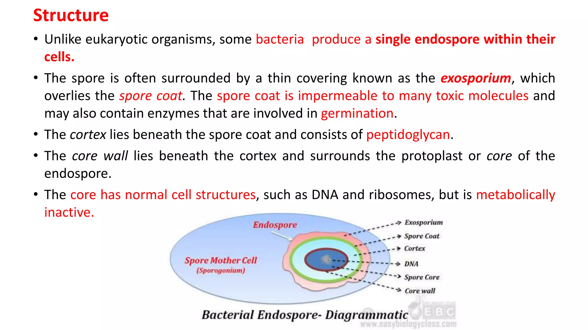
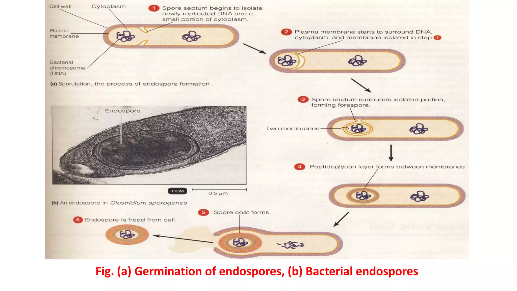
Chapter Four
Bacteria:
Topic: Subcellular component of Bacteria
Outline:
• Endospore: structure, functions
Teaching and learning methods: Lectures, visual aid, interactive forms,
questions- answer session and group discussion.
At end of the lecture student should be able to answer the following
questions:
• Write the structural composition and function of endospore.
• Summarize the functions of endospore.
Reference books:
1. Microbiology: An Introduction, 12th edition- Tortora GJ & Funke BR, Chapter
Four; Page: 85
Endospores
• An endospore is a dormant, tough, and non-reproductive
structure produced by bacteria from the Firmicute phylum, e.g.
Bacillus and Clostridium.
• The primary function of most endospores is to ensure the survival
of a bacterium through periods of environmental stress.
• They are therefore resistant to ultraviolet and gamma radiation,
desiccation, lysozyme, temperature, starvation, and chemical
disinfectants.
• Endospores are commonly found in soil and water, where they
may survive for long periods of time.
Structure
• Unlike eukaryotic organisms, some bacteria produce a single endospore within their
cells.
• The spore is often surrounded by a thin covering known as the exosporium, which
overlies the spore coat. The spore coat is impermeable to many toxic molecules and
may also contain enzymes that are involved in germination.
• The cortex lies beneath the spore coat and consists of peptidoglycan.
• The core wall lies beneath the cortex and surrounds the protoplast or core of the
endospore.
• The core has normal cell structures, such as DNA and ribosomes, but is metabolically
inactive.
Structure
• Up to 15% of the dry weight of the endospore consists of calcium dipicolinate
(DA) within the core, which is thought to stabilize the DNA. DA forms a
complex with calcium, which is called calcium dipicolinate complex.
• Dipicolinic acid could be responsible for the heat resistance of the spore,
and calcium may aid in resistance to heat and oxidizing agents.
• Small, acid-soluble proteins (SASP) of the a/b type are found in spores (they
prevent denaturation of spore DNA) and thus they contribute to heat
resistance.
• Heat resistance appears to be associated with a contractile cortex that either
reduces the water content of the protoplast or maintains it in a state of
dehydration. That protoplast dehydration and diminution (diminished size)
are major factors of spore thermal resistance.
Location
• The position of the endospore differs among bacterial species and is useful in
identification. The main types within the cell are terminal, subterminal and centrally
placed endospores. Terminal endospores are seen at the poles of cells, whereas
central endospores are more or less in the middle. Subterminal endospores are those
between these two extremes, usually seen far enough towards the poles but close
enough to the center so as not to be considered either terminal or central. Lateral
endospores are seen occasionally. Variation and positions of spores in bacterial cells
are shown in the following figure.
Fig. Variations in endospore morphology: (1& 4) central endospore; (2, 3, 5) terminal endospore; (6) lateral
endospore
• Examples of bacteria having terminal endospores include Clostridium tetani, the
pathogen which causes the disease tetanus.
• Bacteria having a centrally placed endospore include Bacillus cereus, and those
having a subterminal endospore include Bacillus subtilis. Sometimes the
endospore can be so large the cell can be distended around the endospore, this is
typical of Clostridium tetani.
• Visualizing endospores under the light microscope can be difficult due to the
impermeability of the endospore wall to dyes and stains. While the rest of a
bacterial cell may stain, the endospore is left colorless. To combat this, a special
stain technique called a Moeller stain is used. That allows the endospore to show
up as red, while the rest of the cell stains blue. Another staining technique for
endospores is the Schaffer-Fulton stain, which stains endospores green and
bacterial bodies red.
Endospore Formation
• Endospores are formed by vegetative cells in a process called
sporulation. Sporulation is initiated when conditions for growth of
the vegetative cells become harsh (for example, when water or an
essential nutrient is limiting’ sudden change of temperature or
pH). Sporulation is a complex process involving as many as 200
genes. These genes are activated by an environmental trigger,
causing sporulation of the cell. The steps leading to endospore
formation result in the creation of a dry, metabolically inert and
extremely resistant endospore from a moist, metabolically active
vegetative cell.
• Extensive sporulation studies have been done on Bacillus subtilis,
and this microbe can perform the entire sporulation process in
about eight hours.
Endospore Formation
• Endospores can remain dormant for many years, but, when conditions allow
i.e. during favourable conditions, they can convert back to vegetative cells
fairly rapidly. This process is called germination, and it involves three steps:
activation, germination and outgrowth.
• Activation can be accomplished by heating freshly formed endospores at a
high temperature. Activated spores can then be conditioned to germinate by
placing them in the presence of specific nutrients.
• During germination, the spore becomes less resistant. This stage includes loss
of calcium dipicolinate and degradation of small acid-soluble spore
proteins.
• The final step is outgrowth, which involves swelling due to water uptake and
the synthesis of new DNA, RNA and proteins. The cell grows out of the broken
spore coat and eventually resumes normal cell function.
• The vegetative cells continue to grow and divide until harsh environmental
conditions once again trigger the sporulation process. The basic sporulation
and germination cycle is illustrated by the picture below-
Fig. (a) Germination of endospores, (b) Bacterial endospores
• When a bacterium detects environmental conditions are becoming
unfavorable it may start the process of sporulation, which takes about eight
hours. The DNA is replicated and a membrane wall known as a spore septum
begins to form between it and the rest of the cell. The plasma membrane of
the cell surrounds this wall and pinches off to leave a double membrane
around the DNA, and the developing structure is now known as a forespore.
Calcium dipicolinate is incorporated into the forespore during this time. Next
the peptidoglycan cortex forms between the two layers and the bacterium
add a spore coat to the outside of the forespore. Sporulation is now
complete, and the mature endospore will be released when the surrounding
vegetative cell is degraded.
• Endospores are resistant to most agents which would normally kill the
vegetative cells they formed from endospore.
• Household cleaning products generally have no effect, nor do most alcohols,
quaternary ammonium compounds or detergents. Alkylating agents however,
such as ethylene oxide, are effective against endospores.
• While resistant to extreme heat and radiation, endospores can be destroyed
by burning or autoclaving. Exposure to extreme heat for a long enough period
will generally have some effect, though many endospores can survive hours of
boiling or cooking. Prolonged exposure to high energy radiation, such as x-
rays and gamma rays, will also kill most endospores.
Reactivation
• Reactivation of the endospore occurs when conditions are more favorable and
involves activation, germination, and outgrowth. Even if an endospore is
located in plentiful nutrients, it may fail to germinate unless activation has
taken place.
• This may be triggered by heating the endospore. Germination involves the
dormant endospore starting metabolic activity and thus breaking hibernation.
It is commonly characterized by rupture or absorption of the spore coat,
swelling of the endospore, an increase in metabolic activity, and loss of
resistance to environmental stress.
• Outgrowth follows germination and involves the core of the endospore
manufacturing new chemical components and exiting the old spore coat to
develop into a fully functional vegetative bacterial cell, which can divide to
produce more cells.
Importance
• As a simplified model for cellular differentiation, the molecular details of
endospore formation have been extensively studied, specifically in the model
organism Bacillus subtilis.
• These studies have contributed much to our understanding of the regulation
of gene expression, transcription factors, and the sigma factor subunits of RNA
polymerase.
• Endospores of the bacterium Bacillus anthracis were used in the 2001 anthrax
attacks (Microbial weapons). The powder found in contaminated postal letters
was composed of extra cellular anthrax endospores. Inhalation, ingestion or
skin contamination of these endospores, which were technically incorrectly
labeled as "spores", led to a number of deaths.
• Geobacillus stearothermophilus endospores are used as biological indicator
when autoclave is used in sterilization procedures.
• Bacillus stearothermophilus is used as biological indicator in autoclave.

Bacterial Endospore.ppt

  • 1.
    Chapter Four Bacteria: Topic: Subcellularcomponent of Bacteria Outline: • Endospore: structure, functions
  • 2.
    Teaching and learningmethods: Lectures, visual aid, interactive forms, questions- answer session and group discussion. At end of the lecture student should be able to answer the following questions: • Write the structural composition and function of endospore. • Summarize the functions of endospore. Reference books: 1. Microbiology: An Introduction, 12th edition- Tortora GJ & Funke BR, Chapter Four; Page: 85
  • 3.
    Endospores • An endosporeis a dormant, tough, and non-reproductive structure produced by bacteria from the Firmicute phylum, e.g. Bacillus and Clostridium. • The primary function of most endospores is to ensure the survival of a bacterium through periods of environmental stress. • They are therefore resistant to ultraviolet and gamma radiation, desiccation, lysozyme, temperature, starvation, and chemical disinfectants. • Endospores are commonly found in soil and water, where they may survive for long periods of time.
  • 4.
    Structure • Unlike eukaryoticorganisms, some bacteria produce a single endospore within their cells. • The spore is often surrounded by a thin covering known as the exosporium, which overlies the spore coat. The spore coat is impermeable to many toxic molecules and may also contain enzymes that are involved in germination. • The cortex lies beneath the spore coat and consists of peptidoglycan. • The core wall lies beneath the cortex and surrounds the protoplast or core of the endospore. • The core has normal cell structures, such as DNA and ribosomes, but is metabolically inactive.
  • 5.
    Structure • Up to15% of the dry weight of the endospore consists of calcium dipicolinate (DA) within the core, which is thought to stabilize the DNA. DA forms a complex with calcium, which is called calcium dipicolinate complex. • Dipicolinic acid could be responsible for the heat resistance of the spore, and calcium may aid in resistance to heat and oxidizing agents. • Small, acid-soluble proteins (SASP) of the a/b type are found in spores (they prevent denaturation of spore DNA) and thus they contribute to heat resistance. • Heat resistance appears to be associated with a contractile cortex that either reduces the water content of the protoplast or maintains it in a state of dehydration. That protoplast dehydration and diminution (diminished size) are major factors of spore thermal resistance.
  • 6.
    Location • The positionof the endospore differs among bacterial species and is useful in identification. The main types within the cell are terminal, subterminal and centrally placed endospores. Terminal endospores are seen at the poles of cells, whereas central endospores are more or less in the middle. Subterminal endospores are those between these two extremes, usually seen far enough towards the poles but close enough to the center so as not to be considered either terminal or central. Lateral endospores are seen occasionally. Variation and positions of spores in bacterial cells are shown in the following figure. Fig. Variations in endospore morphology: (1& 4) central endospore; (2, 3, 5) terminal endospore; (6) lateral endospore
  • 7.
    • Examples ofbacteria having terminal endospores include Clostridium tetani, the pathogen which causes the disease tetanus. • Bacteria having a centrally placed endospore include Bacillus cereus, and those having a subterminal endospore include Bacillus subtilis. Sometimes the endospore can be so large the cell can be distended around the endospore, this is typical of Clostridium tetani. • Visualizing endospores under the light microscope can be difficult due to the impermeability of the endospore wall to dyes and stains. While the rest of a bacterial cell may stain, the endospore is left colorless. To combat this, a special stain technique called a Moeller stain is used. That allows the endospore to show up as red, while the rest of the cell stains blue. Another staining technique for endospores is the Schaffer-Fulton stain, which stains endospores green and bacterial bodies red.
  • 8.
    Endospore Formation • Endosporesare formed by vegetative cells in a process called sporulation. Sporulation is initiated when conditions for growth of the vegetative cells become harsh (for example, when water or an essential nutrient is limiting’ sudden change of temperature or pH). Sporulation is a complex process involving as many as 200 genes. These genes are activated by an environmental trigger, causing sporulation of the cell. The steps leading to endospore formation result in the creation of a dry, metabolically inert and extremely resistant endospore from a moist, metabolically active vegetative cell. • Extensive sporulation studies have been done on Bacillus subtilis, and this microbe can perform the entire sporulation process in about eight hours.
  • 9.
    Endospore Formation • Endosporescan remain dormant for many years, but, when conditions allow i.e. during favourable conditions, they can convert back to vegetative cells fairly rapidly. This process is called germination, and it involves three steps: activation, germination and outgrowth. • Activation can be accomplished by heating freshly formed endospores at a high temperature. Activated spores can then be conditioned to germinate by placing them in the presence of specific nutrients. • During germination, the spore becomes less resistant. This stage includes loss of calcium dipicolinate and degradation of small acid-soluble spore proteins. • The final step is outgrowth, which involves swelling due to water uptake and the synthesis of new DNA, RNA and proteins. The cell grows out of the broken spore coat and eventually resumes normal cell function. • The vegetative cells continue to grow and divide until harsh environmental conditions once again trigger the sporulation process. The basic sporulation and germination cycle is illustrated by the picture below-
  • 11.
    Fig. (a) Germinationof endospores, (b) Bacterial endospores
  • 12.
    • When abacterium detects environmental conditions are becoming unfavorable it may start the process of sporulation, which takes about eight hours. The DNA is replicated and a membrane wall known as a spore septum begins to form between it and the rest of the cell. The plasma membrane of the cell surrounds this wall and pinches off to leave a double membrane around the DNA, and the developing structure is now known as a forespore. Calcium dipicolinate is incorporated into the forespore during this time. Next the peptidoglycan cortex forms between the two layers and the bacterium add a spore coat to the outside of the forespore. Sporulation is now complete, and the mature endospore will be released when the surrounding vegetative cell is degraded. • Endospores are resistant to most agents which would normally kill the vegetative cells they formed from endospore.
  • 13.
    • Household cleaningproducts generally have no effect, nor do most alcohols, quaternary ammonium compounds or detergents. Alkylating agents however, such as ethylene oxide, are effective against endospores. • While resistant to extreme heat and radiation, endospores can be destroyed by burning or autoclaving. Exposure to extreme heat for a long enough period will generally have some effect, though many endospores can survive hours of boiling or cooking. Prolonged exposure to high energy radiation, such as x- rays and gamma rays, will also kill most endospores.
  • 14.
    Reactivation • Reactivation ofthe endospore occurs when conditions are more favorable and involves activation, germination, and outgrowth. Even if an endospore is located in plentiful nutrients, it may fail to germinate unless activation has taken place. • This may be triggered by heating the endospore. Germination involves the dormant endospore starting metabolic activity and thus breaking hibernation. It is commonly characterized by rupture or absorption of the spore coat, swelling of the endospore, an increase in metabolic activity, and loss of resistance to environmental stress. • Outgrowth follows germination and involves the core of the endospore manufacturing new chemical components and exiting the old spore coat to develop into a fully functional vegetative bacterial cell, which can divide to produce more cells.
  • 15.
    Importance • As asimplified model for cellular differentiation, the molecular details of endospore formation have been extensively studied, specifically in the model organism Bacillus subtilis. • These studies have contributed much to our understanding of the regulation of gene expression, transcription factors, and the sigma factor subunits of RNA polymerase. • Endospores of the bacterium Bacillus anthracis were used in the 2001 anthrax attacks (Microbial weapons). The powder found in contaminated postal letters was composed of extra cellular anthrax endospores. Inhalation, ingestion or skin contamination of these endospores, which were technically incorrectly labeled as "spores", led to a number of deaths. • Geobacillus stearothermophilus endospores are used as biological indicator when autoclave is used in sterilization procedures. • Bacillus stearothermophilus is used as biological indicator in autoclave.