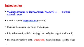
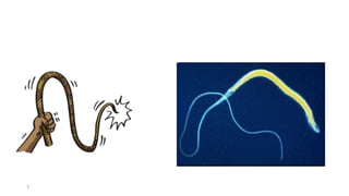
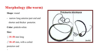
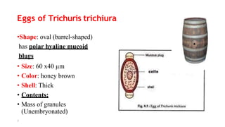
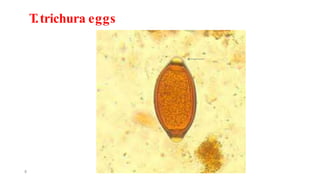
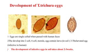
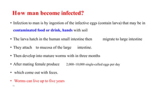
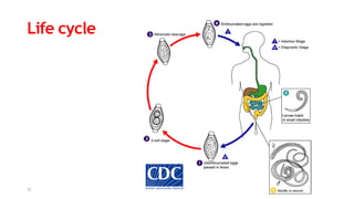
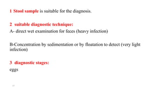
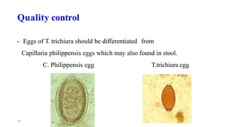
Trichuris trichiura
The whipworm
Lecture by: Osman H. Ali
2
Introduction
• Trichuris trichiura or Trichocephalus trichiuris is intestinal
nematode worm
• Inhabit a human large intestine (ceacum)
• Causing the disease known as trichiuriasis
• It is soil transmitted infection (eggs are infective stage found in soil)
• Is commonly known as the whipworm because it looks like the whip
3
• The worms has thin anterior and thick posterior part
• They attach to intestinal mucosa by embedding their anterior part.
• They feed on tissue fluid (not blood).
4
5
Geographical distribution:
• Worldwide distribution
• It is more common in Asia
• Less in Africa and South America.
• Rare in the United states.
Morphology (the worm)
Shape: round
• narrow long anterior part end and
shorter and thicker posterior.
Color: pinkish-white
Size:
♀ 35–50 mm long
♂ 30–45 mm, with a coiled
posterior end
6
Eggs of Trichuris trichiura
•Shape: oval (barrel-shaped)
has polar hyaline mucoid
blugs
• Size: 60 x40 µm
• Color: honey brown
• Shell: Thick
• Contents:
• Mass of granules
(Unembryonated)
7
T.trichura eggs
8
Development of T.trichura eggs
1 Eggs are single celled when passed with human feces
2The develop into 2 cell, 4 cell, morula, egg contain larva (in soil ) 3-Thelarvated egg
(infective to human)
• The development of infective eggs in soil takes about 2-3weeks,
9
10
How man become infected?
• Infection to man is by ingestion of the infective eggs (contain larva) that may be in
contaminated food or drink, hands with soil
• The larva hatch in the human small intestine then migrate to large intestine
• They attach to mucosa of the large intestine.
• Then develop into mature worms with in three months
• After mating female produce 2,000–10,000 single-celled eggs per day
• which come out with feces.
• Worms can live up to five years
Life cycle
11
12
Pathology & Symptomatology
People with light infections usually have no symptoms.
 People with heavy infection show symptoms like:
• Frequent defecation
• Painful passage of stool
• Stool appearance: contains a mixture of mucus and blood (dysentery).
• The clinical feature is identical to amebic dysentery.
13
Complications:
• Colonic obstruction because of the tangle of worms
• Ulceration of large intestine which result in blood loss.
• Iron deficiency anemia
• Rectal prolapse
Rectal prolapse
14
15
Laboratory Diagnosis
1 what is /are suitable sample/s?
2what is are suitable diagnostic test/s or technique/s?
3- what is/are the diagnostic stage/s?
Summary:
• T. trichuria worm inhabit human large intestine
• The female produces large number of eggs 2,000–10,000 per day
• The worms are not found stool because they attach to the mucosa of
large intestine.
16
17
1 Stool sample is suitable for the diagnosis.
2 suitable diagnostic technique:
A- direct wet examination for feces (heavy infection)
B-Concentration by sedimentation or by floatation to detect (very light
infection)
3 diagnostic stages:
eggs
Quality control
• Eggs of T. trichiura should be differentiated from
Capillaria philippensis eggs which may also found in stool.
C. Philippensis egg T.trichiura egg
18
19
Treatment
Several anthelmintics drugs of choice:
• Albindazole
• Mebendozle
Prognosis
The prognosis is good in light infection Poor in heavy chronic
infection
20
Prevention & control
• Prevent contamination of soil with human feces
• Construction of latrines
• Washing hands before eating (children, soil worker).
• Washing vegetables & fruits
• Do not use the night soil as fertilizer

trichuristrichiura.pptx

  • 1.
  • 2.
    2 Introduction • Trichuris trichiuraor Trichocephalus trichiuris is intestinal nematode worm • Inhabit a human large intestine (ceacum) • Causing the disease known as trichiuriasis • It is soil transmitted infection (eggs are infective stage found in soil) • Is commonly known as the whipworm because it looks like the whip
  • 3.
  • 4.
    • The wormshas thin anterior and thick posterior part • They attach to intestinal mucosa by embedding their anterior part. • They feed on tissue fluid (not blood). 4
  • 5.
    5 Geographical distribution: • Worldwidedistribution • It is more common in Asia • Less in Africa and South America. • Rare in the United states.
  • 6.
    Morphology (the worm) Shape:round • narrow long anterior part end and shorter and thicker posterior. Color: pinkish-white Size: ♀ 35–50 mm long ♂ 30–45 mm, with a coiled posterior end 6
  • 7.
    Eggs of Trichuristrichiura •Shape: oval (barrel-shaped) has polar hyaline mucoid blugs • Size: 60 x40 µm • Color: honey brown • Shell: Thick • Contents: • Mass of granules (Unembryonated) 7
  • 8.
  • 9.
    Development of T.trichuraeggs 1 Eggs are single celled when passed with human feces 2The develop into 2 cell, 4 cell, morula, egg contain larva (in soil ) 3-Thelarvated egg (infective to human) • The development of infective eggs in soil takes about 2-3weeks, 9
  • 10.
    10 How man becomeinfected? • Infection to man is by ingestion of the infective eggs (contain larva) that may be in contaminated food or drink, hands with soil • The larva hatch in the human small intestine then migrate to large intestine • They attach to mucosa of the large intestine. • Then develop into mature worms with in three months • After mating female produce 2,000–10,000 single-celled eggs per day • which come out with feces. • Worms can live up to five years
  • 11.
  • 12.
    12 Pathology & Symptomatology Peoplewith light infections usually have no symptoms.  People with heavy infection show symptoms like: • Frequent defecation • Painful passage of stool • Stool appearance: contains a mixture of mucus and blood (dysentery). • The clinical feature is identical to amebic dysentery.
  • 13.
    13 Complications: • Colonic obstructionbecause of the tangle of worms • Ulceration of large intestine which result in blood loss. • Iron deficiency anemia • Rectal prolapse
  • 14.
  • 15.
    15 Laboratory Diagnosis 1 whatis /are suitable sample/s? 2what is are suitable diagnostic test/s or technique/s? 3- what is/are the diagnostic stage/s?
  • 16.
    Summary: • T. trichuriaworm inhabit human large intestine • The female produces large number of eggs 2,000–10,000 per day • The worms are not found stool because they attach to the mucosa of large intestine. 16
  • 17.
    17 1 Stool sampleis suitable for the diagnosis. 2 suitable diagnostic technique: A- direct wet examination for feces (heavy infection) B-Concentration by sedimentation or by floatation to detect (very light infection) 3 diagnostic stages: eggs
  • 18.
    Quality control • Eggsof T. trichiura should be differentiated from Capillaria philippensis eggs which may also found in stool. C. Philippensis egg T.trichiura egg 18
  • 19.
    19 Treatment Several anthelmintics drugsof choice: • Albindazole • Mebendozle Prognosis The prognosis is good in light infection Poor in heavy chronic infection
  • 20.
    20 Prevention & control •Prevent contamination of soil with human feces • Construction of latrines • Washing hands before eating (children, soil worker). • Washing vegetables & fruits • Do not use the night soil as fertilizer