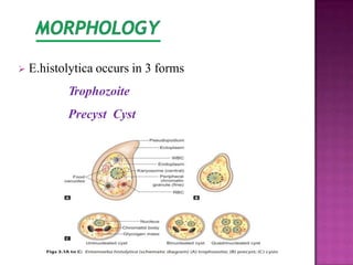
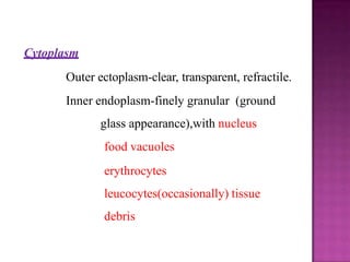
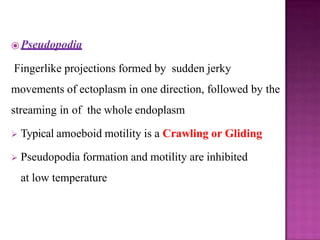
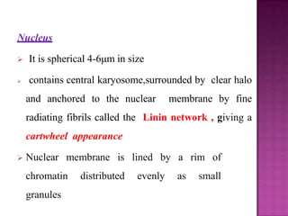
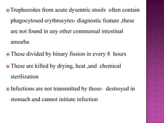
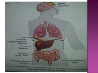
Mr. Osman H. Ali
B.SC Microbiology, MPH, HDCH
⦿Amoeba are structurally simple protozoans which have
no fixed shape
⦿ Phylum : Sarcomastigophora
⦿ Subphylum :Sarcodina
⦿ Super class :Rhizopoda
⦿ Order :Amoebida
Introduction
⦿Amoeba
 Free living
 Intestinal
⦿ Entamoeba histolytica is an intestinal amoeba
⦿All intestinal amoebae are non pathogenic except
Entamoeba histolytica
⦿All free living amoeba are oppurtunistic pathogens.
⦿ E. histolytica was discovered by Losch in 1875
⦿Demonstrated the parasite in the dysenteric feces of a
patient in St.Petersburg in Russia.
⦿ Morphology
⦿ Life Cycle
⦿ Pathogenesis & Clinical Features
⦿ Laboratory Diagnosis
⦿ Treatment
⦿ Prevention
 E.histolytica occurs in 3 forms
Trophozoite
Precyst Cyst
⦿ Vegetative or growing stage of the parasite
⦿ Only form present in tissues
⦿ Irregular in shape
⦿ Size: 12-60 μm(Average 20μm)
⦿ Large and actively motile in freshly passed dysenteric
stool, while smaller in convalescents and carriers.
⦿ In the lumen, commensal and small in size(15-
20 μm)-MINUTAFORM
Cytoplasm
Outer ectoplasm-clear, transparent, refractile.
Inner endoplasm-finely granular (ground
glass appearance),with nucleus
food vacuoles
erythrocytes
leucocytes(occasionally) tissue
debris
⦿ Pseudopodia
Fingerlike projections formed by sudden jerky
movements of ectoplasm in one direction, followed by the
streaming in of the whole endoplasm
 Typical amoeboid motility is a Crawling or Gliding
 Pseudopodia formation and motility are inhibited
at low temperature
Nucleus
 It is spherical 4-6μm in size
 contains central karyosome,surrounded by clear halo
and anchored to the nuclear membrane by fine
radiating fibrils called the Linin network , giving a
cartwheel appearance
 Nuclear membrane is lined by a rim of
chromatin distributed evenly as small
granules
⦿Trophozoites from acute dysentric stools often contain
phagocytosed erythrocytes- diagnostic feature ,these
are not found in any other commensal intestinal
amoeba
⦿ These divided by binary fission in every 8 hours
⦿ These are killed by drying, heat ,and chemical
sterilization
⦿ Infections are not transmitted by these- destroyed in
stomach and cannot initiate infection
⦿Trophozoites undergo encystment in the lumen
⦿ Before encystment,the trophozoites extrudesits food
vacuoles and become round or oval,10-20µm in size-
precyst
⦿ Contains a large glycogen vacuole and two
chromatid bars
⦿ It then secretes a highly retractile cyst wall around it
and become cyst
⦿ Spherical ,10-20µm
⦿ 3 types of cyst
Early cyst
Binucleate cyst
mature quadrinucleate cyst
Early cyst
contains a single nucleus and two other
structures-a mass of glycogen and 1-4
chromatid bodies or chromadial bars
⦿ As the cyst mature, the glycogen mass and chromidial
bars disappear and the nucleus undergoes 2 succesive
mitotic divisions to form 2 and then 4 nuclei .
⦿The cyst wall is a highly resistant to gastric juice and
unfavorable environmental conditions
⦿Infective form :mature quadrinucleate cyst passed in
feces of convalscents and carriers
⦿Mode of transmission :man acquires infection by
swallowing food and water contaminated with cyst .
⦿ Stomach –cyst wall is resistant to gastric juice
⦿ Exystation :cyst reaches the caecum or lower part of
ileum ,due to alkaline medium ,cyst wall damaged by
trypsin ,leading to exystation
⦿ E.histolytica causes intestinal and extra
intestinal amoebiasis
Intestinal amoebiasis -PATHOGENESIS Lumen
dwelling amoeba do not cause any
illness .They causes disease only when they
invade the intestinal tissues .
10 % -symptomatic
90% -asymptomatic
⦿Not all strains of E.histolytica are pathogenic or invasive .
⦿Differentiation between pathogenic and non
pathogenic strains can be made by





susceptibility to complement-mediated lysis
Phagocytic activity
by the use of genetic markers
monoclonal antibodies
Zymodeme analysis
 The metacystic trophozoites penetrate the columnar epithelial
cells of crypts of Liberkuhn
 Penetration is facilitated by
motility of the trophozoites tissue lytic
enzyme –histolysin
amoebic lectin -mediate adherence
 Mucosal penetration by amoeba produce discrete ulcers with
pinhead center and raised edges .
 Sometimes invasion remains superficial and heal
spontaneously.
 More often, the amoeba penetrates to submucosal layer and
multiplies rapidly- lytic necrosis- abscess- ulcer
⦿Ulcer appear initially on the mucosa as raised nodules
with pouting edges .
⦿ They breakdown discharging brownish necrotic
material contains large numbers of trophozoites.
⦿ The typical amoebic ulcer –flask shaped
⦿ Multiple ulcers may coalesce to form large necrotic
lesions with ragged and undermined edges,covered
with brownish slough
⦿The ulcers generally do not extend deeper than
submucosal layer.
⦿ Occassionally, a granulamatous pseudotumoral
growth may develop on the intestinal wall from a
chronic ulcer.This amoebic granuloma or amoeboma
may be mistaken for are malignant tumor
⦿ Small superficial ulcers involving only the mucosa
⦿ Round or oval shaped with ragged and undermined
margin and flask-shaped in cross section
⦿ Marked scarring of intestinal wall with thining ,dilatation
,and sacculation
⦿ Extensive adhesions with neighboring viscera
⦿ Formation of tumor-like masses of granulation tissue
amoeba
⦿The incubation period is highly variable from 1-4 months
⦿ The clinical course is characterized by prolonged
latency,relapses and intermissions.
⦿ Typical manifestation is amoebic dysentry
⦿ Compared to bacillary desentry ,it is usually insidious in
onset and the abdominal tenderness is less and localised.
⦿ The stools are large, foul-smelling,and brownish
black, often with bloodstreaked mucus intermingled
with feces.
⦿ The RBCs in stools are clumped and reddish brown
in color.
⦿ Cellular exudate is scanty.
⦿Charcot-leyden crystals are often present.
⦿ The patient is usually afebrile and non toxic.
⦿ In fulminant colitis, there is confluent ulceration
and necrosis of colon-patient is febrile and toxic.
⦿ Intestinal amoebiasis not always result in
dysentry.quite often there may be only diarrhea or
vagueabdominal symptoms popularly called
uncomfortable belly or growling abdomen.
⦿ Chronic involvement of the caecum causes a
condition simulating appendicitis.
⦿Fulminant amoebic colitis toxic megacolon
perianal ulceration Amoeboma
⦿Extra intestinal Amoebiasis
amoebic hepatitis
amoebic liver abscess
amoebic appendicitis and peritonitis pulmonary
amoebiasis
cerebral amoebiasis
splenic abscess cutaneous amoebiasis
genitourinary amoebiasis
⦿ HepaticAmoebiasis
⦿ PulmonaryAmoebiasis
⦿ MetastaticAmoebiasis
⦿ CutaneousAmoebiasis
⦿ GenitourinaryAmoebiasis
⦿ Most common extra intestinal amoebiasis .
⦿ The history of amoebic dysentry is absent in more than 50%
cases
⦿Several patients with amoebic colitis develop an enlarged tender
liver without detectable impairment of liver function or fever.
⦿ This acute hepatic involvement (amoebic hepatitis) may be due
to repeated invasion by amoeba from an active colonic
infection or to toxic substance from the colon reaching the liver.
⦿ In about 5-10% of person with intestinal amoebiasis
,liver abscesses may ensue.
⦿ The center of the abscess contains thick chocolate brown
pus(anchovy pus),which is liqeefied necrotic liver tissue.
⦿At the periphery, there is almost normal liver
tissue,which contain invading amoeba.
⦿ Liver abscess may
multiple
solitary
⦿Usually located on right lobe of liver
⦿ Jaundice develops
only when lesions are multiple OR when
they press on the biliary tract
 Incidenca of liver abscess is less common in women
and rare in children under age of 10
⦿ It may occure by direct hematogenous spread from colon
bypassing the liver,but it most often follows extension of
hepatic abscess through the diaphragm
⦿ Hepatobronchial fistula usually results with expectoration
of chocolate brown sputum
⦿ Patient presents with
severe pleuritic chest pain dyspnea
non-productive cough
⦿ Involvement of distant organs is by
hematogenous spread and through lymphatics
⦿Abscess in
Kidney Brain
Spleen
Adrenals
Spread to brain leads to severe destruction of brain tissues
and is fatal
⦿ It occurs by direct extension around anus,
colostomy site,or discharging sinuses from
amoebic abscess .
⦿ Extensive gangrenous destruction of the skin occurs .
⦿ The lesion may be mistaken for condyloma or
epithelioma
⦿ Penile amoebiasis
prepuce and glans are affected
acquired through anal intercourse
In females
⦿ vulva ,vagina, or cervix by
spread from perineum
destructive ulcerative lesions resemble
carcinoma
⦿
⦿
Preventive measures are as follows:
1. Avoidance of the ingestion of food and water contaminated
with human feces
2. Treatment of asymptomatic persons who pass E.
histolytica cysts in the stool may help to reduce
opportunities for disease transmission.
3. Vaccination Till now, there is no eff active vaccine licensed
for human use.
Prevention
THANK OU

Entamoeba histolytica.pptx

  • 1.
    Mr. Osman H.Ali B.SC Microbiology, MPH, HDCH
  • 2.
    ⦿Amoeba are structurallysimple protozoans which have no fixed shape ⦿ Phylum : Sarcomastigophora ⦿ Subphylum :Sarcodina ⦿ Super class :Rhizopoda ⦿ Order :Amoebida Introduction
  • 3.
    ⦿Amoeba  Free living Intestinal ⦿ Entamoeba histolytica is an intestinal amoeba ⦿All intestinal amoebae are non pathogenic except Entamoeba histolytica ⦿All free living amoeba are oppurtunistic pathogens.
  • 4.
    ⦿ E. histolyticawas discovered by Losch in 1875 ⦿Demonstrated the parasite in the dysenteric feces of a patient in St.Petersburg in Russia.
  • 5.
    ⦿ Morphology ⦿ LifeCycle ⦿ Pathogenesis & Clinical Features ⦿ Laboratory Diagnosis ⦿ Treatment ⦿ Prevention
  • 6.
     E.histolytica occursin 3 forms Trophozoite Precyst Cyst
  • 7.
    ⦿ Vegetative orgrowing stage of the parasite ⦿ Only form present in tissues ⦿ Irregular in shape ⦿ Size: 12-60 μm(Average 20μm) ⦿ Large and actively motile in freshly passed dysenteric stool, while smaller in convalescents and carriers. ⦿ In the lumen, commensal and small in size(15- 20 μm)-MINUTAFORM
  • 9.
    Cytoplasm Outer ectoplasm-clear, transparent,refractile. Inner endoplasm-finely granular (ground glass appearance),with nucleus food vacuoles erythrocytes leucocytes(occasionally) tissue debris
  • 10.
    ⦿ Pseudopodia Fingerlike projectionsformed by sudden jerky movements of ectoplasm in one direction, followed by the streaming in of the whole endoplasm  Typical amoeboid motility is a Crawling or Gliding  Pseudopodia formation and motility are inhibited at low temperature
  • 11.
    Nucleus  It isspherical 4-6μm in size  contains central karyosome,surrounded by clear halo and anchored to the nuclear membrane by fine radiating fibrils called the Linin network , giving a cartwheel appearance  Nuclear membrane is lined by a rim of chromatin distributed evenly as small granules
  • 12.
    ⦿Trophozoites from acutedysentric stools often contain phagocytosed erythrocytes- diagnostic feature ,these are not found in any other commensal intestinal amoeba ⦿ These divided by binary fission in every 8 hours ⦿ These are killed by drying, heat ,and chemical sterilization ⦿ Infections are not transmitted by these- destroyed in stomach and cannot initiate infection
  • 13.
    ⦿Trophozoites undergo encystmentin the lumen ⦿ Before encystment,the trophozoites extrudesits food vacuoles and become round or oval,10-20µm in size- precyst ⦿ Contains a large glycogen vacuole and two chromatid bars ⦿ It then secretes a highly retractile cyst wall around it and become cyst
  • 15.
    ⦿ Spherical ,10-20µm ⦿3 types of cyst Early cyst Binucleate cyst mature quadrinucleate cyst Early cyst contains a single nucleus and two other structures-a mass of glycogen and 1-4 chromatid bodies or chromadial bars
  • 16.
    ⦿ As thecyst mature, the glycogen mass and chromidial bars disappear and the nucleus undergoes 2 succesive mitotic divisions to form 2 and then 4 nuclei . ⦿The cyst wall is a highly resistant to gastric juice and unfavorable environmental conditions
  • 17.
    ⦿Infective form :maturequadrinucleate cyst passed in feces of convalscents and carriers ⦿Mode of transmission :man acquires infection by swallowing food and water contaminated with cyst . ⦿ Stomach –cyst wall is resistant to gastric juice ⦿ Exystation :cyst reaches the caecum or lower part of ileum ,due to alkaline medium ,cyst wall damaged by trypsin ,leading to exystation
  • 20.
    ⦿ E.histolytica causesintestinal and extra intestinal amoebiasis Intestinal amoebiasis -PATHOGENESIS Lumen dwelling amoeba do not cause any illness .They causes disease only when they invade the intestinal tissues . 10 % -symptomatic 90% -asymptomatic
  • 21.
    ⦿Not all strainsof E.histolytica are pathogenic or invasive . ⦿Differentiation between pathogenic and non pathogenic strains can be made by      susceptibility to complement-mediated lysis Phagocytic activity by the use of genetic markers monoclonal antibodies Zymodeme analysis
  • 22.
     The metacystictrophozoites penetrate the columnar epithelial cells of crypts of Liberkuhn  Penetration is facilitated by motility of the trophozoites tissue lytic enzyme –histolysin amoebic lectin -mediate adherence  Mucosal penetration by amoeba produce discrete ulcers with pinhead center and raised edges .  Sometimes invasion remains superficial and heal spontaneously.  More often, the amoeba penetrates to submucosal layer and multiplies rapidly- lytic necrosis- abscess- ulcer
  • 23.
    ⦿Ulcer appear initiallyon the mucosa as raised nodules with pouting edges . ⦿ They breakdown discharging brownish necrotic material contains large numbers of trophozoites. ⦿ The typical amoebic ulcer –flask shaped ⦿ Multiple ulcers may coalesce to form large necrotic lesions with ragged and undermined edges,covered with brownish slough
  • 24.
    ⦿The ulcers generallydo not extend deeper than submucosal layer. ⦿ Occassionally, a granulamatous pseudotumoral growth may develop on the intestinal wall from a chronic ulcer.This amoebic granuloma or amoeboma may be mistaken for are malignant tumor
  • 25.
    ⦿ Small superficialulcers involving only the mucosa ⦿ Round or oval shaped with ragged and undermined margin and flask-shaped in cross section ⦿ Marked scarring of intestinal wall with thining ,dilatation ,and sacculation ⦿ Extensive adhesions with neighboring viscera ⦿ Formation of tumor-like masses of granulation tissue amoeba
  • 26.
    ⦿The incubation periodis highly variable from 1-4 months ⦿ The clinical course is characterized by prolonged latency,relapses and intermissions. ⦿ Typical manifestation is amoebic dysentry ⦿ Compared to bacillary desentry ,it is usually insidious in onset and the abdominal tenderness is less and localised. ⦿ The stools are large, foul-smelling,and brownish black, often with bloodstreaked mucus intermingled with feces.
  • 28.
    ⦿ The RBCsin stools are clumped and reddish brown in color. ⦿ Cellular exudate is scanty. ⦿Charcot-leyden crystals are often present. ⦿ The patient is usually afebrile and non toxic. ⦿ In fulminant colitis, there is confluent ulceration and necrosis of colon-patient is febrile and toxic.
  • 29.
    ⦿ Intestinal amoebiasisnot always result in dysentry.quite often there may be only diarrhea or vagueabdominal symptoms popularly called uncomfortable belly or growling abdomen. ⦿ Chronic involvement of the caecum causes a condition simulating appendicitis.
  • 30.
    ⦿Fulminant amoebic colitistoxic megacolon perianal ulceration Amoeboma ⦿Extra intestinal Amoebiasis amoebic hepatitis amoebic liver abscess amoebic appendicitis and peritonitis pulmonary amoebiasis cerebral amoebiasis splenic abscess cutaneous amoebiasis genitourinary amoebiasis
  • 33.
    ⦿ HepaticAmoebiasis ⦿ PulmonaryAmoebiasis ⦿MetastaticAmoebiasis ⦿ CutaneousAmoebiasis ⦿ GenitourinaryAmoebiasis
  • 34.
    ⦿ Most commonextra intestinal amoebiasis . ⦿ The history of amoebic dysentry is absent in more than 50% cases ⦿Several patients with amoebic colitis develop an enlarged tender liver without detectable impairment of liver function or fever. ⦿ This acute hepatic involvement (amoebic hepatitis) may be due to repeated invasion by amoeba from an active colonic infection or to toxic substance from the colon reaching the liver.
  • 35.
    ⦿ In about5-10% of person with intestinal amoebiasis ,liver abscesses may ensue. ⦿ The center of the abscess contains thick chocolate brown pus(anchovy pus),which is liqeefied necrotic liver tissue. ⦿At the periphery, there is almost normal liver tissue,which contain invading amoeba. ⦿ Liver abscess may multiple solitary
  • 36.
    ⦿Usually located onright lobe of liver ⦿ Jaundice develops only when lesions are multiple OR when they press on the biliary tract  Incidenca of liver abscess is less common in women and rare in children under age of 10
  • 37.
    ⦿ It mayoccure by direct hematogenous spread from colon bypassing the liver,but it most often follows extension of hepatic abscess through the diaphragm ⦿ Hepatobronchial fistula usually results with expectoration of chocolate brown sputum ⦿ Patient presents with severe pleuritic chest pain dyspnea non-productive cough
  • 38.
    ⦿ Involvement ofdistant organs is by hematogenous spread and through lymphatics ⦿Abscess in Kidney Brain Spleen Adrenals Spread to brain leads to severe destruction of brain tissues and is fatal
  • 39.
    ⦿ It occursby direct extension around anus, colostomy site,or discharging sinuses from amoebic abscess . ⦿ Extensive gangrenous destruction of the skin occurs . ⦿ The lesion may be mistaken for condyloma or epithelioma
  • 40.
    ⦿ Penile amoebiasis prepuceand glans are affected acquired through anal intercourse In females ⦿ vulva ,vagina, or cervix by spread from perineum destructive ulcerative lesions resemble carcinoma ⦿ ⦿
  • 42.
    Preventive measures areas follows: 1. Avoidance of the ingestion of food and water contaminated with human feces 2. Treatment of asymptomatic persons who pass E. histolytica cysts in the stool may help to reduce opportunities for disease transmission. 3. Vaccination Till now, there is no eff active vaccine licensed for human use. Prevention
  • 43.