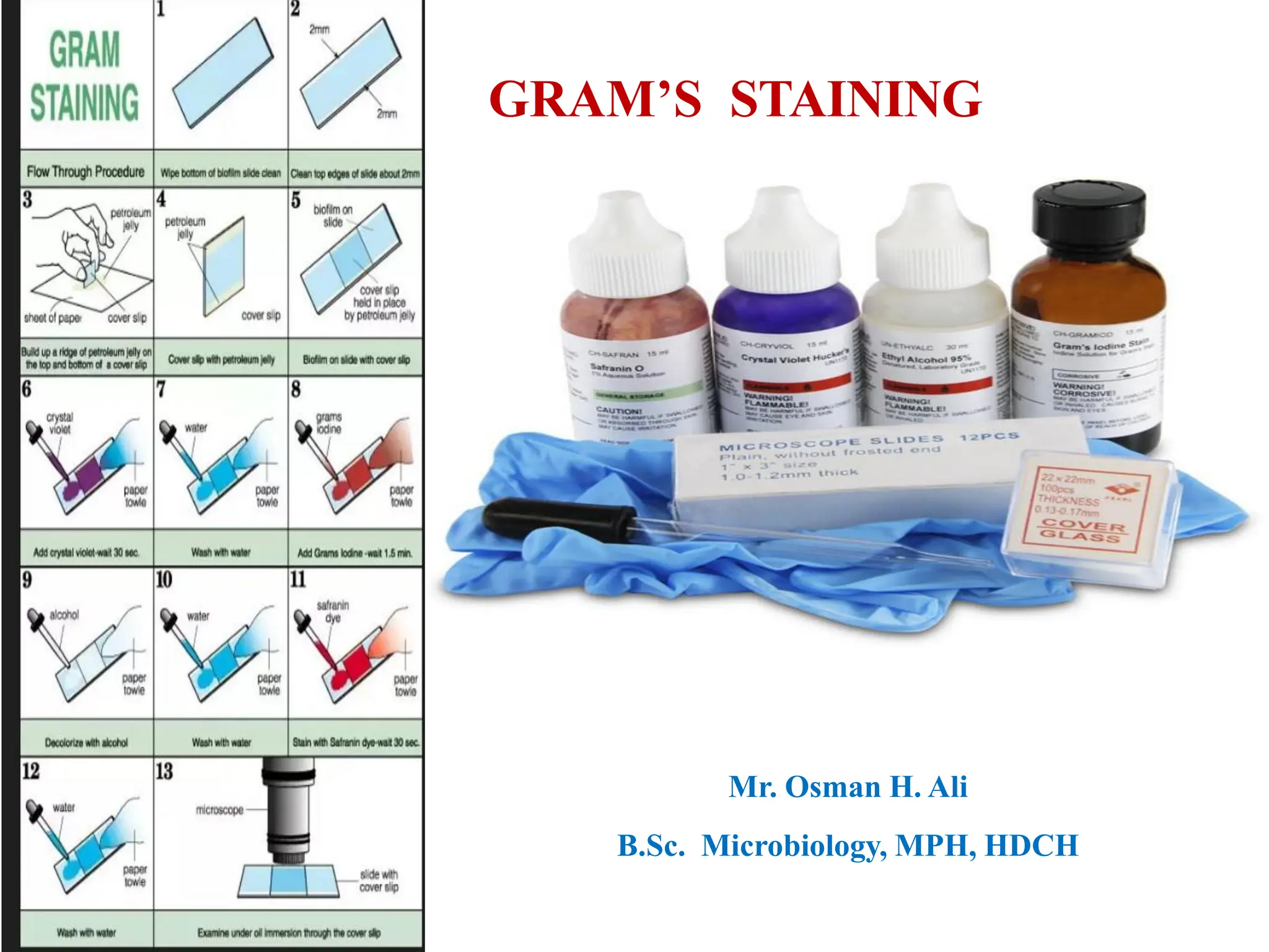
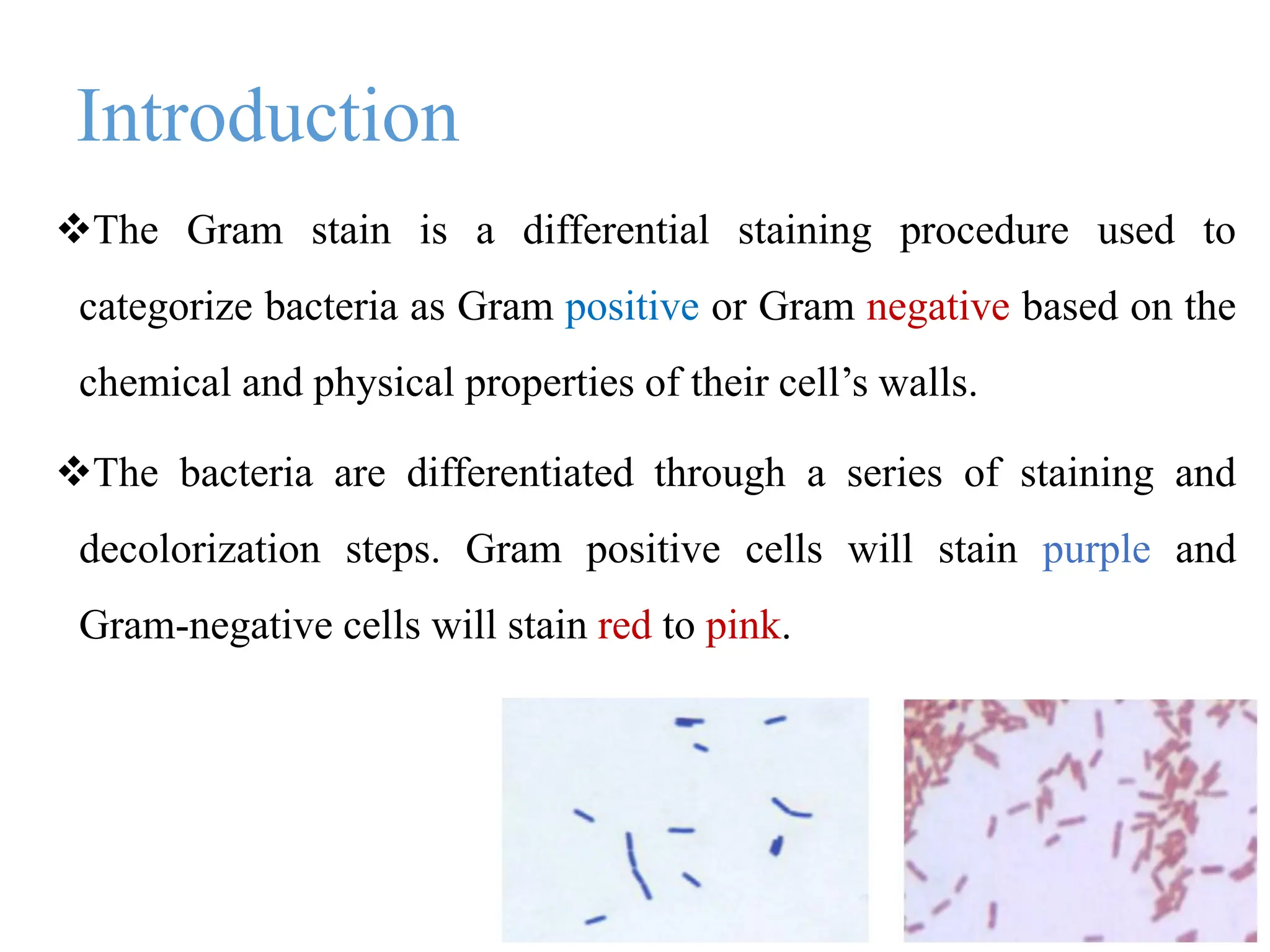
2. Introduction
The Gram stain is a differential staining procedure used to
categorize bacteria as Gram positive or Gram negative based on the
chemical and physical properties of their cell’s walls.
The bacteria are differentiated through a series of staining and
decolorization steps. Gram positive cells will stain purple and
Gram-negative cells will stain red to pink.
3. Conti…….
The Gram stain was devised by the Danish physician Hans
Christian Gram, while working in Berlin in 1883.
He later published this procedure in 1884.
4. Purpose
The Gram stain is fundamental to the phenotypic characterization of
bacteria.
The staining procedure differentiates organisms of the domain
Bacteria according to cell wall structure.
Gram-positive cells have a thick peptidoglycan layer and stain blue
to purple.
Gram-negative cells have a thin peptidoglycan layer and stain red to
pink.
5. GRAM’S STAIN
Gram staining (or Gram's method) is a method of
differentiating bacterial species into two large groups
Gram Positive Gram Negative
Gram staining differentiates bacteria by the chemical and
physical properties of their cell walls.
6. GRAM POSITIVE BACTERIA
• Gram positive bacteria have a thick
cell wall of peptidoglycan.
• Peptidoglycan is a polymer consisting
of sugar amino acids that form a mesh
like outside the plasma membrane of
bacteria forming cell wall.
• In Gram positive bacteria, between the
cell wall and cell membrane, there is a
"membrane teichoic acid".
7. GRAM NEGATIVE BACTERIA
Gram negative bacteria have an outer
membrane of phospholipids and
bacterial Lipopolysaccharides outside of
their thin peptidoglycan layer.
The space between the outer membrane
and the peptidoglycan layer is called the
periplasmic space.
8.
9. Principle of Gram’s Staining
The structure of the organism ‘s cell wall determines whether
the organism is gram positive or negative.
When stained with a primary stain and fixed by a mordant, some
bacteria are able to retain the primary stain by resisting
declorization while other get decolorized by decolorizer.
Those bacteria which retain the primary stain are called Gram
positive.
Those bacteria which get decolorized and then get counterstained
are called Gram negative.
10. 1. Crystal violet - all bacteria take crystal violet- so all appears
violet.
2.Iodine – Crystal Violet-iodine(CV-I) complex is formed.
3.Acetone- bacteria with high lipid content loose CV-I
complex(appear colourless) but bacteria with less lipid content
retains CV-I complex ( appear violet).
4.Safranine/ basic fuchsin – only colourless bacteria takes –
appear pink.
11.
12. Preparation of a slide smear:
1. Inoculation loop is used to transfer a drop of suspended culture to the
microscope slide.
2. A few loopful of water is added to facilitate a minimal amount of
colony transfer to the examination slide.
3. spread with an inoculation loop to an even thin film over a circle of
15mm in diameter.
4. The slide can be either air-dried or dried with the help of heat over a
gentle flame.
13.
14. Procedure of gram stain
• Preparation of smear
• Air dry and Heat fixation