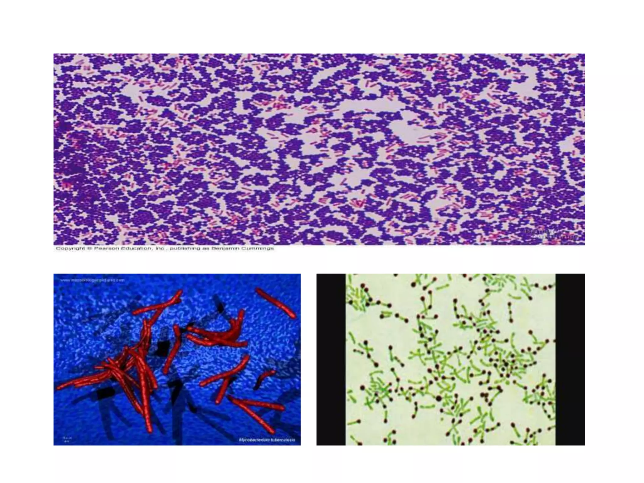
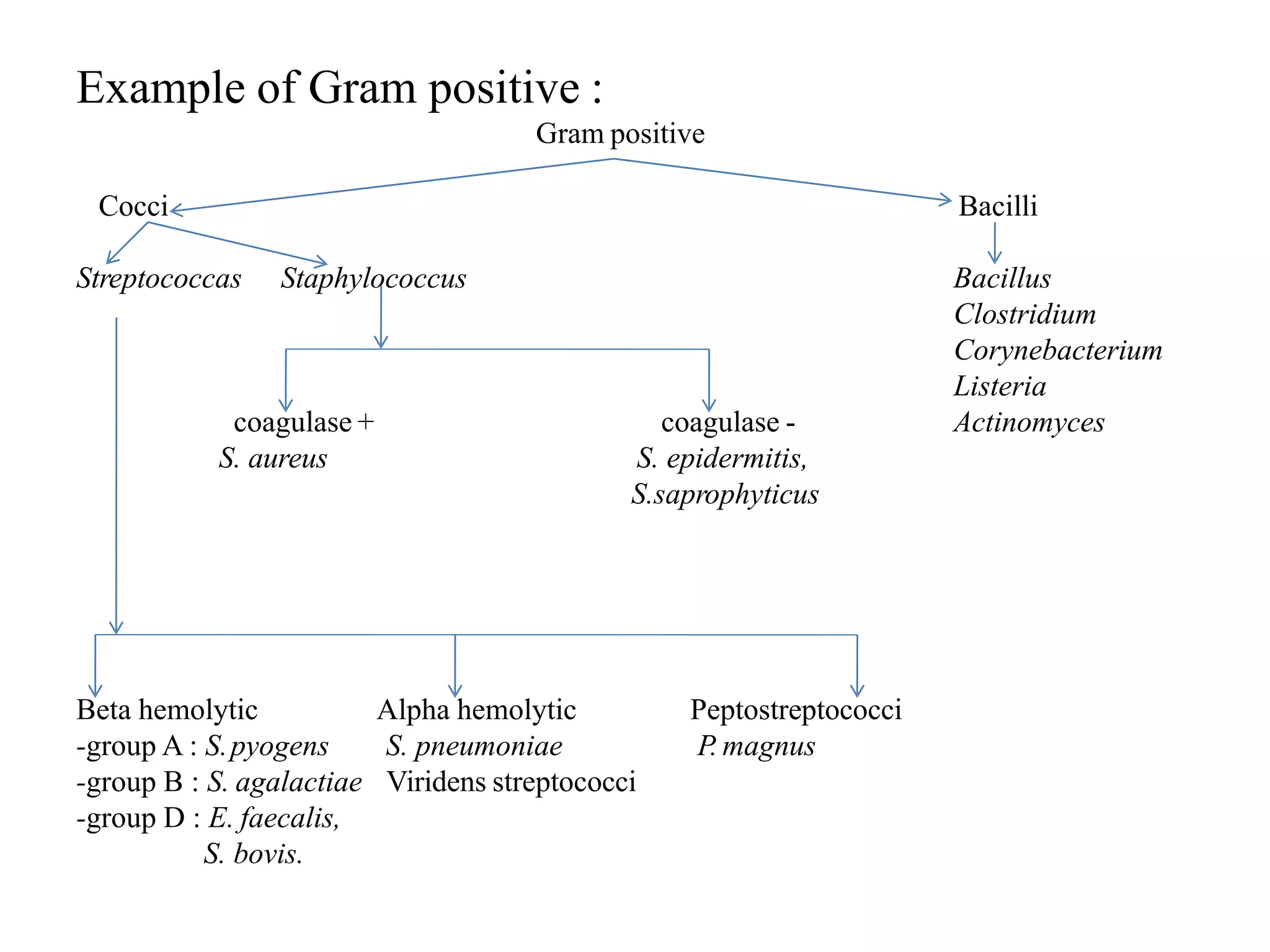
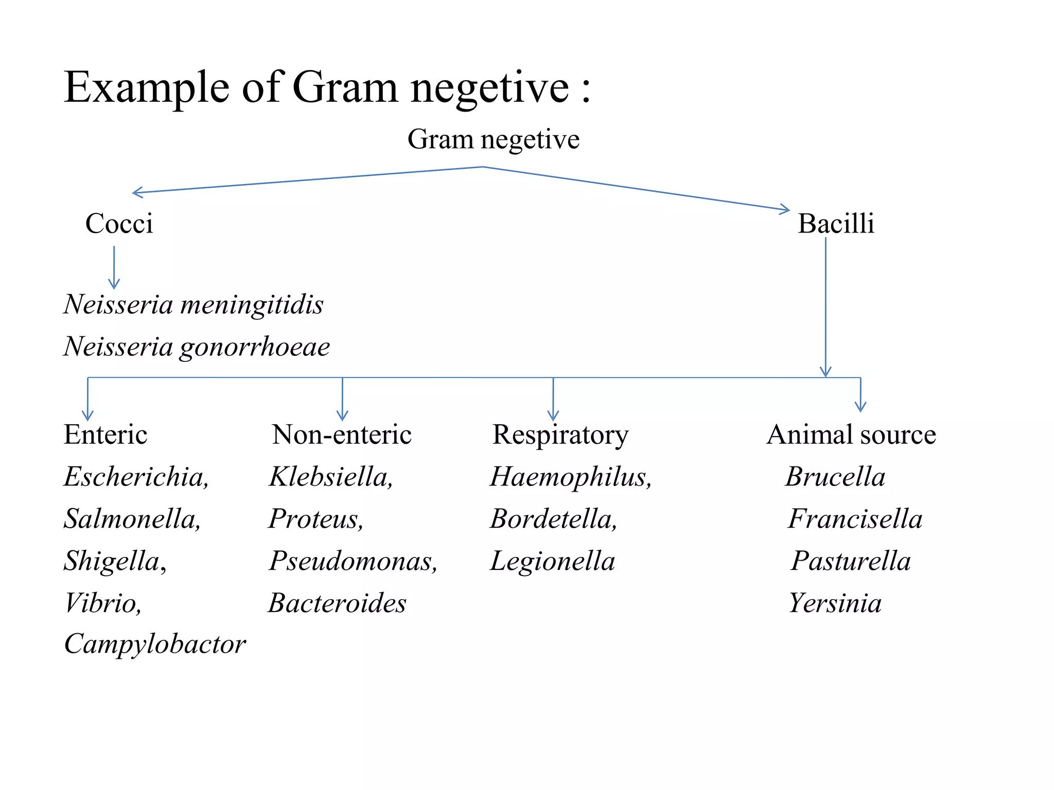
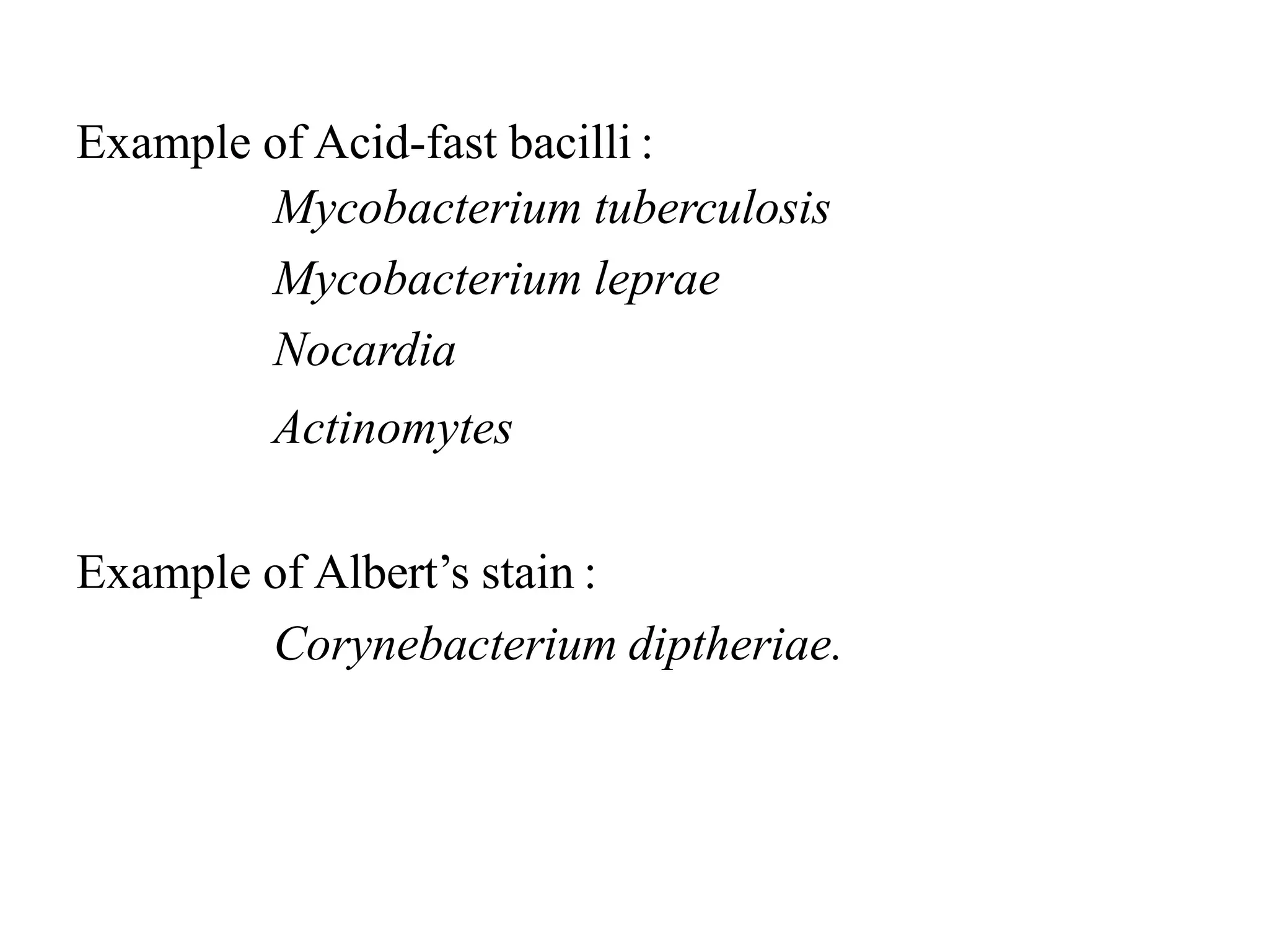
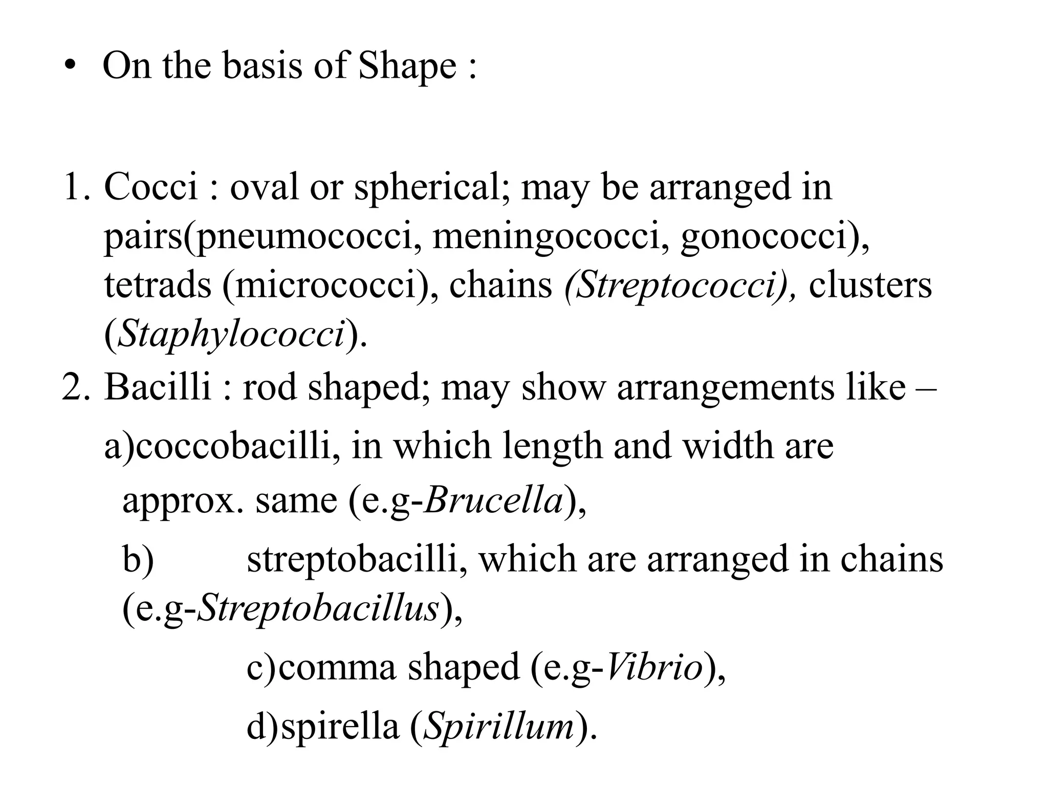
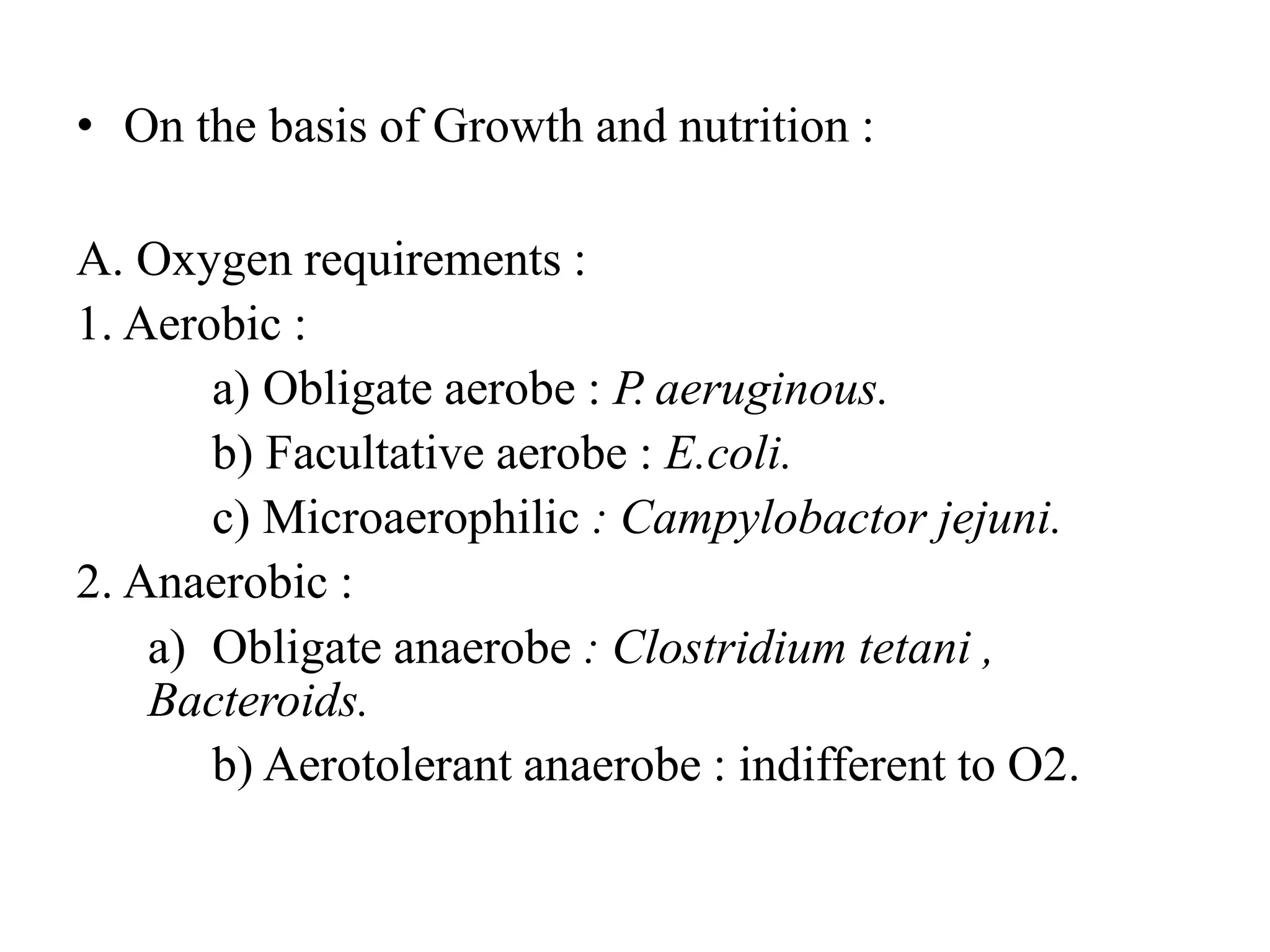
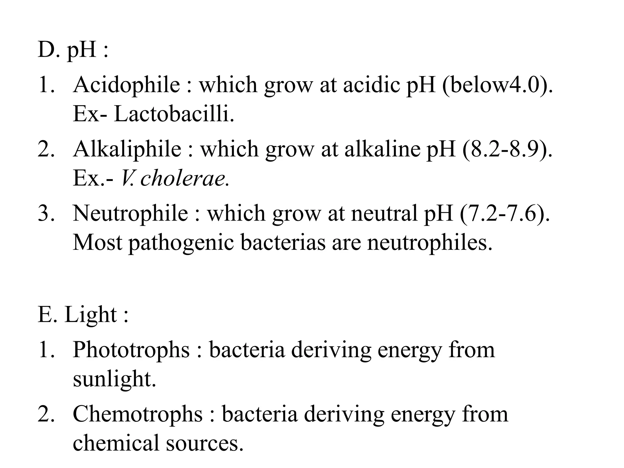
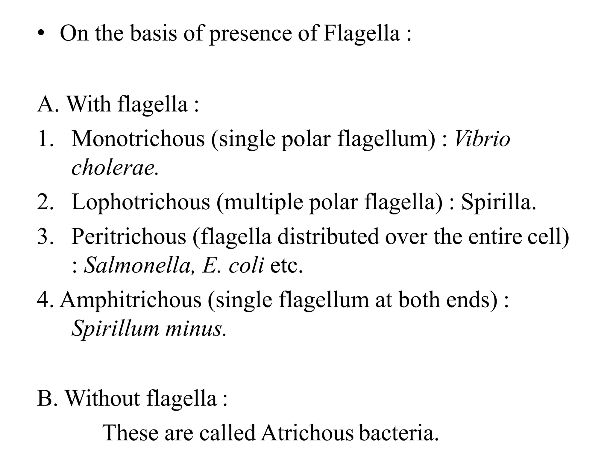
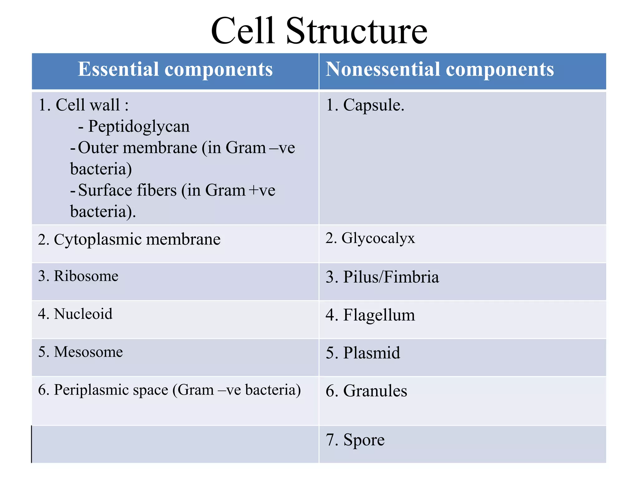
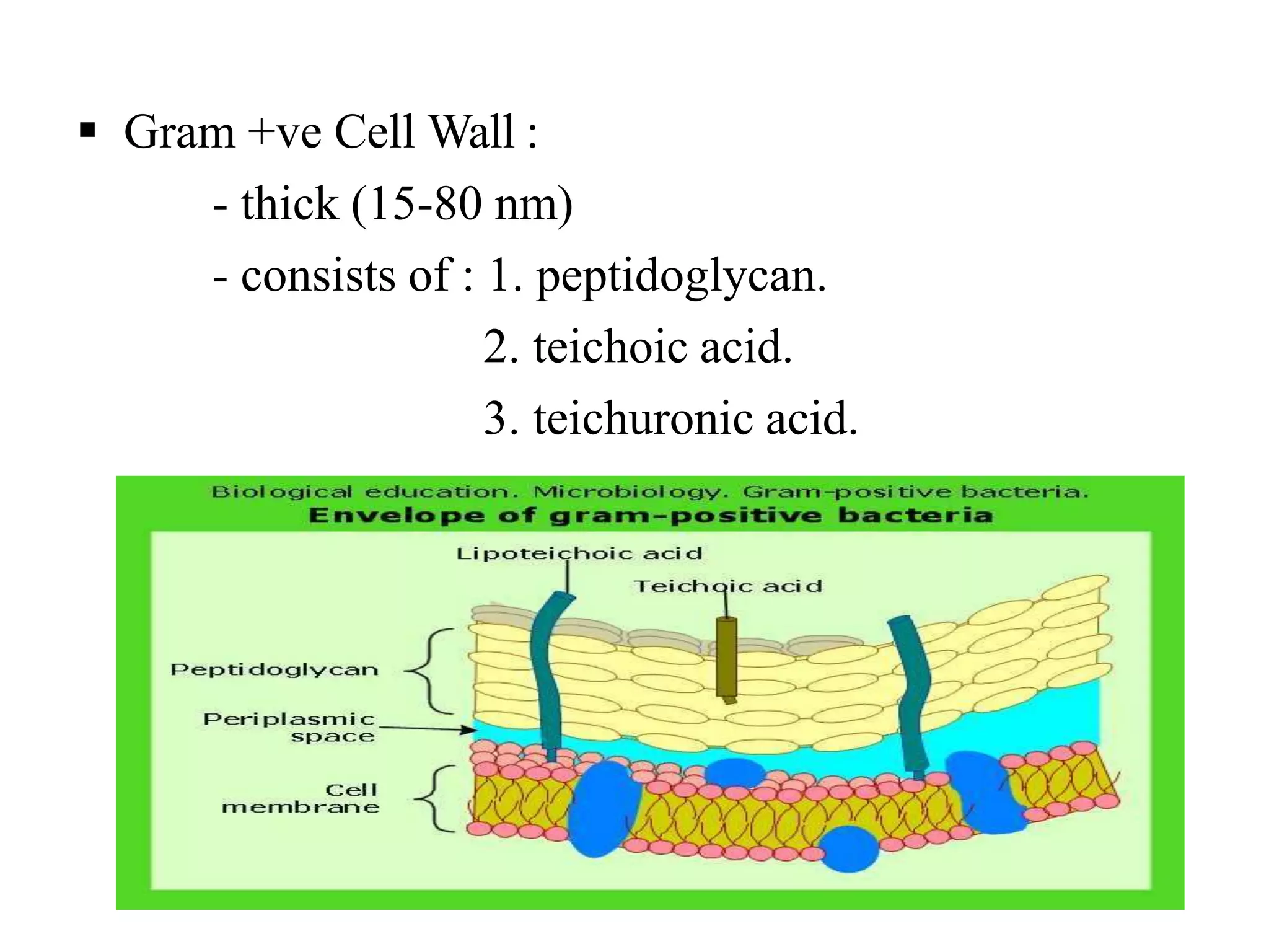
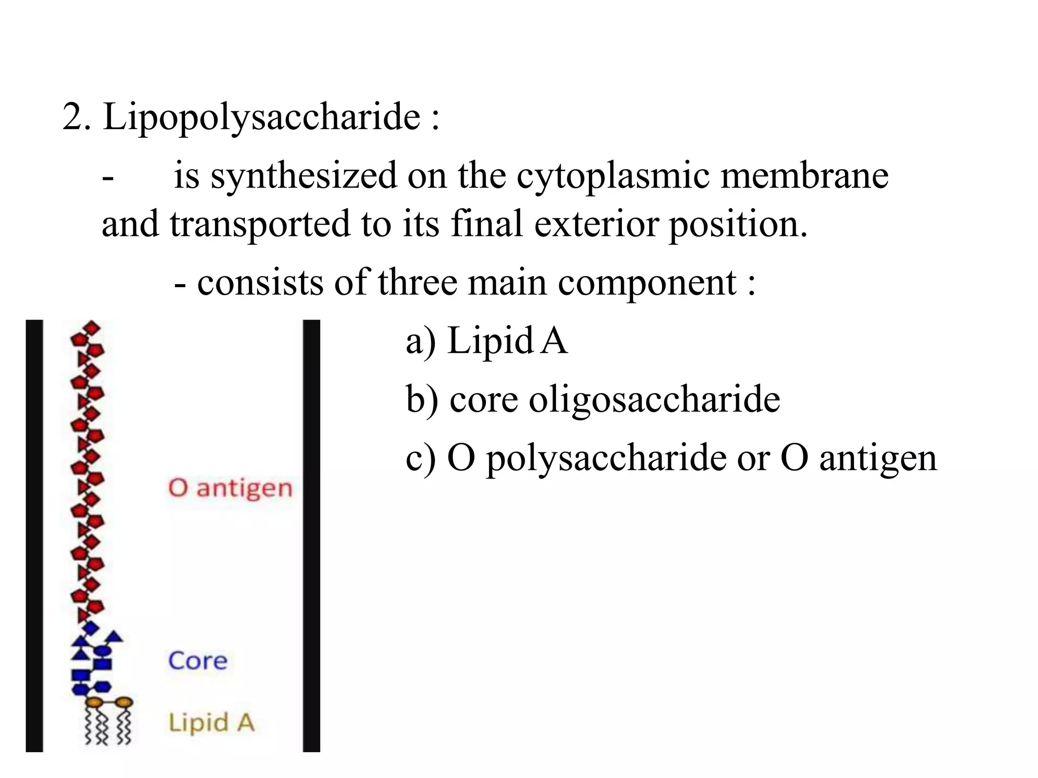
SlideShare a Scribd company logo
CLASSIFICATION OF BACTERIA
& IT’S STRUCTURE
PRESENTED BY
MISS TASMIA ZEB
Topics
Classification of bacteria
Baterial cell structure
Difference between prokaryotes and
eukaryotes,fungus,bacteria,virus
Obligate intracellular organisms.
Classification
• On the basis of Staining :
1. Gram staining :
a) Gram positive
b) Gram negetive.
2. Acid-fast staining.
3. Albert’s staining.
Example of Gram positive :
Gram positive
Cocci Bacilli
Streptococcas Staphylococcus Bacillus
Clostridium
Corynebacterium
Listeria
Actinomycescoagulase +
S. aureus
coagulase -
S. epidermitis,
S.saprophyticus
Peptostreptococci
P. magnus
Alpha hemolytic
S. pneumoniae
Viridens streptococci
Beta hemolytic
-group A : S.pyogens
-group B : S. agalactiae
-group D : E. faecalis,
S. bovis.
Example of Gram negetive :
Gram negetive
Cocci Bacilli
Neisseria meningitidis
Neisseria gonorrhoeae
Enteric Non-enteric Respiratory Animal source
Escherichia, Klebsiella, Haemophilus, Brucella
Salmonella, Proteus, Bordetella, Francisella
Shigella, Pseudomonas, Legionella Pasturella
Vibrio, Bacteroides Yersinia
Campylobactor
Example of Acid-fast bacilli :
Mycobacterium tuberculosis
Mycobacterium leprae
Nocardia
Actinomytes
Example of Albert’s stain :
Corynebacterium diptheriae.
• On the basis of Shape :
1. Cocci : oval or spherical; may be arranged in
pairs(pneumococci, meningococci, gonococci),
tetrads (micrococci), chains (Streptococci), clusters
(Staphylococci).
2. Bacilli : rod shaped; may show arrangements like –
a)coccobacilli, in which length and width are
approx. same (e.g-Brucella),
b) streptobacilli, which are arranged in chains
(e.g-Streptobacillus),
c)comma shaped (e.g-Vibrio),
d)spirella (Spirillum).
3. Spirochetes : slender,flexious,spiral (Treponema).
4. Actinomytes : branching filamentous.
• On the basis of Growth and nutrition :
A. Oxygen requirements :
1. Aerobic :
a) Obligate aerobe : P. aeruginous.
b) Facultative aerobe : E.coli.
c) Microaerophilic : Campylobactor jejuni.
2. Anaerobic :
a) Obligate anaerobe : Clostridium tetani ,
Bacteroids.
b) Aerotolerant anaerobe : indifferent to O2.
B. Carbon dioxide requirement :
1. Capnophilic bacteria : Which require higher amount
of CO2 for their growth (5-10% CO2 and 15% O2).
Ex – H. influenzae, Brucella abortius.
C. Temperature :
1. Psychrophiles : microbes that grow within 0-20°C.
2. Mesophiles : microbes that grow within 25-40°C.
3. Thermophiles : microbes that grow within 55-80°C.
Ex- B. stearothermophilus.
D. pH :
1. Acidophile : which grow at acidic pH (below4.0).
Ex- Lactobacilli.
2. Alkaliphile : which grow at alkaline pH (8.2-8.9).
Ex.- V. cholerae.
3. Neutrophile : which grow at neutral pH (7.2-7.6).
Most pathogenic bacterias are neutrophiles.
E. Light :
1. Phototrophs : bacteria deriving energy from
sunlight.
2. Chemotrophs : bacteria deriving energy from
chemical sources.
F. Osmotic pressure :
1. Halophiles : which can survive at high salt
concentration.
2. Osmophiles : which can survive at high sugar
concentration.
G. Carbon source :
1. Autotrophs : which reduce inorganic carbon into
organic compounds, such as through photosynthesis
2. Heterotrophs : bacteria that grow by using the carbon
that has been reduced by the autotrophs.
H. Method of obtaining nutrition :
1. Heterotropic bacteria- Obtain their food from other
living organisms, as they cannot synthesize it on their
own.
2. Symbiotic Bacteria - Obtain nutrition from host
organism by offering something in return. Establish a
mutual give-and-take relationship with host.
3. Pathogenic Bacteria - Obtain food from host but are
harmful to the host, generally causing diseases.
4. Saprophytic Bacteria - Obtain Nutrition from dead
and decaying matter.
• On the basis of presence of Flagella :
A. With flagella :
1. Monotrichous (single polar flagellum) : Vibrio
cholerae.
2. Lophotrichous (multiple polar flagella) : Spirilla.
3. Peritrichous (flagella distributed over the entire cell)
: Salmonella, E. coli etc.
4. Amphitrichous (single flagellum at both ends) :
Spirillum minus.
B. Without flagella :
These are called Atrichous bacteria.
• On the basis of Motility :
1. Motile :
- Salmonella
- Vibrio cholerae
- Pseudomonas etc.
2. Non motile :
- Staphylococcas,
- Shigella.
Cell Structure
Essential components Nonessential components
1. Cell wall :
- Peptidoglycan
-Outer membrane (in Gram –ve
bacteria)
-Surface fibers (in Gram +ve
bacteria).
1. Capsule.
2. Cytoplasmic membrane 2. Glycocalyx
3. Ribosome 3. Pilus/Fimbria
4. Nucleoid 4. Flagellum
5. Mesosome 5. Plasmid
6. Periplasmic space (Gram –ve bacteria) 6. Granules
7. Spore
• Cell wall :
- fairly rigid and chemically complexed
outermost component, common to all bacteria
except Mycoplasma species.
- multilayerd,located external to the
cytoplasmic membrane.
- composed of : 1. Peptidoglycan
2. Outer membrane
3. Surface fibers.
Illustration of cell wall of Gram +ve and Gram –ve
Bacteria
 Gram +ve Cell Wall :
- thick (15-80 nm)
- consists of : 1. peptidoglycan.
2. teichoic acid.
3. teichuronic acid.
1. Peptidoglycan :
- synonym : murein and mucopeptide.
- thick; 40 sheets which comprises 50% of cell
wall.
- complexed polymer consisting
a) a backbone, composed of alternating two
sugar derivatives N-acetyleglucosamine and N-
acetylemuramic acid connected by β-1→4 linkage.
b) a tetrapeptide side chain, consisting of
D- and L- amino acid with L-lysine.
c) a pentaglycine bridge, which connects
the tetrapeptide side chains.
Importance :
1. provides rigid support for the cell
maintain its characteristic shape and allows it to
withstand media of low osmotic pressure.
2. good target for antimicrobial which
inhibit transpeptidase thus inhibit synthesis
peptidoglycan.
3. lysozyme, a natural resistance of host,
cleaves disaccharide backbone by breaking
glycosyle bond (β-1→4 linkage).
2. Teichoic acid :
- polymer of polyribitol phosphate or
polyglycerol phosphate which are connected by
phosphodiester linkage.
- two types : a) wall teichoic acid (WTA)
b) membrane teichoic acid or
lipoteichoic acid (LTA).
- most teichoic acids contain large amount of D-
alanine .
Importance :
1.togather with peptidoglycan it provides elasticity,
porosity, tensile strength, electrostatic properties of
the envelop.
2. used as antigen for serological classification.
3. binds Mg and supply this ion to the cell.
4. induce inflammation and septic shock.
5. mediate the attachment of staph. to mucosal cell.
3. Teichoronic acid :
- consists of repeat units of sugar acids (such as
N-acetylemannuronic acid or D-glucosuronic acid).
- they are synthesized when phosphate is
limiting.
4.Polysaccharides :
-also contains neutral sugars; such as, mannose,
arabinose, rhamnose, glucosamine and acidic sugars;
such as glucoronic and mannuronic acid.
 Gram –ve cell wall :
- more complexed than Gram +ve bacteria.
- consists of : 1. peptidoglycan
2. lipopolysaccharides
3. outer membrane
4. lipoprotein layer.
1. Peptidoglycan :
- thin (2-8 nm); 1-2 sheets which comprises 5-
10% of the cell wall.
- complexed polymer consisting
a) a backbone, composed of alternating two
sugar derivatives N-acetyleglucosamine and N-
acetylemuramic acid connected by β-1→4 linkage.
b) a tetrapeptide side chain, consisting of D-
and L- amino acid with mesodiaminopimelic acid.
c) pentaglycine bridge is absent.
2. Lipopolysaccharide :
- is synthesized on the cytoplasmic membrane
and transported to its final exterior position.
- consists of three main component :
a) LipidA
b) core oligosaccharide
c) O polysaccharide or O antigen
a) Lipid A:
- consists of phosphorylated glucosamine
disaccharide to which a number of long chain fatty
acids are attached.
- also consists of hydroxymyristic acid, a unique
fatty acid.
b) Core oligosaccharide :
- includes two characteristic sugars :
a) ketodeoxyoctulonate acid
b) heptose
- both joined togather by lipidA.
c) O antigen :
- an outer polysaccharide consisting of up to 25
repeating units of 3-5 sugars.
Importance :
1.LPS is the endotoxin which causes fever, shock,
DIC by activating macrophage, complement and
coagulation system.
2. LOS is an important virulent factor.
3.O antigen is used to identify certain organism in
the clinical lab.
1. Outer membrane :
- bilayered structure; outer membrane contains
LPS and inner one resembles cell membrane.
- porins : make special channels of outer
membrane.
- outer membrane protiens : includes Omp C,D,F
and PhoE and LamB, Tsx, Omp A.
- minor protiens:
enzymes,phospholipases,protease.
Importance :
1.large antibiotics penetrate it slowly which makes
high antibiotic resistance of gram-ve bacteria.
2.permit passive diffusion of hydrophilic
compounds, such as, sugar, amino acid and certain
ions. Some special molecules are also transported,
such as vitamin B12.
3. exclude hydrophobic molecules, such as bile salt.
4. serve to protect the cell.
4. Lipoprotein layer :
- contains : a) 57 amino acids, representing 15
amino acid sequence.
b) diglyceride thioether linked to a
terminal cysteine.
Impotance :
stabilize the outer membrane and anchor it to the
peptidoglycan layer.
5. Periplasmic space :
- space between the inner and outer membrane.
- contains peptidoglycan layer and 20-40% of cell
volume.
Importance :
1. has binding protein for specific substance.
2. has hydrolytic enzymes that break down
nontransportable substance to transportable one.
3. has detoxifying enzymes that inactivate certain
antibiotic.
 The acid fast cell wall :
- contains large amount of waxes,named Mycolic
acid.
- composed of peptidoglycan and asymmetric
lipid bilayer.
- inner lipid bilayer contains mycolic acid linked
to arabinoglycan and outer layer contains extractable
lipids.
Importance :
protect the acid fast bacilli from harsh chemicals
and detergents.
 Cell wall deficient bacteria :
1. Protoplast : produced from gram +ve bacteria
on treatment with lysozyme,in hypertonic media.
2. Spheroplast : derived from gram-ve bacteria
treated with lysozyme in presence of EDTA.
3. L-form : produced more easily with penicillin
than with lysozyme. They are able to grow and
devide.
4. Mycoplasma : wall lacking bacteria measuring
50-300nm.
• Cytoplasmic membrane :
- thin (5-10nm) semipermiable membrane.
- composed of a phospholipid bilayer and 200
different kinds of protein.
Importance :
1.acts as osmotic barrier and regulate the
tranportation of metabolites to and from protoplasm.
2. electron transport and oxidative phosphorylation.
3.excretion of hydrolytic exoenzyme and
pathogenicity protein.
4. Biosynthetic function.
5. Chemotactic systems.
• Cytoplasm :
- matrix is largely formed with 70% water.
- lack endoplasmic reticulam, mitocondria.
- consists : 1. Ribosome
2. mesosome
3. Intracytoplasmic inclusion bodies.
1. Ribosome :
- look like small featureless particle.
- two subunits of 30S and 50S, giving a net 70S.
Importance :
1. serve as a site for protein synthesis .
2.site of action of antibiotic such as
aminoglycosides, macrolids and tetracycline.
1. Mesosome :
- vesicular and multilaminated structure formed
as invagination of plasma membrane into the
cytoplasm.
- two types : 1. septal
2. lateral.
Importance :
1.septal mesosome coordinate nuclear and
cytoplasmic divisions during binary fission.
2. principal sites of respiratory enzymes in bacteria.
3. Intracytoplasmic inclusion bodies :
- two types :
i) organic : contains glycogen or
polyhydroxybutyrate.
ii) inorganic : polyphosphate granules or
sulfer granules.
Importance:
1. used for storage; they are the source of carbon,
inorganic substance and energy.
2. some func to reduce osmotic pressure.
• Nucleoid :
- absence of nuclear membrane and mitotic
apparatus.
- consists of a single circular molecule of loosely
organised DNA (.58-10million base pairs).
- exception : V.cholerae and Brucellamelitensis
have two dissimilar chromosome.
- bacterial DNA has no introns.
• Capsule :
- a condensed,well defined layer closely
surrounds the cell, which excludes particle like india
ink.
-composed of polysaccharide, collectively
reffered as exopolysaccharide, which are sometimes
homopolysaccharide or negetively charged (except in
Bacillus anthracis, in which capsule comprises of
polypeptide, such as D-glutamic acid.)
Importance :
1.It is a determinant of virulence of many bacteria
for its antiphagocytic property.
2.Specific identification of an organism can be
made by ‘Quellung reaction’.
3.Used as vaccines due to their capability of
eliciting protective antibodies.
4.May play a role in the adherence of bacteria to
human tissue.
• Glycocalyx :
- is defined as polysaccharide-containing
material lying outside the cell.
-if it is loosely associated with the cell and does
not exclude particles, it is refered to as Slime layer.
Importance :
1. helps in adherence with foreign body to host cell.
2.facilitates Streptococcas mutens to adhere tightly
with tooth enamel and the acid excreted by this
bacteria causes dental carries.
• Flagella :
- thread like appendages intricately embedded in cell
envelop.
- 3-20μm in length and .01-.03μm in diameter.
- consists of :
a) filament, made up of protein flagellin.
b) hook, acts as propeller.
c)basal body, a set of rings;
one pair in gram +ve
and two pairs in
gram –ve bacteria.
Function :
1.primarily responsible for motility of bacteriam by
chemotaxis.
2.may play a role in bacterial survival
pathogenesis.
3.highly antigenic, possess H antigens, which are
different in different bacterias. Important in
serological diagnosis.
• Pili/Fimbria :
- hair-like filaments that extend from cell surface
and exclusively present in gram –ve bacteria.
- composed of structural protein subunit pilin
arranged in helical structure..
- .5μm long, 10nm thick.
- antigenic in nature.
Importance :
1. adherence of symbiotic and pathogenic bacteria to
host cells.
2.Transfer of bacterial DNA takes place through
sex pili during the process of conjugation.
3.Antigenic variation of pili of Neisseria
gonorrhoeae makes it to survive even after the
presence of antibody against the original type of pili.
4. Inhibit phagocytic ability of leukocyte.
5. Helps in identification by haemagglitination.
• Difference between Flagella and Pili :
Characteristics Flagella pili
1. Definition Long, whip like, helical
appendages, made up of
flagellin, that protrude
through the cell membrane.
Short, straight, hair like,
filamentous appendages,
made up of pilin, that
protrude from the surface
of cell.
2. Length, width 3-20μm long .5μm long
3. Thickness 0.01-0.03μm 10nm thick
5. Number Can also be one in number numerous
6.Occurance Polar, lateral or peritrichous Thouthout the surface
7. Found in Both Gram +ve and Gram
–ve bac.
Exclusively on Gram –ve
bac.
10. Required for
conjugation
No Yes
12. Function Motility Attachment, transfer of
DNA.
13. Motion Wave like, undulating. Twitching motility.
• Plasmid :
- extrachromosomal, double-standed, circular DNA
molecules.
- capable of replicating independently of the
bacterial chromosome.
- occurs both in gram +ve and gram -ve bacteria.
- types :
a) transmissible
b) non-transmissible.
Importance :
they carry genes for –
a) resistance to antibiotic, heavy metal,
ultraviolet light.
b) synthesis of pili.
c) enzymes and toxins- exotoxin, enterotoxin.
• Spore :
- highly resistance, metabolically inart structure.
- contains bacterial DNA, a small amount of
cytoplasm, cell membrane, peptidoglycan, very little
water and a thick keratin coat.
- composed of : 1. core.
2. spore wall
3. cortex
4. protein coat (keratin)
- resistance of spore is due to dipicolinic acid, a
calcium chelator.
- spores may be central, subterminal or terminal
and oval or spherical, bulging or non bulging.
Difference Between Prokaryotes
& Eukaryotes
Structure Prokaryotes Eukaryotes
Nucleus
1. Nuclear membrane Absent Present
2. Nucleolus Absent Present
3. Chromosome One, circular More than one,linear
4.Location Free in cytoplasm, Attached tomesosome. Contained in membrane bound structure
6. Replication Binary fission Mitotic division
6. Extrachromosomal DNA Plasmid Inside the mitochondria
Cytoplasm
1. Cytoplasmic organalle Absent Present
2.Cytoplasmic streaming Absent Present
3. Lysosomes Absent Present
4. Ribosome 70S(50S+30S),free in cytoplasm or boundto
cell membrane
80S(60S+40S),attached to rough ER
Chemical composition
1. Cell wall Present Absent,except for fungi
2. Sterols Absent Present
3. Muramic acid Present Absent
4. Energy production site Electron transport chain located in the cell
membrane.
Within membrane bound mitochondria
Characteristics Viruses Bacteria Fungi Protozoa and
Helminths
1. Cell No yes Yes Yes
2. Approximatediameter
(μm)
.02-.2 1-5 3-10 15-25
3. Nucleic acid Either DNA orRNA Both DNA andRNA Both DNA andRNA Both DNA andRNA
4. Type of nucleus None Prokaryotic Eukaryotic Eukaryotic
5. Ribosomes Absent 70S 80S 80S
6. Mitochondria Absent Absent Present Present
7. Nature of outer
surface
Protein capsid and
lipoprotein envelop
Rigid wall
containing
peptidoglycan
Rigid wall
containing chitin
Flexible membrane
8.Motility None Some None Most
9. Method of replication Not binary fission Binary fission Budding or mitosis Mitosis
• Obligate Intracellular Organism :
- organisms that can not reproduce outside the
cell.
- includes :
a) viruses
b) bacteria: Chlamydia, Rickettsia,
Coxiella, Mycobacterium leprae.
c) protozoa: Plasmodium spp., Toxoplasma
gondi, leishmania spp., trypanosoma cruzi.
d) fungi: Pneumocystis jirovecii
- people with T-cell deficiencies are particularly
susceptible to intracellular pathogen.
THANK YOU

More Related Content

What's hot

Medical Parasitology Laboratory
Medical Parasitology LaboratoryMedical Parasitology Laboratory
Medical Parasitology Laboratory
Tapeshwar Yadav
 
Introduction mycology
Introduction mycologyIntroduction mycology
Introduction mycology
Sk. Mizanur Rahman
 
Clssification of parasites
Clssification of parasitesClssification of parasites
Clssification of parasites
Dr Choudhry Abdul Sami
 
Fungi
FungiFungi
Viruses, structure, classification and characteristics
Viruses, structure, classification and characteristicsViruses, structure, classification and characteristics
Viruses, structure, classification and characteristics
Bahauddin Zakariya University lahore
 
Viruses structure and classification
Viruses structure and classificationViruses structure and classification
Viruses structure and classification
HARINATHA REDDY ASWARTHA
 
SYSTEMIC MYCOSES `
SYSTEMIC MYCOSES `SYSTEMIC MYCOSES `
SYSTEMIC MYCOSES `
THILAKAR MANI
 
Bacterial morphology
Bacterial morphology Bacterial morphology
Bacterial morphology
MAULIK CHAUDHARI
 
Virus
VirusVirus
Introduction mycology
Introduction mycologyIntroduction mycology
Introduction mycology
Dr.Dinesh Jain
 
6 - Mycology and Fungal Pathogens
6 - Mycology and Fungal Pathogens6 - Mycology and Fungal Pathogens
6 - Mycology and Fungal PathogensRachel Belton
 
Introduction to medical parasitology
Introduction to medical parasitologyIntroduction to medical parasitology
Introduction to medical parasitology
Aman Ullah
 
Classification of fungi
Classification of fungiClassification of fungi
Classification of fungi
John Patrick Carl Hermosura
 
Virus morphology & classification
Virus morphology & classificationVirus morphology & classification
Virus morphology & classificationMD Specialclass
 
Classification of Bacteria
Classification  of BacteriaClassification  of Bacteria
Classification of Bacteria
Mahtab Rashid
 
Actinomycetes
ActinomycetesActinomycetes
Actinomycetes
DR. ANKUR KUMAR
 
Classification of viruses
Classification of virusesClassification of viruses
Gram positive and gram negative bacteria
Gram positive and gram negative bacteriaGram positive and gram negative bacteria
Gram positive and gram negative bacteria
Mohit Hinsu
 

What's hot (20)

Medical Parasitology Laboratory
Medical Parasitology LaboratoryMedical Parasitology Laboratory
Medical Parasitology Laboratory
 
Introduction mycology
Introduction mycologyIntroduction mycology
Introduction mycology
 
Clssification of parasites
Clssification of parasitesClssification of parasites
Clssification of parasites
 
Fungi
FungiFungi
Fungi
 
Viruses, structure, classification and characteristics
Viruses, structure, classification and characteristicsViruses, structure, classification and characteristics
Viruses, structure, classification and characteristics
 
Viruses structure and classification
Viruses structure and classificationViruses structure and classification
Viruses structure and classification
 
Fungi
FungiFungi
Fungi
 
SYSTEMIC MYCOSES `
SYSTEMIC MYCOSES `SYSTEMIC MYCOSES `
SYSTEMIC MYCOSES `
 
Bacterial morphology
Bacterial morphology Bacterial morphology
Bacterial morphology
 
Virus
VirusVirus
Virus
 
Introduction mycology
Introduction mycologyIntroduction mycology
Introduction mycology
 
6 - Mycology and Fungal Pathogens
6 - Mycology and Fungal Pathogens6 - Mycology and Fungal Pathogens
6 - Mycology and Fungal Pathogens
 
Neisseria Meningitidis
Neisseria MeningitidisNeisseria Meningitidis
Neisseria Meningitidis
 
Introduction to medical parasitology
Introduction to medical parasitologyIntroduction to medical parasitology
Introduction to medical parasitology
 
Classification of fungi
Classification of fungiClassification of fungi
Classification of fungi
 
Virus morphology & classification
Virus morphology & classificationVirus morphology & classification
Virus morphology & classification
 
Classification of Bacteria
Classification  of BacteriaClassification  of Bacteria
Classification of Bacteria
 
Actinomycetes
ActinomycetesActinomycetes
Actinomycetes
 
Classification of viruses
Classification of virusesClassification of viruses
Classification of viruses
 
Gram positive and gram negative bacteria
Gram positive and gram negative bacteriaGram positive and gram negative bacteria
Gram positive and gram negative bacteria
 

Similar to Classification of bacteria

CLASSIFICATION OF BACTERIA & IT’S STRUCTURE
CLASSIFICATION OF BACTERIA & IT’S STRUCTURECLASSIFICATION OF BACTERIA & IT’S STRUCTURE
CLASSIFICATION OF BACTERIA & IT’S STRUCTURErubaiya kabir
 
Microbiology Fundamentals
Microbiology FundamentalsMicrobiology Fundamentals
Microbiology Fundamentals
Kamindu Gayashan
 
3 morphology & cell biology of bacteria
3 morphology & cell biology of bacteria3 morphology & cell biology of bacteria
3 morphology & cell biology of bacteriaUiTM Jasin
 
Classification of Bacteria microbiology
Classification of Bacteria microbiologyClassification of Bacteria microbiology
Classification of Bacteria microbiology
Vinay Dhiman
 
Basic Microbiology
Basic MicrobiologyBasic Microbiology
Basic Microbiology
DJ CrissCross
 
Bacterial structure.pptx
Bacterial structure.pptxBacterial structure.pptx
Bacterial structure.pptx
ShahriarHabib4
 
Bacteria
BacteriaBacteria
Bacteria
Nursing Path
 
bacteria- lecture 3.pptx
bacteria- lecture 3.pptxbacteria- lecture 3.pptx
bacteria- lecture 3.pptx
OsmanAli92
 
bacteria- lecture 3.pptx microbiology and Immunology
bacteria- lecture 3.pptx microbiology and Immunologybacteria- lecture 3.pptx microbiology and Immunology
bacteria- lecture 3.pptx microbiology and Immunology
osmanolow
 
introduction physiology.pptx
introduction physiology.pptxintroduction physiology.pptx
introduction physiology.pptx
NusratJahan25636
 
Bacteria cell structure.pptx
 Bacteria cell structure.pptx Bacteria cell structure.pptx
Bacteria cell structure.pptx
ShayanAhmad48
 
Bacterial structure
Bacterial structureBacterial structure
Bacterial structure
aliya yasir
 
Classification of bacteria based on temperature, ph and oxygen reqiurement.do...
Classification of bacteria based on temperature, ph and oxygen reqiurement.do...Classification of bacteria based on temperature, ph and oxygen reqiurement.do...
Classification of bacteria based on temperature, ph and oxygen reqiurement.do...
Drx Sharma
 
(Cell structure ) mf
(Cell structure ) mf(Cell structure ) mf
(Cell structure ) mf
mehr92
 
B.Sc. Microbiology II Bacteriology Unit II Morphology of Bacterial Cell
B.Sc. Microbiology II Bacteriology Unit II Morphology of Bacterial CellB.Sc. Microbiology II Bacteriology Unit II Morphology of Bacterial Cell
B.Sc. Microbiology II Bacteriology Unit II Morphology of Bacterial Cell
Rai University
 
B.sc. microbiology II Bacteriology Unit II Morphology of Bacterial Cell
B.sc. microbiology II Bacteriology Unit II Morphology of Bacterial CellB.sc. microbiology II Bacteriology Unit II Morphology of Bacterial Cell
B.sc. microbiology II Bacteriology Unit II Morphology of Bacterial Cell
Rai University
 

Similar to Classification of bacteria (20)

CLASSIFICATION OF BACTERIA & IT’S STRUCTURE
CLASSIFICATION OF BACTERIA & IT’S STRUCTURECLASSIFICATION OF BACTERIA & IT’S STRUCTURE
CLASSIFICATION OF BACTERIA & IT’S STRUCTURE
 
Microbiology Fundamentals
Microbiology FundamentalsMicrobiology Fundamentals
Microbiology Fundamentals
 
3 morphology & cell biology of bacteria
3 morphology & cell biology of bacteria3 morphology & cell biology of bacteria
3 morphology & cell biology of bacteria
 
Classification of Bacteria microbiology
Classification of Bacteria microbiologyClassification of Bacteria microbiology
Classification of Bacteria microbiology
 
Basic Microbiology
Basic MicrobiologyBasic Microbiology
Basic Microbiology
 
Bacterial structure.pptx
Bacterial structure.pptxBacterial structure.pptx
Bacterial structure.pptx
 
Bacteria
BacteriaBacteria
Bacteria
 
Bacterial classification and its structure
Bacterial classification and its structureBacterial classification and its structure
Bacterial classification and its structure
 
bacteria- lecture 3.pptx
bacteria- lecture 3.pptxbacteria- lecture 3.pptx
bacteria- lecture 3.pptx
 
bacteria- lecture 3.pptx microbiology and Immunology
bacteria- lecture 3.pptx microbiology and Immunologybacteria- lecture 3.pptx microbiology and Immunology
bacteria- lecture 3.pptx microbiology and Immunology
 
Morphologyy
MorphologyyMorphologyy
Morphologyy
 
Morphologyy
MorphologyyMorphologyy
Morphologyy
 
introduction physiology.pptx
introduction physiology.pptxintroduction physiology.pptx
introduction physiology.pptx
 
Quiz by yp-with answers
Quiz by yp-with answersQuiz by yp-with answers
Quiz by yp-with answers
 
Bacteria cell structure.pptx
 Bacteria cell structure.pptx Bacteria cell structure.pptx
Bacteria cell structure.pptx
 
Bacterial structure
Bacterial structureBacterial structure
Bacterial structure
 
Classification of bacteria based on temperature, ph and oxygen reqiurement.do...
Classification of bacteria based on temperature, ph and oxygen reqiurement.do...Classification of bacteria based on temperature, ph and oxygen reqiurement.do...
Classification of bacteria based on temperature, ph and oxygen reqiurement.do...
 
(Cell structure ) mf
(Cell structure ) mf(Cell structure ) mf
(Cell structure ) mf
 
B.Sc. Microbiology II Bacteriology Unit II Morphology of Bacterial Cell
B.Sc. Microbiology II Bacteriology Unit II Morphology of Bacterial CellB.Sc. Microbiology II Bacteriology Unit II Morphology of Bacterial Cell
B.Sc. Microbiology II Bacteriology Unit II Morphology of Bacterial Cell
 
B.sc. microbiology II Bacteriology Unit II Morphology of Bacterial Cell
B.sc. microbiology II Bacteriology Unit II Morphology of Bacterial CellB.sc. microbiology II Bacteriology Unit II Morphology of Bacterial Cell
B.sc. microbiology II Bacteriology Unit II Morphology of Bacterial Cell
 

More from TasmiaZeb1

Quality control & quality assurance presentation
Quality control & quality assurance presentationQuality control & quality assurance presentation
Quality control & quality assurance presentation
TasmiaZeb1
 
Basic methods in haematology
Basic methods in haematologyBasic methods in haematology
Basic methods in haematology
TasmiaZeb1
 
Haemocytometry
HaemocytometryHaemocytometry
Haemocytometry
TasmiaZeb1
 
Blood collection and presevation
Blood collection and presevationBlood collection and presevation
Blood collection and presevation
TasmiaZeb1
 
Calculation of red cell indices
Calculation of red cell indicesCalculation of red cell indices
Calculation of red cell indices
TasmiaZeb1
 
Haemostasis
HaemostasisHaemostasis
Haemostasis
TasmiaZeb1
 
Leukaemias
LeukaemiasLeukaemias
Leukaemias
TasmiaZeb1
 
Blood cell morphology
Blood cell morphologyBlood cell morphology
Blood cell morphology
TasmiaZeb1
 
Types of anemias
Types of anemiasTypes of anemias
Types of anemias
TasmiaZeb1
 
Anemias
AnemiasAnemias
Anemias
TasmiaZeb1
 
Anticoagulant
AnticoagulantAnticoagulant
Anticoagulant
TasmiaZeb1
 
Haemoglobin
HaemoglobinHaemoglobin
Haemoglobin
TasmiaZeb1
 
Blood cells and hematopoesis
Blood cells and hematopoesis Blood cells and hematopoesis
Blood cells and hematopoesis
TasmiaZeb1
 
Blood 140906133857-phpapp02
Blood 140906133857-phpapp02Blood 140906133857-phpapp02
Blood 140906133857-phpapp02
TasmiaZeb1
 
Blood composition-190117102520 (1)
Blood composition-190117102520 (1)Blood composition-190117102520 (1)
Blood composition-190117102520 (1)
TasmiaZeb1
 
Bacteria cell structure
Bacteria cell structureBacteria cell structure
Bacteria cell structure
TasmiaZeb1
 
Micoscope introduction
Micoscope introductionMicoscope introduction
Micoscope introduction
TasmiaZeb1
 
Introductuion to microbiology
Introductuion to microbiologyIntroductuion to microbiology
Introductuion to microbiology
TasmiaZeb1
 
Introduction to microscope
Introduction to microscopeIntroduction to microscope
Introduction to microscope
TasmiaZeb1
 

More from TasmiaZeb1 (19)

Quality control & quality assurance presentation
Quality control & quality assurance presentationQuality control & quality assurance presentation
Quality control & quality assurance presentation
 
Basic methods in haematology
Basic methods in haematologyBasic methods in haematology
Basic methods in haematology
 
Haemocytometry
HaemocytometryHaemocytometry
Haemocytometry
 
Blood collection and presevation
Blood collection and presevationBlood collection and presevation
Blood collection and presevation
 
Calculation of red cell indices
Calculation of red cell indicesCalculation of red cell indices
Calculation of red cell indices
 
Haemostasis
HaemostasisHaemostasis
Haemostasis
 
Leukaemias
LeukaemiasLeukaemias
Leukaemias
 
Blood cell morphology
Blood cell morphologyBlood cell morphology
Blood cell morphology
 
Types of anemias
Types of anemiasTypes of anemias
Types of anemias
 
Anemias
AnemiasAnemias
Anemias
 
Anticoagulant
AnticoagulantAnticoagulant
Anticoagulant
 
Haemoglobin
HaemoglobinHaemoglobin
Haemoglobin
 
Blood cells and hematopoesis
Blood cells and hematopoesis Blood cells and hematopoesis
Blood cells and hematopoesis
 
Blood 140906133857-phpapp02
Blood 140906133857-phpapp02Blood 140906133857-phpapp02
Blood 140906133857-phpapp02
 
Blood composition-190117102520 (1)
Blood composition-190117102520 (1)Blood composition-190117102520 (1)
Blood composition-190117102520 (1)
 
Bacteria cell structure
Bacteria cell structureBacteria cell structure
Bacteria cell structure
 
Micoscope introduction
Micoscope introductionMicoscope introduction
Micoscope introduction
 
Introductuion to microbiology
Introductuion to microbiologyIntroductuion to microbiology
Introductuion to microbiology
 
Introduction to microscope
Introduction to microscopeIntroduction to microscope
Introduction to microscope
 

Recently uploaded

Lab report on liquid viscosity of glycerin
Lab report on liquid viscosity of glycerinLab report on liquid viscosity of glycerin
Lab report on liquid viscosity of glycerin
ossaicprecious19
 
erythropoiesis-I_mechanism& clinical significance.pptx
erythropoiesis-I_mechanism& clinical significance.pptxerythropoiesis-I_mechanism& clinical significance.pptx
erythropoiesis-I_mechanism& clinical significance.pptx
muralinath2
 
Structures and textures of metamorphic rocks
Structures and textures of metamorphic rocksStructures and textures of metamorphic rocks
Structures and textures of metamorphic rocks
kumarmathi863
 
Mammalian Pineal Body Structure and Also Functions
Mammalian Pineal Body Structure and Also FunctionsMammalian Pineal Body Structure and Also Functions
Mammalian Pineal Body Structure and Also Functions
YOGESH DOGRA
 
Body fluids_tonicity_dehydration_hypovolemia_hypervolemia.pptx
Body fluids_tonicity_dehydration_hypovolemia_hypervolemia.pptxBody fluids_tonicity_dehydration_hypovolemia_hypervolemia.pptx
Body fluids_tonicity_dehydration_hypovolemia_hypervolemia.pptx
muralinath2
 
Unveiling the Energy Potential of Marshmallow Deposits.pdf
Unveiling the Energy Potential of Marshmallow Deposits.pdfUnveiling the Energy Potential of Marshmallow Deposits.pdf
Unveiling the Energy Potential of Marshmallow Deposits.pdf
Erdal Coalmaker
 
Viksit bharat till 2047 India@2047.pptx
Viksit bharat till 2047  India@2047.pptxViksit bharat till 2047  India@2047.pptx
Viksit bharat till 2047 India@2047.pptx
rakeshsharma20142015
 
The ASGCT Annual Meeting was packed with exciting progress in the field advan...
The ASGCT Annual Meeting was packed with exciting progress in the field advan...The ASGCT Annual Meeting was packed with exciting progress in the field advan...
The ASGCT Annual Meeting was packed with exciting progress in the field advan...
Health Advances
 
ESR_factors_affect-clinic significance-Pathysiology.pptx
ESR_factors_affect-clinic significance-Pathysiology.pptxESR_factors_affect-clinic significance-Pathysiology.pptx
ESR_factors_affect-clinic significance-Pathysiology.pptx
muralinath2
 
GBSN - Biochemistry (Unit 5) Chemistry of Lipids
GBSN - Biochemistry (Unit 5) Chemistry of LipidsGBSN - Biochemistry (Unit 5) Chemistry of Lipids
GBSN - Biochemistry (Unit 5) Chemistry of Lipids
Areesha Ahmad
 
Large scale production of streptomycin.pptx
Large scale production of streptomycin.pptxLarge scale production of streptomycin.pptx
Large scale production of streptomycin.pptx
Cherry
 
Penicillin...........................pptx
Penicillin...........................pptxPenicillin...........................pptx
Penicillin...........................pptx
Cherry
 
RNA INTERFERENCE: UNRAVELING GENETIC SILENCING
RNA INTERFERENCE: UNRAVELING GENETIC SILENCINGRNA INTERFERENCE: UNRAVELING GENETIC SILENCING
RNA INTERFERENCE: UNRAVELING GENETIC SILENCING
AADYARAJPANDEY1
 
Seminar of U.V. Spectroscopy by SAMIR PANDA
 Seminar of U.V. Spectroscopy by SAMIR PANDA Seminar of U.V. Spectroscopy by SAMIR PANDA
Seminar of U.V. Spectroscopy by SAMIR PANDA
SAMIR PANDA
 
platelets_clotting_biogenesis.clot retractionpptx
platelets_clotting_biogenesis.clot retractionpptxplatelets_clotting_biogenesis.clot retractionpptx
platelets_clotting_biogenesis.clot retractionpptx
muralinath2
 
extra-chromosomal-inheritance[1].pptx.pdfpdf
extra-chromosomal-inheritance[1].pptx.pdfpdfextra-chromosomal-inheritance[1].pptx.pdfpdf
extra-chromosomal-inheritance[1].pptx.pdfpdf
DiyaBiswas10
 
plant biotechnology Lecture note ppt.pptx
plant biotechnology Lecture note ppt.pptxplant biotechnology Lecture note ppt.pptx
plant biotechnology Lecture note ppt.pptx
yusufzako14
 
SCHIZOPHRENIA Disorder/ Brain Disorder.pdf
SCHIZOPHRENIA Disorder/ Brain Disorder.pdfSCHIZOPHRENIA Disorder/ Brain Disorder.pdf
SCHIZOPHRENIA Disorder/ Brain Disorder.pdf
SELF-EXPLANATORY
 
Hemoglobin metabolism_pathophysiology.pptx
Hemoglobin metabolism_pathophysiology.pptxHemoglobin metabolism_pathophysiology.pptx
Hemoglobin metabolism_pathophysiology.pptx
muralinath2
 
justice-and-fairness-ethics with example
justice-and-fairness-ethics with examplejustice-and-fairness-ethics with example
justice-and-fairness-ethics with example
azzyixes
 

Recently uploaded (20)

Lab report on liquid viscosity of glycerin
Lab report on liquid viscosity of glycerinLab report on liquid viscosity of glycerin
Lab report on liquid viscosity of glycerin
 
erythropoiesis-I_mechanism& clinical significance.pptx
erythropoiesis-I_mechanism& clinical significance.pptxerythropoiesis-I_mechanism& clinical significance.pptx
erythropoiesis-I_mechanism& clinical significance.pptx
 
Structures and textures of metamorphic rocks
Structures and textures of metamorphic rocksStructures and textures of metamorphic rocks
Structures and textures of metamorphic rocks
 
Mammalian Pineal Body Structure and Also Functions
Mammalian Pineal Body Structure and Also FunctionsMammalian Pineal Body Structure and Also Functions
Mammalian Pineal Body Structure and Also Functions
 
Body fluids_tonicity_dehydration_hypovolemia_hypervolemia.pptx
Body fluids_tonicity_dehydration_hypovolemia_hypervolemia.pptxBody fluids_tonicity_dehydration_hypovolemia_hypervolemia.pptx
Body fluids_tonicity_dehydration_hypovolemia_hypervolemia.pptx
 
Unveiling the Energy Potential of Marshmallow Deposits.pdf
Unveiling the Energy Potential of Marshmallow Deposits.pdfUnveiling the Energy Potential of Marshmallow Deposits.pdf
Unveiling the Energy Potential of Marshmallow Deposits.pdf
 
Viksit bharat till 2047 India@2047.pptx
Viksit bharat till 2047  India@2047.pptxViksit bharat till 2047  India@2047.pptx
Viksit bharat till 2047 India@2047.pptx
 
The ASGCT Annual Meeting was packed with exciting progress in the field advan...
The ASGCT Annual Meeting was packed with exciting progress in the field advan...The ASGCT Annual Meeting was packed with exciting progress in the field advan...
The ASGCT Annual Meeting was packed with exciting progress in the field advan...
 
ESR_factors_affect-clinic significance-Pathysiology.pptx
ESR_factors_affect-clinic significance-Pathysiology.pptxESR_factors_affect-clinic significance-Pathysiology.pptx
ESR_factors_affect-clinic significance-Pathysiology.pptx
 
GBSN - Biochemistry (Unit 5) Chemistry of Lipids
GBSN - Biochemistry (Unit 5) Chemistry of LipidsGBSN - Biochemistry (Unit 5) Chemistry of Lipids
GBSN - Biochemistry (Unit 5) Chemistry of Lipids
 
Large scale production of streptomycin.pptx
Large scale production of streptomycin.pptxLarge scale production of streptomycin.pptx
Large scale production of streptomycin.pptx
 
Penicillin...........................pptx
Penicillin...........................pptxPenicillin...........................pptx
Penicillin...........................pptx
 
RNA INTERFERENCE: UNRAVELING GENETIC SILENCING
RNA INTERFERENCE: UNRAVELING GENETIC SILENCINGRNA INTERFERENCE: UNRAVELING GENETIC SILENCING
RNA INTERFERENCE: UNRAVELING GENETIC SILENCING
 
Seminar of U.V. Spectroscopy by SAMIR PANDA
 Seminar of U.V. Spectroscopy by SAMIR PANDA Seminar of U.V. Spectroscopy by SAMIR PANDA
Seminar of U.V. Spectroscopy by SAMIR PANDA
 
platelets_clotting_biogenesis.clot retractionpptx
platelets_clotting_biogenesis.clot retractionpptxplatelets_clotting_biogenesis.clot retractionpptx
platelets_clotting_biogenesis.clot retractionpptx
 
extra-chromosomal-inheritance[1].pptx.pdfpdf
extra-chromosomal-inheritance[1].pptx.pdfpdfextra-chromosomal-inheritance[1].pptx.pdfpdf
extra-chromosomal-inheritance[1].pptx.pdfpdf
 
plant biotechnology Lecture note ppt.pptx
plant biotechnology Lecture note ppt.pptxplant biotechnology Lecture note ppt.pptx
plant biotechnology Lecture note ppt.pptx
 
SCHIZOPHRENIA Disorder/ Brain Disorder.pdf
SCHIZOPHRENIA Disorder/ Brain Disorder.pdfSCHIZOPHRENIA Disorder/ Brain Disorder.pdf
SCHIZOPHRENIA Disorder/ Brain Disorder.pdf
 
Hemoglobin metabolism_pathophysiology.pptx
Hemoglobin metabolism_pathophysiology.pptxHemoglobin metabolism_pathophysiology.pptx
Hemoglobin metabolism_pathophysiology.pptx
 
justice-and-fairness-ethics with example
justice-and-fairness-ethics with examplejustice-and-fairness-ethics with example
justice-and-fairness-ethics with example
 

Classification of bacteria

  • 1. CLASSIFICATION OF BACTERIA & IT’S STRUCTURE PRESENTED BY MISS TASMIA ZEB
  • 2. Topics Classification of bacteria Baterial cell structure Difference between prokaryotes and eukaryotes,fungus,bacteria,virus Obligate intracellular organisms.
  • 3. Classification • On the basis of Staining : 1. Gram staining : a) Gram positive b) Gram negetive. 2. Acid-fast staining. 3. Albert’s staining.
  • 4.
  • 5. Example of Gram positive : Gram positive Cocci Bacilli Streptococcas Staphylococcus Bacillus Clostridium Corynebacterium Listeria Actinomycescoagulase + S. aureus coagulase - S. epidermitis, S.saprophyticus Peptostreptococci P. magnus Alpha hemolytic S. pneumoniae Viridens streptococci Beta hemolytic -group A : S.pyogens -group B : S. agalactiae -group D : E. faecalis, S. bovis.
  • 6. Example of Gram negetive : Gram negetive Cocci Bacilli Neisseria meningitidis Neisseria gonorrhoeae Enteric Non-enteric Respiratory Animal source Escherichia, Klebsiella, Haemophilus, Brucella Salmonella, Proteus, Bordetella, Francisella Shigella, Pseudomonas, Legionella Pasturella Vibrio, Bacteroides Yersinia Campylobactor
  • 7. Example of Acid-fast bacilli : Mycobacterium tuberculosis Mycobacterium leprae Nocardia Actinomytes Example of Albert’s stain : Corynebacterium diptheriae.
  • 8. • On the basis of Shape : 1. Cocci : oval or spherical; may be arranged in pairs(pneumococci, meningococci, gonococci), tetrads (micrococci), chains (Streptococci), clusters (Staphylococci). 2. Bacilli : rod shaped; may show arrangements like – a)coccobacilli, in which length and width are approx. same (e.g-Brucella), b) streptobacilli, which are arranged in chains (e.g-Streptobacillus), c)comma shaped (e.g-Vibrio), d)spirella (Spirillum).
  • 9. 3. Spirochetes : slender,flexious,spiral (Treponema). 4. Actinomytes : branching filamentous.
  • 10.
  • 11. • On the basis of Growth and nutrition : A. Oxygen requirements : 1. Aerobic : a) Obligate aerobe : P. aeruginous. b) Facultative aerobe : E.coli. c) Microaerophilic : Campylobactor jejuni. 2. Anaerobic : a) Obligate anaerobe : Clostridium tetani , Bacteroids. b) Aerotolerant anaerobe : indifferent to O2.
  • 12. B. Carbon dioxide requirement : 1. Capnophilic bacteria : Which require higher amount of CO2 for their growth (5-10% CO2 and 15% O2). Ex – H. influenzae, Brucella abortius. C. Temperature : 1. Psychrophiles : microbes that grow within 0-20°C. 2. Mesophiles : microbes that grow within 25-40°C. 3. Thermophiles : microbes that grow within 55-80°C. Ex- B. stearothermophilus.
  • 13. D. pH : 1. Acidophile : which grow at acidic pH (below4.0). Ex- Lactobacilli. 2. Alkaliphile : which grow at alkaline pH (8.2-8.9). Ex.- V. cholerae. 3. Neutrophile : which grow at neutral pH (7.2-7.6). Most pathogenic bacterias are neutrophiles. E. Light : 1. Phototrophs : bacteria deriving energy from sunlight. 2. Chemotrophs : bacteria deriving energy from chemical sources.
  • 14. F. Osmotic pressure : 1. Halophiles : which can survive at high salt concentration. 2. Osmophiles : which can survive at high sugar concentration. G. Carbon source : 1. Autotrophs : which reduce inorganic carbon into organic compounds, such as through photosynthesis 2. Heterotrophs : bacteria that grow by using the carbon that has been reduced by the autotrophs.
  • 15. H. Method of obtaining nutrition : 1. Heterotropic bacteria- Obtain their food from other living organisms, as they cannot synthesize it on their own. 2. Symbiotic Bacteria - Obtain nutrition from host organism by offering something in return. Establish a mutual give-and-take relationship with host. 3. Pathogenic Bacteria - Obtain food from host but are harmful to the host, generally causing diseases. 4. Saprophytic Bacteria - Obtain Nutrition from dead and decaying matter.
  • 16. • On the basis of presence of Flagella : A. With flagella : 1. Monotrichous (single polar flagellum) : Vibrio cholerae. 2. Lophotrichous (multiple polar flagella) : Spirilla. 3. Peritrichous (flagella distributed over the entire cell) : Salmonella, E. coli etc. 4. Amphitrichous (single flagellum at both ends) : Spirillum minus. B. Without flagella : These are called Atrichous bacteria.
  • 17. • On the basis of Motility : 1. Motile : - Salmonella - Vibrio cholerae - Pseudomonas etc. 2. Non motile : - Staphylococcas, - Shigella.
  • 18.
  • 19. Cell Structure Essential components Nonessential components 1. Cell wall : - Peptidoglycan -Outer membrane (in Gram –ve bacteria) -Surface fibers (in Gram +ve bacteria). 1. Capsule. 2. Cytoplasmic membrane 2. Glycocalyx 3. Ribosome 3. Pilus/Fimbria 4. Nucleoid 4. Flagellum 5. Mesosome 5. Plasmid 6. Periplasmic space (Gram –ve bacteria) 6. Granules 7. Spore
  • 20.
  • 21. • Cell wall : - fairly rigid and chemically complexed outermost component, common to all bacteria except Mycoplasma species. - multilayerd,located external to the cytoplasmic membrane. - composed of : 1. Peptidoglycan 2. Outer membrane 3. Surface fibers.
  • 22. Illustration of cell wall of Gram +ve and Gram –ve Bacteria
  • 23.  Gram +ve Cell Wall : - thick (15-80 nm) - consists of : 1. peptidoglycan. 2. teichoic acid. 3. teichuronic acid.
  • 24. 1. Peptidoglycan : - synonym : murein and mucopeptide. - thick; 40 sheets which comprises 50% of cell wall. - complexed polymer consisting a) a backbone, composed of alternating two sugar derivatives N-acetyleglucosamine and N- acetylemuramic acid connected by β-1→4 linkage. b) a tetrapeptide side chain, consisting of D- and L- amino acid with L-lysine. c) a pentaglycine bridge, which connects the tetrapeptide side chains.
  • 25.
  • 26. Importance : 1. provides rigid support for the cell maintain its characteristic shape and allows it to withstand media of low osmotic pressure. 2. good target for antimicrobial which inhibit transpeptidase thus inhibit synthesis peptidoglycan. 3. lysozyme, a natural resistance of host, cleaves disaccharide backbone by breaking glycosyle bond (β-1→4 linkage).
  • 27. 2. Teichoic acid : - polymer of polyribitol phosphate or polyglycerol phosphate which are connected by phosphodiester linkage. - two types : a) wall teichoic acid (WTA) b) membrane teichoic acid or lipoteichoic acid (LTA). - most teichoic acids contain large amount of D- alanine .
  • 28. Importance : 1.togather with peptidoglycan it provides elasticity, porosity, tensile strength, electrostatic properties of the envelop. 2. used as antigen for serological classification. 3. binds Mg and supply this ion to the cell. 4. induce inflammation and septic shock. 5. mediate the attachment of staph. to mucosal cell.
  • 29. 3. Teichoronic acid : - consists of repeat units of sugar acids (such as N-acetylemannuronic acid or D-glucosuronic acid). - they are synthesized when phosphate is limiting. 4.Polysaccharides : -also contains neutral sugars; such as, mannose, arabinose, rhamnose, glucosamine and acidic sugars; such as glucoronic and mannuronic acid.
  • 30.  Gram –ve cell wall : - more complexed than Gram +ve bacteria. - consists of : 1. peptidoglycan 2. lipopolysaccharides 3. outer membrane 4. lipoprotein layer.
  • 31. 1. Peptidoglycan : - thin (2-8 nm); 1-2 sheets which comprises 5- 10% of the cell wall. - complexed polymer consisting a) a backbone, composed of alternating two sugar derivatives N-acetyleglucosamine and N- acetylemuramic acid connected by β-1→4 linkage. b) a tetrapeptide side chain, consisting of D- and L- amino acid with mesodiaminopimelic acid. c) pentaglycine bridge is absent.
  • 32.
  • 33. 2. Lipopolysaccharide : - is synthesized on the cytoplasmic membrane and transported to its final exterior position. - consists of three main component : a) LipidA b) core oligosaccharide c) O polysaccharide or O antigen
  • 34.
  • 35. a) Lipid A: - consists of phosphorylated glucosamine disaccharide to which a number of long chain fatty acids are attached. - also consists of hydroxymyristic acid, a unique fatty acid. b) Core oligosaccharide : - includes two characteristic sugars : a) ketodeoxyoctulonate acid b) heptose - both joined togather by lipidA.
  • 36. c) O antigen : - an outer polysaccharide consisting of up to 25 repeating units of 3-5 sugars. Importance : 1.LPS is the endotoxin which causes fever, shock, DIC by activating macrophage, complement and coagulation system. 2. LOS is an important virulent factor. 3.O antigen is used to identify certain organism in the clinical lab.
  • 37. 1. Outer membrane : - bilayered structure; outer membrane contains LPS and inner one resembles cell membrane. - porins : make special channels of outer membrane. - outer membrane protiens : includes Omp C,D,F and PhoE and LamB, Tsx, Omp A. - minor protiens: enzymes,phospholipases,protease.
  • 38. Importance : 1.large antibiotics penetrate it slowly which makes high antibiotic resistance of gram-ve bacteria. 2.permit passive diffusion of hydrophilic compounds, such as, sugar, amino acid and certain ions. Some special molecules are also transported, such as vitamin B12. 3. exclude hydrophobic molecules, such as bile salt. 4. serve to protect the cell.
  • 39. 4. Lipoprotein layer : - contains : a) 57 amino acids, representing 15 amino acid sequence. b) diglyceride thioether linked to a terminal cysteine. Impotance : stabilize the outer membrane and anchor it to the peptidoglycan layer.
  • 40. 5. Periplasmic space : - space between the inner and outer membrane. - contains peptidoglycan layer and 20-40% of cell volume. Importance : 1. has binding protein for specific substance. 2. has hydrolytic enzymes that break down nontransportable substance to transportable one. 3. has detoxifying enzymes that inactivate certain antibiotic.
  • 41.  The acid fast cell wall : - contains large amount of waxes,named Mycolic acid. - composed of peptidoglycan and asymmetric lipid bilayer. - inner lipid bilayer contains mycolic acid linked to arabinoglycan and outer layer contains extractable lipids. Importance : protect the acid fast bacilli from harsh chemicals and detergents.
  • 42.  Cell wall deficient bacteria : 1. Protoplast : produced from gram +ve bacteria on treatment with lysozyme,in hypertonic media. 2. Spheroplast : derived from gram-ve bacteria treated with lysozyme in presence of EDTA. 3. L-form : produced more easily with penicillin than with lysozyme. They are able to grow and devide. 4. Mycoplasma : wall lacking bacteria measuring 50-300nm.
  • 43. • Cytoplasmic membrane : - thin (5-10nm) semipermiable membrane. - composed of a phospholipid bilayer and 200 different kinds of protein.
  • 44. Importance : 1.acts as osmotic barrier and regulate the tranportation of metabolites to and from protoplasm. 2. electron transport and oxidative phosphorylation. 3.excretion of hydrolytic exoenzyme and pathogenicity protein. 4. Biosynthetic function. 5. Chemotactic systems.
  • 45. • Cytoplasm : - matrix is largely formed with 70% water. - lack endoplasmic reticulam, mitocondria. - consists : 1. Ribosome 2. mesosome 3. Intracytoplasmic inclusion bodies.
  • 46. 1. Ribosome : - look like small featureless particle. - two subunits of 30S and 50S, giving a net 70S. Importance : 1. serve as a site for protein synthesis . 2.site of action of antibiotic such as aminoglycosides, macrolids and tetracycline.
  • 47. 1. Mesosome : - vesicular and multilaminated structure formed as invagination of plasma membrane into the cytoplasm. - two types : 1. septal 2. lateral. Importance : 1.septal mesosome coordinate nuclear and cytoplasmic divisions during binary fission. 2. principal sites of respiratory enzymes in bacteria.
  • 48. 3. Intracytoplasmic inclusion bodies : - two types : i) organic : contains glycogen or polyhydroxybutyrate. ii) inorganic : polyphosphate granules or sulfer granules. Importance: 1. used for storage; they are the source of carbon, inorganic substance and energy. 2. some func to reduce osmotic pressure.
  • 49. • Nucleoid : - absence of nuclear membrane and mitotic apparatus. - consists of a single circular molecule of loosely organised DNA (.58-10million base pairs). - exception : V.cholerae and Brucellamelitensis have two dissimilar chromosome. - bacterial DNA has no introns.
  • 50. • Capsule : - a condensed,well defined layer closely surrounds the cell, which excludes particle like india ink. -composed of polysaccharide, collectively reffered as exopolysaccharide, which are sometimes homopolysaccharide or negetively charged (except in Bacillus anthracis, in which capsule comprises of polypeptide, such as D-glutamic acid.)
  • 51. Importance : 1.It is a determinant of virulence of many bacteria for its antiphagocytic property. 2.Specific identification of an organism can be made by ‘Quellung reaction’. 3.Used as vaccines due to their capability of eliciting protective antibodies. 4.May play a role in the adherence of bacteria to human tissue.
  • 52. • Glycocalyx : - is defined as polysaccharide-containing material lying outside the cell. -if it is loosely associated with the cell and does not exclude particles, it is refered to as Slime layer. Importance : 1. helps in adherence with foreign body to host cell. 2.facilitates Streptococcas mutens to adhere tightly with tooth enamel and the acid excreted by this bacteria causes dental carries.
  • 53. • Flagella : - thread like appendages intricately embedded in cell envelop. - 3-20μm in length and .01-.03μm in diameter. - consists of : a) filament, made up of protein flagellin. b) hook, acts as propeller. c)basal body, a set of rings; one pair in gram +ve and two pairs in gram –ve bacteria.
  • 54. Function : 1.primarily responsible for motility of bacteriam by chemotaxis. 2.may play a role in bacterial survival pathogenesis. 3.highly antigenic, possess H antigens, which are different in different bacterias. Important in serological diagnosis.
  • 55. • Pili/Fimbria : - hair-like filaments that extend from cell surface and exclusively present in gram –ve bacteria. - composed of structural protein subunit pilin arranged in helical structure.. - .5μm long, 10nm thick. - antigenic in nature. Importance : 1. adherence of symbiotic and pathogenic bacteria to host cells.
  • 56. 2.Transfer of bacterial DNA takes place through sex pili during the process of conjugation. 3.Antigenic variation of pili of Neisseria gonorrhoeae makes it to survive even after the presence of antibody against the original type of pili. 4. Inhibit phagocytic ability of leukocyte. 5. Helps in identification by haemagglitination.
  • 57. • Difference between Flagella and Pili : Characteristics Flagella pili 1. Definition Long, whip like, helical appendages, made up of flagellin, that protrude through the cell membrane. Short, straight, hair like, filamentous appendages, made up of pilin, that protrude from the surface of cell. 2. Length, width 3-20μm long .5μm long 3. Thickness 0.01-0.03μm 10nm thick 5. Number Can also be one in number numerous 6.Occurance Polar, lateral or peritrichous Thouthout the surface 7. Found in Both Gram +ve and Gram –ve bac. Exclusively on Gram –ve bac. 10. Required for conjugation No Yes 12. Function Motility Attachment, transfer of DNA. 13. Motion Wave like, undulating. Twitching motility.
  • 58. • Plasmid : - extrachromosomal, double-standed, circular DNA molecules. - capable of replicating independently of the bacterial chromosome. - occurs both in gram +ve and gram -ve bacteria. - types : a) transmissible b) non-transmissible.
  • 59. Importance : they carry genes for – a) resistance to antibiotic, heavy metal, ultraviolet light. b) synthesis of pili. c) enzymes and toxins- exotoxin, enterotoxin.
  • 60. • Spore : - highly resistance, metabolically inart structure. - contains bacterial DNA, a small amount of cytoplasm, cell membrane, peptidoglycan, very little water and a thick keratin coat. - composed of : 1. core. 2. spore wall 3. cortex 4. protein coat (keratin) - resistance of spore is due to dipicolinic acid, a calcium chelator. - spores may be central, subterminal or terminal and oval or spherical, bulging or non bulging.
  • 61.
  • 63.
  • 64. Structure Prokaryotes Eukaryotes Nucleus 1. Nuclear membrane Absent Present 2. Nucleolus Absent Present 3. Chromosome One, circular More than one,linear 4.Location Free in cytoplasm, Attached tomesosome. Contained in membrane bound structure 6. Replication Binary fission Mitotic division 6. Extrachromosomal DNA Plasmid Inside the mitochondria Cytoplasm 1. Cytoplasmic organalle Absent Present 2.Cytoplasmic streaming Absent Present 3. Lysosomes Absent Present 4. Ribosome 70S(50S+30S),free in cytoplasm or boundto cell membrane 80S(60S+40S),attached to rough ER Chemical composition 1. Cell wall Present Absent,except for fungi 2. Sterols Absent Present 3. Muramic acid Present Absent 4. Energy production site Electron transport chain located in the cell membrane. Within membrane bound mitochondria
  • 65. Characteristics Viruses Bacteria Fungi Protozoa and Helminths 1. Cell No yes Yes Yes 2. Approximatediameter (μm) .02-.2 1-5 3-10 15-25 3. Nucleic acid Either DNA orRNA Both DNA andRNA Both DNA andRNA Both DNA andRNA 4. Type of nucleus None Prokaryotic Eukaryotic Eukaryotic 5. Ribosomes Absent 70S 80S 80S 6. Mitochondria Absent Absent Present Present 7. Nature of outer surface Protein capsid and lipoprotein envelop Rigid wall containing peptidoglycan Rigid wall containing chitin Flexible membrane 8.Motility None Some None Most 9. Method of replication Not binary fission Binary fission Budding or mitosis Mitosis
  • 66. • Obligate Intracellular Organism : - organisms that can not reproduce outside the cell. - includes : a) viruses b) bacteria: Chlamydia, Rickettsia, Coxiella, Mycobacterium leprae. c) protozoa: Plasmodium spp., Toxoplasma gondi, leishmania spp., trypanosoma cruzi. d) fungi: Pneumocystis jirovecii - people with T-cell deficiencies are particularly susceptible to intracellular pathogen.