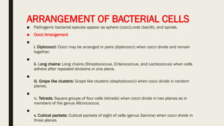
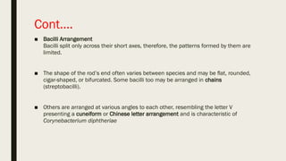
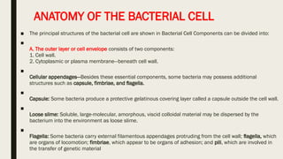
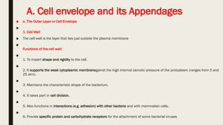
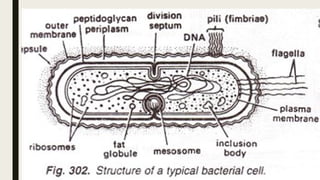
BACTERIA MORPHOLOGY
LAMECK ONTWEKA
INTRODUCTION
■ Size of Bacteria
■
Bacteria are very small in size. The unit of measurement in bacteriology is the micron or micrometer .
■ Shape of Bacteria
■
1. Cocci: Cocci (from kokkos meaning berry) are spherical, or nearly spherical.
■
2. Bacilli: Bacilli (from baculus meaning rod) are relatively straight, rod shaped (cylindrical) cells.
■
3. Vibrios: Vibrios are curved or comma-shaped rods and derive the name from their characteristic
vibratory motility.
■
4. Spirilla: Spirilla are rigid spiral or helical forms.
■
5. Spirochetes: Spirochetes (from speira meaning coil and chaite meaning hair) are flexuous spiral forms.
■
6. Mycoplasma: Mycoplasma are cell wall deficient bacteria and hence do not possess a stable
morphology. They occur as round or oval bodies and interlacing filaments
ARRANGEMENT OF BACTERIAL CELLS
■ Pathogenic bacterial species appear as sphere (cocci),rods (bacilli), and spirals.
■ Cocci Arrangement
■
i. Diplococci: Cocci may be arranged in pairs (diplococci) when cocci divide and remain
together.
■
ii. Long chains: Long chains (Streptococcus, Enterococcus, and Lactococcus) when cells
adhere after repeated divisions in one plane.
■
iii. Grape like clusters: Grape like clusters (staphylococci) when cocci divide in random
planes.
■
iv. Tetrads: Square groups of four cells (tetrads) when cocci divide in two planes as in
members of the genus Micrococcus.
■
v. Cubical packets: Cubical packets of eight of cells (genus Sarcina) when cocci divide in
three planes
Cont….
■ Bacilli Arrangement
Bacilli split only across their short axes, therefore, the patterns formed by them are
limited.
■ The shape of the rod’s end often varies between species and may be flat, rounded,
cigar-shaped, or bifurcated. Some bacilli too may be arranged in chains
(streptobacilli).
■ Others are arranged at various angles to each other, resembling the letter V
presenting a cuneiform or Chinese letter arrangement and is characteristic of
Corynebacterium diphtheriae
ANATOMY OF THE BACTERIAL CELL
■ The principal structures of the bacterial cell are shown in Bacterial Cell Components can be divided into:
■
A. The outer layer or cell envelope consists of two components:
1. Cell wall.
2. Cytoplasmic or plasma membrane—beneath cell wall.
■
Cellular appendages—Besides these essential components, some bacteria may possess additional
structures such as capsule, fimbriae, and flagella.
■
Capsule: Some bacteria produce a protective gelatinous covering layer called a capsule outside the cell wall.
■
Loose slime: Soluble, large-molecular, amorphous, viscid colloidal material may be dispersed by the
bacterium into the environment as loose slime.
■
Flagella: Some bacteria carry external filamentous appendages protruding from the cell wall; flagella, which
are organs of locomotion; fimbriae, which appear to be organs of adhesion; and pili, which are involved in
the transfer of genetic material
Cont….
■ B. Cell Interior
■
Those structures and substances that are bounded by the cytoplasmic membrane, compose
the cell interior and include cytoplasm,
■ cytoplasmic inclusions (mesosomes, ribosomes, inclusion granules, vacuoles)
■ and a single circular chromosome of deoxyribonucleic acid (DNA).
A. Cell envelope and its Appendages
■ a. The Outer Layer or Cell Envelope
■
1. Cell Wall
■ The cell wall is the layer that lies just outside the plasma membrane
■
Functions of the cell wall:
■
1. To impart shape and rigidity to the cell.
■
2. It supports the weak cytoplasmic membranegainst the high internal osmotic pressure of the protoplasm (ranges from 5 and
25 atm).
■
3. Maintains the characteristic shape of the bacterium.
■
4. It takes part in cell division.
■
5. Also functions in interactions (e.g. adhesion) with other bacteria and with mammalian cells.
■
6. Provide specific protein and carbohydrate receptors for the attachment of some bacterial viruses
Cont…
■ 2. Cytoplasmic (Plasma) Membrane
■
Structure: The cytoplasmic (plasma) membrane limits the bacterial protoplast
■
Functions of Cytoplasmic Membrane
■
i. Semipermeable membrane—controlling the inflow and outflow of metabolites to and from the protoplasm.
■
ii. Housing enzymes—involved in outer membrane synthesis, cell wall synthesis, and in the assembly and secretion
of extracytoplasmic and extracellular substances.
■
iii. Housing many sensory and chemotaxis proteins that monitor chemical and physical changes in the environment.
■
iv. Generation of chemical energy (i.e, ATP).
■
v. Cell motility.
■
vi. Mediation of chromosomal segregation during replication.
CONT…
■ b. Cellular Appendages
■
i. Capsule or Slime Layer
■
Structure: Many bacteria synthesize large amount of extracellular polymer in their natural environments.When
the polymer forms a condensed, well defined layer closely surrounding the cell, it is called the capsule as in the
pneumococcus.
■ If the polymer is easily washed off and does not appear to be associated with the cell in any definite fashion, it
is referred as a slime layer as in Leuconostoc
■ A glycocalyx is a network of polysaccharide extending from the surface of bacteria and other cells. Capsules too
thin to be seen under the light microscope are called microcapsules.
■
Capsulated bacteria: Streptococcus pneumoniae, several groups of streptococci, Neisseria meningitidis,
Klebsiella, Haemophilus influenzae, Yersinia and Bacillus.
Cont…
■ Functions of Capsule
i. Virulence factor: Capsules often act as a virulence factor by protecting the bacterium from ingestion by
phagocytosis, and noncapsulate mutant of these bacteria are nonvirulent.
■ ii. Protection of the cell wall: In protecting the cell wall attack by various kinds of antibacterial agents,
e.g. bacteriophages, colicines, complement, lysozyme and other lytic enzymes.
■
iii. Identifcation and typing of bacteria: Capsular antigen is specifc for bacteria and can be used for
identifcation and typing of bacteria.
■ Arrangement/Types
The number and location of flagella are distinctive for each genus. There are four types of flagella
arrangement:
■
• Monotrichous—Single polar flagellum (e.g. Cholera vibrio).
• Amphitrichous—Single flagellum at both ends (e.g. Alcaligenes faecalis).
• Lophotrichous—Tuft of flagella at one or both ends (e.g. spirilla).
• Peritrichous—Flagella surrounding the cell (e.g. Typhoid bacilli).
CONT…
■ ii. Flagella
■
Motile bacteria, except spirochetes, possess one or more unbranched, long, sinuous filaments
called flagella, which are the organs of locomotion.
■
Structure: They are long, hollow, helical filaments, usually several times the length of the cell.
■ Parts and Composition
■ Each flagellum consists of three parts
i. Filament
ii. Hook
iii. Basal body.
CONT…
■ iii. Fimbria or Pili
Structure and synthesis: Many gram-negative bacteria have short, fine, hair like surface appendages
called fimbriae or pili depending on their function.
■ They originate in the cytoplasmic membrane and are composed of structural protein subunits termed
pilins like flagella. They occur in non-motile, as well as in motile strains.
■ Functions of Pili
Two classes can be distinguished on the basis of their function: ordinary (common) pili and sex pili.
■
A. Ordinary (common) pili: Fimbriae probably function as organs of adhesions that allow attachment of
a bacterial cell to other cells or surfaces.
■ B. Sex pili: They are genetically determined by sex factors or conjugative plasmids and appear to be
involved in the transfer of DNA during conjugation.
■ They are found on ‘male’ bacteria and help in the attachment of those cells to ‘female’ bacteria,
forming hollow conjugation tubes through which, it is assumed, genetic material is transferred from
the donor to the recipient cell.
B. Cell interior
■ 1. Cytoplasm
The cytoplasm of the bacterial cell is a viscous watery solution or soft gel, containing a variety of organic or
inorganic solutes, and numerous ribosomes and polysomes.
■ 2. Ribosomes
The ribosomes are the location for all bacterial protein synthesis.
■ 3. Mesosomes (Chondroids)
Strucure: These are convoluted or multilaminated membranous bodies formed as invaginations of the
plasma membrane into the cytoplasm.
■ Types of Mesosomes
These are of two types:
i. Septal mesosomes: They function in the formation of cross-walls during cell division.
ii. Lateral mesosomes: They are present in a more random fashion.
■
Functions of Mesosomes
i. Compartmenting of DNA: These are thought to be involved in mechanisms responsible for the
compartmenting of DNA at cell division and sporulation.
■
ii. Sites of the respiratory enzymes: They provide increased membrane surface and are the principle sites of
the respiratory enzymes in bacteria and are analogous to mitochondria of eukaryotes.
Cont..
■ 5. Bacterial Nucleus
■
Structure: The genetic material of a bacterial cell is contained in a single, long molecule of double-
stranded deoxyribonucleic acid (DNA) .
■ Plasmids
Bacteria may possess extranuclear genetic elements in the cytoplasm consisting of DNA, termed
plasmids or episomes.These can exist and replicate independently of the chromosome or may be
integrated with it.
■ Function of plasmids: may confer on it certain properties such as drug resistance, bacteriocin
production, resistance to toxic metaons, enterotoxin production, enhanced pathogenicity, reduced
sensitivity to mutagens, or to the ability to degrade complex organic molecules which may constitute
a survival advantage
Bacterial Spore
A number of gram-positive bacteria, such as those of the genera Clostridium and Bacillus can form a special
resistant dormant structure called an endospore or, simply, spores. Endospore develop when essential nutrients
are depleted.
Properties of Endospores
■ 1. Core: The fully developed spore has the core which is the spore protoplast containing the normal
cell structures but is metabolically inactive.
■
2. Spore wall: The innermost layer surrounding the inner spore membrane is called the spore wall.
It contains normal peptidoglycan and becomes the cell wall of the germinating vegetative cell.
■
3. Cortex: The cortex is the thickest layer of the spore envelope. Cortex peptidoglycan is extremely
sensitive to lysozyme, and its autolysins plays a role in spore germination.
■
4. Spore coat: Cortex, in turn, is enclosed by fairly thick spore coat.
■
5. Exosporium: Spores of some species have an additional, apparently rather loose covering known
as the exosporium, which may have distinctive ridges and grooves.

BACTERIA MORPHOLOGY and characterization

  • 1.
  • 2.
    INTRODUCTION ■ Size ofBacteria ■ Bacteria are very small in size. The unit of measurement in bacteriology is the micron or micrometer . ■ Shape of Bacteria ■ 1. Cocci: Cocci (from kokkos meaning berry) are spherical, or nearly spherical. ■ 2. Bacilli: Bacilli (from baculus meaning rod) are relatively straight, rod shaped (cylindrical) cells. ■ 3. Vibrios: Vibrios are curved or comma-shaped rods and derive the name from their characteristic vibratory motility. ■ 4. Spirilla: Spirilla are rigid spiral or helical forms. ■ 5. Spirochetes: Spirochetes (from speira meaning coil and chaite meaning hair) are flexuous spiral forms. ■ 6. Mycoplasma: Mycoplasma are cell wall deficient bacteria and hence do not possess a stable morphology. They occur as round or oval bodies and interlacing filaments
  • 3.
    ARRANGEMENT OF BACTERIALCELLS ■ Pathogenic bacterial species appear as sphere (cocci),rods (bacilli), and spirals. ■ Cocci Arrangement ■ i. Diplococci: Cocci may be arranged in pairs (diplococci) when cocci divide and remain together. ■ ii. Long chains: Long chains (Streptococcus, Enterococcus, and Lactococcus) when cells adhere after repeated divisions in one plane. ■ iii. Grape like clusters: Grape like clusters (staphylococci) when cocci divide in random planes. ■ iv. Tetrads: Square groups of four cells (tetrads) when cocci divide in two planes as in members of the genus Micrococcus. ■ v. Cubical packets: Cubical packets of eight of cells (genus Sarcina) when cocci divide in three planes
  • 4.
    Cont…. ■ Bacilli Arrangement Bacillisplit only across their short axes, therefore, the patterns formed by them are limited. ■ The shape of the rod’s end often varies between species and may be flat, rounded, cigar-shaped, or bifurcated. Some bacilli too may be arranged in chains (streptobacilli). ■ Others are arranged at various angles to each other, resembling the letter V presenting a cuneiform or Chinese letter arrangement and is characteristic of Corynebacterium diphtheriae
  • 6.
    ANATOMY OF THEBACTERIAL CELL ■ The principal structures of the bacterial cell are shown in Bacterial Cell Components can be divided into: ■ A. The outer layer or cell envelope consists of two components: 1. Cell wall. 2. Cytoplasmic or plasma membrane—beneath cell wall. ■ Cellular appendages—Besides these essential components, some bacteria may possess additional structures such as capsule, fimbriae, and flagella. ■ Capsule: Some bacteria produce a protective gelatinous covering layer called a capsule outside the cell wall. ■ Loose slime: Soluble, large-molecular, amorphous, viscid colloidal material may be dispersed by the bacterium into the environment as loose slime. ■ Flagella: Some bacteria carry external filamentous appendages protruding from the cell wall; flagella, which are organs of locomotion; fimbriae, which appear to be organs of adhesion; and pili, which are involved in the transfer of genetic material
  • 7.
    Cont…. ■ B. CellInterior ■ Those structures and substances that are bounded by the cytoplasmic membrane, compose the cell interior and include cytoplasm, ■ cytoplasmic inclusions (mesosomes, ribosomes, inclusion granules, vacuoles) ■ and a single circular chromosome of deoxyribonucleic acid (DNA).
  • 8.
    A. Cell envelopeand its Appendages ■ a. The Outer Layer or Cell Envelope ■ 1. Cell Wall ■ The cell wall is the layer that lies just outside the plasma membrane ■ Functions of the cell wall: ■ 1. To impart shape and rigidity to the cell. ■ 2. It supports the weak cytoplasmic membranegainst the high internal osmotic pressure of the protoplasm (ranges from 5 and 25 atm). ■ 3. Maintains the characteristic shape of the bacterium. ■ 4. It takes part in cell division. ■ 5. Also functions in interactions (e.g. adhesion) with other bacteria and with mammalian cells. ■ 6. Provide specific protein and carbohydrate receptors for the attachment of some bacterial viruses
  • 10.
    Cont… ■ 2. Cytoplasmic(Plasma) Membrane ■ Structure: The cytoplasmic (plasma) membrane limits the bacterial protoplast ■ Functions of Cytoplasmic Membrane ■ i. Semipermeable membrane—controlling the inflow and outflow of metabolites to and from the protoplasm. ■ ii. Housing enzymes—involved in outer membrane synthesis, cell wall synthesis, and in the assembly and secretion of extracytoplasmic and extracellular substances. ■ iii. Housing many sensory and chemotaxis proteins that monitor chemical and physical changes in the environment. ■ iv. Generation of chemical energy (i.e, ATP). ■ v. Cell motility. ■ vi. Mediation of chromosomal segregation during replication.
  • 11.
    CONT… ■ b. CellularAppendages ■ i. Capsule or Slime Layer ■ Structure: Many bacteria synthesize large amount of extracellular polymer in their natural environments.When the polymer forms a condensed, well defined layer closely surrounding the cell, it is called the capsule as in the pneumococcus. ■ If the polymer is easily washed off and does not appear to be associated with the cell in any definite fashion, it is referred as a slime layer as in Leuconostoc ■ A glycocalyx is a network of polysaccharide extending from the surface of bacteria and other cells. Capsules too thin to be seen under the light microscope are called microcapsules. ■ Capsulated bacteria: Streptococcus pneumoniae, several groups of streptococci, Neisseria meningitidis, Klebsiella, Haemophilus influenzae, Yersinia and Bacillus.
  • 12.
    Cont… ■ Functions ofCapsule i. Virulence factor: Capsules often act as a virulence factor by protecting the bacterium from ingestion by phagocytosis, and noncapsulate mutant of these bacteria are nonvirulent. ■ ii. Protection of the cell wall: In protecting the cell wall attack by various kinds of antibacterial agents, e.g. bacteriophages, colicines, complement, lysozyme and other lytic enzymes. ■ iii. Identifcation and typing of bacteria: Capsular antigen is specifc for bacteria and can be used for identifcation and typing of bacteria. ■ Arrangement/Types The number and location of flagella are distinctive for each genus. There are four types of flagella arrangement: ■ • Monotrichous—Single polar flagellum (e.g. Cholera vibrio). • Amphitrichous—Single flagellum at both ends (e.g. Alcaligenes faecalis). • Lophotrichous—Tuft of flagella at one or both ends (e.g. spirilla). • Peritrichous—Flagella surrounding the cell (e.g. Typhoid bacilli).
  • 13.
    CONT… ■ ii. Flagella ■ Motilebacteria, except spirochetes, possess one or more unbranched, long, sinuous filaments called flagella, which are the organs of locomotion. ■ Structure: They are long, hollow, helical filaments, usually several times the length of the cell. ■ Parts and Composition ■ Each flagellum consists of three parts i. Filament ii. Hook iii. Basal body.
  • 15.
    CONT… ■ iii. Fimbriaor Pili Structure and synthesis: Many gram-negative bacteria have short, fine, hair like surface appendages called fimbriae or pili depending on their function. ■ They originate in the cytoplasmic membrane and are composed of structural protein subunits termed pilins like flagella. They occur in non-motile, as well as in motile strains. ■ Functions of Pili Two classes can be distinguished on the basis of their function: ordinary (common) pili and sex pili. ■ A. Ordinary (common) pili: Fimbriae probably function as organs of adhesions that allow attachment of a bacterial cell to other cells or surfaces. ■ B. Sex pili: They are genetically determined by sex factors or conjugative plasmids and appear to be involved in the transfer of DNA during conjugation. ■ They are found on ‘male’ bacteria and help in the attachment of those cells to ‘female’ bacteria, forming hollow conjugation tubes through which, it is assumed, genetic material is transferred from the donor to the recipient cell.
  • 16.
    B. Cell interior ■1. Cytoplasm The cytoplasm of the bacterial cell is a viscous watery solution or soft gel, containing a variety of organic or inorganic solutes, and numerous ribosomes and polysomes. ■ 2. Ribosomes The ribosomes are the location for all bacterial protein synthesis. ■ 3. Mesosomes (Chondroids) Strucure: These are convoluted or multilaminated membranous bodies formed as invaginations of the plasma membrane into the cytoplasm. ■ Types of Mesosomes These are of two types: i. Septal mesosomes: They function in the formation of cross-walls during cell division. ii. Lateral mesosomes: They are present in a more random fashion. ■ Functions of Mesosomes i. Compartmenting of DNA: These are thought to be involved in mechanisms responsible for the compartmenting of DNA at cell division and sporulation. ■ ii. Sites of the respiratory enzymes: They provide increased membrane surface and are the principle sites of the respiratory enzymes in bacteria and are analogous to mitochondria of eukaryotes.
  • 17.
    Cont.. ■ 5. BacterialNucleus ■ Structure: The genetic material of a bacterial cell is contained in a single, long molecule of double- stranded deoxyribonucleic acid (DNA) . ■ Plasmids Bacteria may possess extranuclear genetic elements in the cytoplasm consisting of DNA, termed plasmids or episomes.These can exist and replicate independently of the chromosome or may be integrated with it. ■ Function of plasmids: may confer on it certain properties such as drug resistance, bacteriocin production, resistance to toxic metaons, enterotoxin production, enhanced pathogenicity, reduced sensitivity to mutagens, or to the ability to degrade complex organic molecules which may constitute a survival advantage
  • 18.
    Bacterial Spore A numberof gram-positive bacteria, such as those of the genera Clostridium and Bacillus can form a special resistant dormant structure called an endospore or, simply, spores. Endospore develop when essential nutrients are depleted.
  • 19.
    Properties of Endospores ■1. Core: The fully developed spore has the core which is the spore protoplast containing the normal cell structures but is metabolically inactive. ■ 2. Spore wall: The innermost layer surrounding the inner spore membrane is called the spore wall. It contains normal peptidoglycan and becomes the cell wall of the germinating vegetative cell. ■ 3. Cortex: The cortex is the thickest layer of the spore envelope. Cortex peptidoglycan is extremely sensitive to lysozyme, and its autolysins plays a role in spore germination. ■ 4. Spore coat: Cortex, in turn, is enclosed by fairly thick spore coat. ■ 5. Exosporium: Spores of some species have an additional, apparently rather loose covering known as the exosporium, which may have distinctive ridges and grooves.