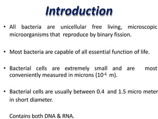
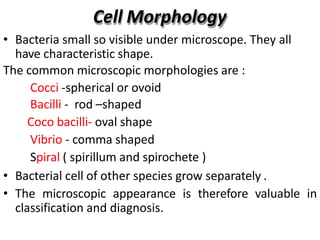
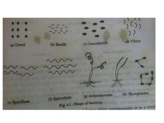
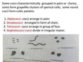
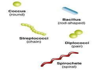
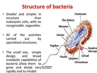
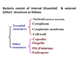
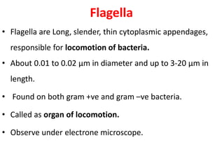
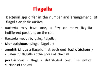
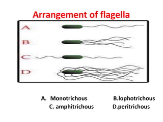
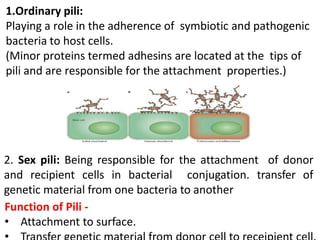
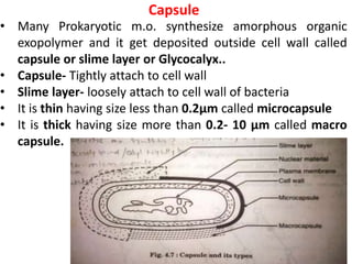
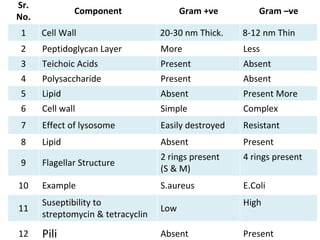
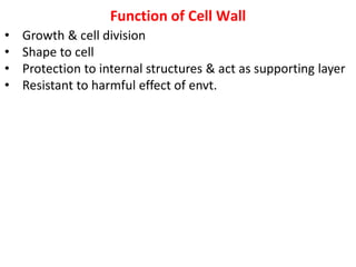
Bacterial
Ultrastructure
Introduction
• All bacteria are unicellular free living, microscopic
microorganisms that reproduce by binary fission.
• Most bacteria are capable of all essential function of life.
• Bacterial cells are extremely small and are most
conveniently measured in microns (10-6 m).
• Bacterial cells are usually between 0.4 and 1.5 micro meter
in short diameter.
Contains both DNA & RNA.
Cell Morphology
• Bacteria small so visible under microscope. They all
have characteristic shape.
The common microscopic morphologies are :
Cocci -spherical or ovoid
Bacilli - rod –shaped
Coco bacilli- oval shape
Vibrio - comma shaped
Spiral ( spirillum and spirochete )
• Bacterial cell of other species grow separately .
• The microscopic appearance is therefore valuable in
classification and diagnosis.
Some cocci characterristically grouped in pairs or chains;
some form grapelike clusters of sperical cells; some round
cocci form cubic packets.
1. Diplococci- cocci arrange in pairs
2. Streptococci- Arranged in form of chain.
3. Tetracocci- cocci arrange in group of four.
4. Staphylococci-cocci divide in irregular maner.
Structure of bacteria
• Smaller and simpler in
structure than
eukaryotic cells, with no
recognizable organelles.
• All of the activities
carried out by
specialized structures.
• The small size, simple
design, and broad
metabolic capabilities of
bacteria allow them to
grow and divide very
rapidly and to inhabit
Bacteria consist of internal (Essential) & external
(other) structures as follows
Flagella
• Flagella are Long, slender, thin cytoplasmic appendages,
responsible for locomotion of bacteria.
• About 0.01 to 0.02 µm in diameter and up to 3-20 µm in
length.
• Found on both gram +ve and gram –ve bacteria.
• Called as organ of locomotion.
• Observe under electrone microscope.
Flagella
• Bacterial spp differ in the number and arrangement of
flagella on their surface.
• Bacteria may have one, a few, or many flagella
indifferent positions on the cell.
• Bacteria moves by using flagella.
• Monotrichous - single flagellum
• amphitrichous a flagellum at each end lophotrichous -
clusters of flagella at the poles of the cell
• peritrichous - flagella distributed over the entire
surface of the cell .
Arrangement of flagella
A. Monotrichous B.lophotrichous
C. amphitrichous D.peritrichous
• Made up of protein subunits called flagellin responsible
for bacterial motility.
• Motility of bacteria observe by Hanging drop technique.
• Some bacteria posses swimming motility in viscous
media due to presence of flagella called as Periplasmic
flagella.
• Movement of bacteria on solid surface called gliding
movement.
• Movement of bacteria towards or away from specific
stimulus called taxis
• Towards chemicals - chemotaxis,
• Towards light- phototaxis, and
• Towards air- aerotaxis.
Structure of Flagella
Flagellum has three basic parts1. Filament 2. Hook
3.Basalbody
•Filament is thin ,cylindrical, long outermost region.
•Filament is made up of protein flagellin with M.W. 20,000-
40,000 dalton.
•The filament attached to wider hook consist of different
proteins.
The basal body composed of rod inserted into rings.
•Gram -ve bacteria contains four rings L-ring, P-ring, S-ring
and M-ring .
L-ring embedded in lipopolysaccharide layer
P-ring embedded in Peptidoglycan layer
S-ring embedded in above cytoplasmic membrane
M-ring embedded within cytoplasmic membrane
•Gram +ve bacteria have only S & M ring.
Pili (Fimbriae)
• Many gram-negative bacteria possess rigid surface
appendages called pili (L "hairs") or fimbriae ("fringes").
• Pili known as organ of Adhesion.
• They are shorter and finer than flagella;
like flagella, they are composed of structural
protein subunits termed pilins.
• Pili are thin, short, more in no. than flagella.
• Present only on gram –ve bacteria. Seen only under electrone
microscope.
• Each bacteria (gram –ve) contains 100-200 pili.
• They join bacterial cells for transfer of DNA in between cells
hence called s sex pili or fertility pili.
1.Ordinary pili:
Playing a role in the adherence of symbiotic and pathogenic
bacteria to host cells.
(Minor proteins termed adhesins are located at the tips of
pili and are responsible for the attachment properties.)
2. Sex pili: Being responsible for the attachment of donor
and recipient cells in bacterial conjugation. transfer of
genetic material from one bacteria to another
Function of Pili -
• Attachment to surface.
Capsule
• Many Prokaryotic m.o. synthesize amorphous organic
exopolymer and it get deposited outside cell wall called
capsule or slime layer or Glycocalyx..
• Capsule- Tightly attach to cell wall
• Slime layer- loosely attach to cell wall of bacteria
• It is thin having size less than 0.2µm called microcapsule
• It is thick having size more than 0.2- 10 µm called macro
capsule.
Function of capsule
• Prevent the cell from desiccation and drying: capsular
polysaccharide bind significant amount of water making
cell resistant to drying
• Protection: it protect from mechanical injury, temperature,
drying etc
• Attachment: capsule helps in attachment on the surface.
Eg. Streptococcus mutants that cause dental carries attach
on teeth surface by its capsule.
• Anti-phagocytic : Capsule resist phagocytosis by WBCs
• Capsule prevent attachment of bacteriophage on cell
surface
• Source of nutrition: capsule is source of nutrition when
nutrient supply is low in cell.
• Repulsion: same charge capsulated bacteria repel each
Cell Wall
• Outer covering of most cells that protects the bacterial cell
and gives it shape (spherical, rod and spiral).
• Composed of peptidoglycan (polysaccharides + protein)
• It is 10~25nm thick, strong and relatively rigid, though
with some elasticity, and openly porous, being freely
permeable to small particles.
• Contains 20-30% dry weight of cell.
• Cell wall of bacteria contains
• Diaminopimelic acid (DAP), Muramic acid and techoic
acid.
• All this three join and give complex structure known as
peptidoglycan.
The peptidoglycan layer
Peptidoglycan is a complex polymer consisting
of three parts:
1. A backbone, composed of alternating N-
acetylglucosamine and N-acetylmuramic acid
2. A set of identical
tetrapeptide side chains
attached to
N-acetylmuramic acid
3. A set of identical
peptide cross bridges.
• Back bone sam for all type of bacteria.
The tetra-peptide side chains and penta-peptide cross
bridges vary from species to species.
But commonly side chain contains four amino acids
L-alanine, D-alanine, D- glutamic acid, Diaminopiemelic
acid (DPA)
Peptidoglycan is major constituent which differ gram +ve
and gram –ve bacteria.
This gives idea and difference between gram +ve and
gram –ve bacteria.
Sr.
No.
Component Gram +ve Gram –ve
1 Cell Wall 20-30 nm Thick. 8-12 nm Thin
2 Peptidoglycan Layer More Less
3 Teichoic Acids Present Absent
4 Polysaccharide Present Absent
5 Lipid Absent Present More
6 Cell wall Simple Complex
7 Effect of lysosome Easily destroyed Resistant
8 Lipid Absent Present
9 Flagellar Structure
2 rings present
(S & M)
4 rings present
10 Example S.aureus E.Coli
11
Suseptibility to
streptomycin & tetracyclin
Low
High
12 Pili Absent Present
Function of Cell Wall
• Growth & cell division
• Shape to cell
• Protection to internal structures & act as supporting layer
• Resistant to harmful effect of envt.
Plasma Membrane
• It is thin layer present in inner surface to cell wall.
• Consists of both phospholipids and proteins.
• Protects the cell from its surroundings.
• The phosphate end of the
molecule is hydrophilic and is
attracted to water.
• The fatty acids are hydrophobic.
• Membrane also contain protein.
 Function :
• The plasma membrane is selectively permeable in
that is helps control what moves into and out of cell.
• Provide mechanical strength, Passage of nutrients.
Cytoplasm
• The cytoplasm of bacterial cell is a viscous watery
solution, or soft gel, containing a variety of organic
and inorganic solutes, and ribosomes.
• Containing plasmids in some species ofbacteria.
• Not containing endoplasmic reticulum.
• Not containing membrane-bearing microsomes.
• Not containing mitochondria.
Mesosomes
• In bacteria depend on growth condition membrane
appears to be in folded at more than one point such
inholdings called as mesosomes.
• It is of two type
A. Central Mesosome
B. Peripheral Mesosome
• Central mesosome located near to middle
of cell.
• Peripheral mesosome not restricted to
middle of cell.
• Central mesosome attached to chromosome and involved
in DNA seperation.
• It form cross wall during cell division cause cell division of
cell.
Mesosome are observe only under electron microscope
Ribosomes- protein synthesis machinery
• Involve in synthesis of protein
• Number varies with rate of protein synthesis
• Abundant in cytoplasm
• Often grouped in long chains called polyribosomes.
• smaller than the ribosomes in eukaryotic cells-but have
a similar function
• Bacterial ribosomes have sedimentation rate of 70S; their
subunits have rates of 30S and 50S.
• The unit used to measure sedimentation velocity is
Svedberg
• When placed in conc. Of magnesium dissociate into two
component 50S and 30S particles.
Nucleoid
• Unlike the eukaryotic (true) cells, bacteria do not have
a membrane enclosed nucleus.
• The nucleoid is a region of cytoplasm where the
chromosomal DNA is located.
• It is not a membrane bound nucleus, but simply an area
of the cytoplasm where the strands of DNA are found.
Endospore
• Bacterial species produce spore inside cell – Endospore
outside cell – Exospore
• Some bacteria devolope a highly resistant layer called
endospore, that’s why the organism can survive in a for
long period in adverse enviromental condition.
1. Core wall, the innermost layer surrounding the inner
spore membrane. It contains normal peptidoglycan and
becomes the cell wall of the germinating vegetative cell.
2. The cortex is the thickest layer of the spore envelope. It
contains an unusual type of peptidoglycan.
Cortex peptidoglycan is extremely sensitive to lysozyme,
and its autolysis plays a role in spore germination.
3. The coat is composed of a keratin-like protein
containing many intramolecular disulfide bonds. The
impermeability of this layer confers on spores their
relative resistance to antibacterial chemical agents.
4. The exosporium is a lipoprotein membrane containing
some carbohydrate.
• Endospores are much more resistant than the
vegetative forms to exposure to disinfectants,
drying and heating.
• Reasons
Impermeability of their cortex and outer coat. High
content of calcium and dipicolinic acid.
Low content of water.
Very low metabolic and enzyme activity.
THANK YOU

Bcterial ultrastructure all

  • 1.
  • 2.
    Introduction • All bacteriaare unicellular free living, microscopic microorganisms that reproduce by binary fission. • Most bacteria are capable of all essential function of life. • Bacterial cells are extremely small and are most conveniently measured in microns (10-6 m). • Bacterial cells are usually between 0.4 and 1.5 micro meter in short diameter. Contains both DNA & RNA.
  • 3.
    Cell Morphology • Bacteriasmall so visible under microscope. They all have characteristic shape. The common microscopic morphologies are : Cocci -spherical or ovoid Bacilli - rod –shaped Coco bacilli- oval shape Vibrio - comma shaped Spiral ( spirillum and spirochete ) • Bacterial cell of other species grow separately . • The microscopic appearance is therefore valuable in classification and diagnosis.
  • 5.
    Some cocci characterristicallygrouped in pairs or chains; some form grapelike clusters of sperical cells; some round cocci form cubic packets. 1. Diplococci- cocci arrange in pairs 2. Streptococci- Arranged in form of chain. 3. Tetracocci- cocci arrange in group of four. 4. Staphylococci-cocci divide in irregular maner.
  • 7.
    Structure of bacteria •Smaller and simpler in structure than eukaryotic cells, with no recognizable organelles. • All of the activities carried out by specialized structures. • The small size, simple design, and broad metabolic capabilities of bacteria allow them to grow and divide very rapidly and to inhabit
  • 8.
    Bacteria consist ofinternal (Essential) & external (other) structures as follows
  • 9.
    Flagella • Flagella areLong, slender, thin cytoplasmic appendages, responsible for locomotion of bacteria. • About 0.01 to 0.02 µm in diameter and up to 3-20 µm in length. • Found on both gram +ve and gram –ve bacteria. • Called as organ of locomotion. • Observe under electrone microscope.
  • 10.
    Flagella • Bacterial sppdiffer in the number and arrangement of flagella on their surface. • Bacteria may have one, a few, or many flagella indifferent positions on the cell. • Bacteria moves by using flagella. • Monotrichous - single flagellum • amphitrichous a flagellum at each end lophotrichous - clusters of flagella at the poles of the cell • peritrichous - flagella distributed over the entire surface of the cell .
  • 11.
    Arrangement of flagella A.Monotrichous B.lophotrichous C. amphitrichous D.peritrichous
  • 12.
    • Made upof protein subunits called flagellin responsible for bacterial motility. • Motility of bacteria observe by Hanging drop technique. • Some bacteria posses swimming motility in viscous media due to presence of flagella called as Periplasmic flagella. • Movement of bacteria on solid surface called gliding movement. • Movement of bacteria towards or away from specific stimulus called taxis • Towards chemicals - chemotaxis, • Towards light- phototaxis, and • Towards air- aerotaxis.
  • 13.
  • 14.
    Flagellum has threebasic parts1. Filament 2. Hook 3.Basalbody •Filament is thin ,cylindrical, long outermost region. •Filament is made up of protein flagellin with M.W. 20,000- 40,000 dalton. •The filament attached to wider hook consist of different proteins. The basal body composed of rod inserted into rings. •Gram -ve bacteria contains four rings L-ring, P-ring, S-ring and M-ring . L-ring embedded in lipopolysaccharide layer P-ring embedded in Peptidoglycan layer S-ring embedded in above cytoplasmic membrane M-ring embedded within cytoplasmic membrane •Gram +ve bacteria have only S & M ring.
  • 15.
    Pili (Fimbriae) • Manygram-negative bacteria possess rigid surface appendages called pili (L "hairs") or fimbriae ("fringes"). • Pili known as organ of Adhesion. • They are shorter and finer than flagella; like flagella, they are composed of structural protein subunits termed pilins. • Pili are thin, short, more in no. than flagella. • Present only on gram –ve bacteria. Seen only under electrone microscope. • Each bacteria (gram –ve) contains 100-200 pili. • They join bacterial cells for transfer of DNA in between cells hence called s sex pili or fertility pili.
  • 16.
    1.Ordinary pili: Playing arole in the adherence of symbiotic and pathogenic bacteria to host cells. (Minor proteins termed adhesins are located at the tips of pili and are responsible for the attachment properties.) 2. Sex pili: Being responsible for the attachment of donor and recipient cells in bacterial conjugation. transfer of genetic material from one bacteria to another Function of Pili - • Attachment to surface.
  • 17.
    Capsule • Many Prokaryoticm.o. synthesize amorphous organic exopolymer and it get deposited outside cell wall called capsule or slime layer or Glycocalyx.. • Capsule- Tightly attach to cell wall • Slime layer- loosely attach to cell wall of bacteria • It is thin having size less than 0.2µm called microcapsule • It is thick having size more than 0.2- 10 µm called macro capsule.
  • 18.
    Function of capsule •Prevent the cell from desiccation and drying: capsular polysaccharide bind significant amount of water making cell resistant to drying • Protection: it protect from mechanical injury, temperature, drying etc • Attachment: capsule helps in attachment on the surface. Eg. Streptococcus mutants that cause dental carries attach on teeth surface by its capsule. • Anti-phagocytic : Capsule resist phagocytosis by WBCs • Capsule prevent attachment of bacteriophage on cell surface • Source of nutrition: capsule is source of nutrition when nutrient supply is low in cell. • Repulsion: same charge capsulated bacteria repel each
  • 19.
    Cell Wall • Outercovering of most cells that protects the bacterial cell and gives it shape (spherical, rod and spiral). • Composed of peptidoglycan (polysaccharides + protein) • It is 10~25nm thick, strong and relatively rigid, though with some elasticity, and openly porous, being freely permeable to small particles. • Contains 20-30% dry weight of cell. • Cell wall of bacteria contains • Diaminopimelic acid (DAP), Muramic acid and techoic acid. • All this three join and give complex structure known as peptidoglycan.
  • 20.
    The peptidoglycan layer Peptidoglycanis a complex polymer consisting of three parts: 1. A backbone, composed of alternating N- acetylglucosamine and N-acetylmuramic acid 2. A set of identical tetrapeptide side chains attached to N-acetylmuramic acid 3. A set of identical peptide cross bridges.
  • 21.
    • Back bonesam for all type of bacteria. The tetra-peptide side chains and penta-peptide cross bridges vary from species to species. But commonly side chain contains four amino acids L-alanine, D-alanine, D- glutamic acid, Diaminopiemelic acid (DPA) Peptidoglycan is major constituent which differ gram +ve and gram –ve bacteria. This gives idea and difference between gram +ve and gram –ve bacteria.
  • 22.
    Sr. No. Component Gram +veGram –ve 1 Cell Wall 20-30 nm Thick. 8-12 nm Thin 2 Peptidoglycan Layer More Less 3 Teichoic Acids Present Absent 4 Polysaccharide Present Absent 5 Lipid Absent Present More 6 Cell wall Simple Complex 7 Effect of lysosome Easily destroyed Resistant 8 Lipid Absent Present 9 Flagellar Structure 2 rings present (S & M) 4 rings present 10 Example S.aureus E.Coli 11 Suseptibility to streptomycin & tetracyclin Low High 12 Pili Absent Present
  • 23.
    Function of CellWall • Growth & cell division • Shape to cell • Protection to internal structures & act as supporting layer • Resistant to harmful effect of envt.
  • 24.
    Plasma Membrane • Itis thin layer present in inner surface to cell wall. • Consists of both phospholipids and proteins. • Protects the cell from its surroundings. • The phosphate end of the molecule is hydrophilic and is attracted to water. • The fatty acids are hydrophobic. • Membrane also contain protein.  Function : • The plasma membrane is selectively permeable in that is helps control what moves into and out of cell. • Provide mechanical strength, Passage of nutrients.
  • 25.
    Cytoplasm • The cytoplasmof bacterial cell is a viscous watery solution, or soft gel, containing a variety of organic and inorganic solutes, and ribosomes. • Containing plasmids in some species ofbacteria. • Not containing endoplasmic reticulum. • Not containing membrane-bearing microsomes. • Not containing mitochondria.
  • 26.
    Mesosomes • In bacteriadepend on growth condition membrane appears to be in folded at more than one point such inholdings called as mesosomes. • It is of two type A. Central Mesosome B. Peripheral Mesosome • Central mesosome located near to middle of cell. • Peripheral mesosome not restricted to middle of cell. • Central mesosome attached to chromosome and involved in DNA seperation. • It form cross wall during cell division cause cell division of cell.
  • 27.
    Mesosome are observeonly under electron microscope
  • 28.
    Ribosomes- protein synthesismachinery • Involve in synthesis of protein • Number varies with rate of protein synthesis • Abundant in cytoplasm • Often grouped in long chains called polyribosomes. • smaller than the ribosomes in eukaryotic cells-but have a similar function • Bacterial ribosomes have sedimentation rate of 70S; their subunits have rates of 30S and 50S. • The unit used to measure sedimentation velocity is Svedberg • When placed in conc. Of magnesium dissociate into two component 50S and 30S particles.
  • 29.
    Nucleoid • Unlike theeukaryotic (true) cells, bacteria do not have a membrane enclosed nucleus. • The nucleoid is a region of cytoplasm where the chromosomal DNA is located. • It is not a membrane bound nucleus, but simply an area of the cytoplasm where the strands of DNA are found.
  • 30.
    Endospore • Bacterial speciesproduce spore inside cell – Endospore outside cell – Exospore • Some bacteria devolope a highly resistant layer called endospore, that’s why the organism can survive in a for long period in adverse enviromental condition.
  • 31.
    1. Core wall,the innermost layer surrounding the inner spore membrane. It contains normal peptidoglycan and becomes the cell wall of the germinating vegetative cell. 2. The cortex is the thickest layer of the spore envelope. It contains an unusual type of peptidoglycan. Cortex peptidoglycan is extremely sensitive to lysozyme, and its autolysis plays a role in spore germination. 3. The coat is composed of a keratin-like protein containing many intramolecular disulfide bonds. The impermeability of this layer confers on spores their relative resistance to antibacterial chemical agents. 4. The exosporium is a lipoprotein membrane containing some carbohydrate.
  • 32.
    • Endospores aremuch more resistant than the vegetative forms to exposure to disinfectants, drying and heating. • Reasons Impermeability of their cortex and outer coat. High content of calcium and dipicolinic acid. Low content of water. Very low metabolic and enzyme activity. THANK YOU