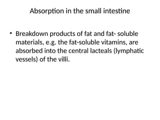
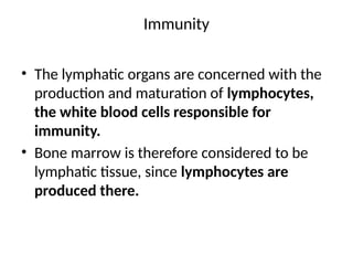
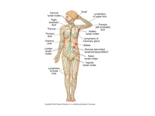
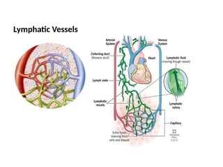
The lymphatic system plays a crucial role in maintaining fluid balance, transporting dietary lipids, and facilitating immune responses by filtering lymph through lymph nodes. It consists of lymph, lymphatic vessels, lymph nodes, and various lymphoid organs such as the spleen and thymus. Lymphatic vessels drain excess interstitial fluid, and the system is essential for both the immune surveillance and absorption of fats from the digestive tract.


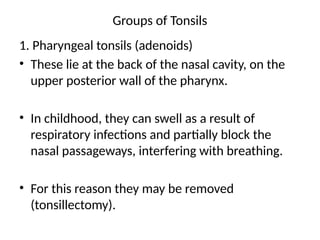
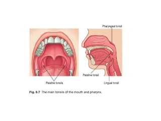


![Lymphatic system[1]](https://cdn.slidesharecdn.com/ss_thumbnails/v8tdil7slo1obvifzera-signature-460517c25b85fc4e63c8080c3e27df73c8dfae9e0c6544cc7ea6d9e8b5e79cc7-poli-180213064029-thumbnail.jpg?width=640&height=640&fit=bounds)


