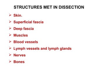
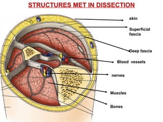
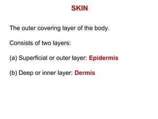
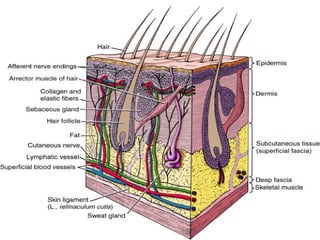
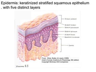
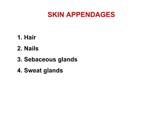
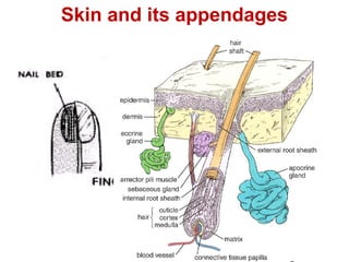
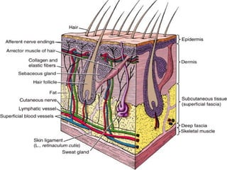
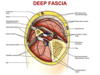
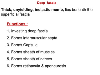
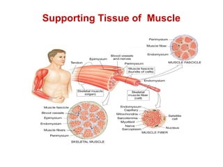
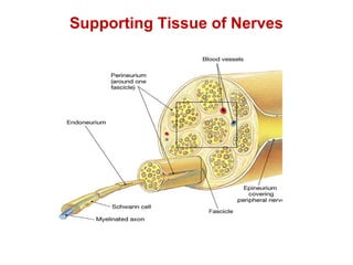
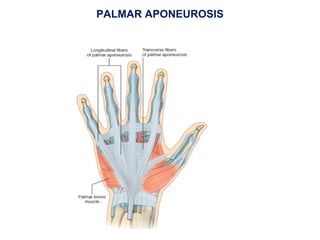
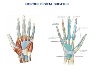
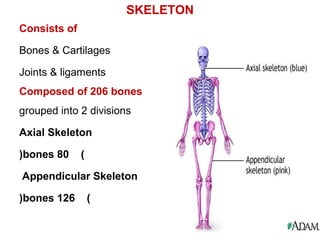
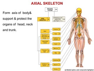
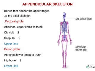
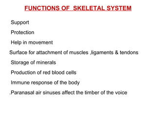
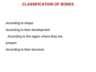
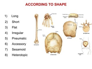
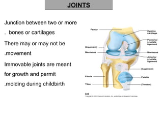
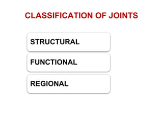
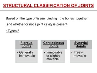
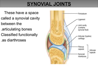
Introduction of structures met in
dissection
 Skin.
 Superficial fascia
 Deep fascia
 Muscles
 Blood vessels
 Lymph vessels and lymph glands
 Nerves
 Bones
STRUCTURES MET IN DISSECTION
skin
Superficial
fascia
Deep fascia
Muscles
Bones
Blood vessels
nerves
STRUCTURES MET IN DISSECTION
The outer covering layer of the body.
Consists of two layers:
(a) Superficial or outer layer: Epidermis
(b) Deep or inner layer: Dermis
SKIN
Epidermis: keratinized stratified squamous epithelium
with five distinct layers.
SKIN APPENDAGES
1. Hair
2. Nails
3. Sebaceous glands
4. Sweat glands
Skin and its appendages
Functions of skin
1. Covers and protect the underlying structures.
2. Regulation of body temperature.
3. Excretion of waste products.
Fibro fatty layer that lies directly under the skin.
It unites the skin to the underlying structures.
It is dense in some places as scalp, palm of hand and sole of
foot and contains collagen bundles
It is thin in the eyelids, auricle, scrotum, penis and clitoris (devoid
of adipose tissue(.
Functions:
Facilitates movement of skin over underlying structures.
Passage for cutaneous vessels, nerves…
Protects the body against heat loss.
SUPERFICIAL FASCIA
SUPERFICIAL FASCIA
Contents:
Cutaneous nerves
Superficial blood vessels
Lymphatics
Parts of sweat glands and hair follicles.
In some regions it may contain superficial muscles
and mammary gland (pectoral region(
Superficial Fascia
Site with Very less fat
-Eyelids
-Pinna
-Penis
Site with more fat
-Breast
-Abdomen
-Gluteal region
DEEP FASCIA
Thick, unyielding, inelastic memb, lies beneath the
superficial fascia
Deep fascia
Functions :
1. Investing deep fascia
2. Forms intermuscular septa
3. Forms Capsule
4. Forms sheath of muscles
5. Forms sheath of nerves
6. Forms retinacula & aponeurosis
Absence of deep fascia
Face
Breast
Penis
Anterior abdominal wall
Supporting Tissue of Muscle
Supporting Tissue of Nerves
PALMAR APONEUROSIS
FIBROUS DIGITAL SHEATHS
NEUROVASCULAR BUNDLE
Blood vessels
Tubular vessels which transport back lymph to the
blood ultimately replacing the volume lost from the
blood during the formation of the interstitial fluid.
Lymph capillaries
Lymph vessels
Lymphatic trunks
Lymphatic ducts
LYMPHATICS
LYMPH
Lymph: Means clear water and it is the colorless fluid and
protein that has been squeezed out of the blood.
(TRANSUDATE(
Similar in composition to blood plasma but protein
concentration is lower than plasma and varies inversely
proportional to rate of formation.
Changes in plasma composition will change lymph
composition.
Formation of lymph
At the arterial end
Hydrostatic pressure=30mm of Hg
Osmotic pressure=25 mm of Hg
Fluid comes out
At the venous end
Hydrostatic pressure=12mm/Hg
Osmotic pressure=25 mm/Hg
Fluid moves in
Most of the fluid returns to its venous
ends and venules; the rest
(10—20%(enters the lymph
capillaries as lymph.
MUSCLES
Latin word : Mus- Mouse
Definition: contractile tissue designed for
movements
CLASSIFICATION
Functionally
Voluntary – can be moved at will
Involuntary – can’t be moved intentionally
Structurally
Striated – have stripes across the fiber
Smooth – no striations
TYPES OF MUSCLES
S k e le ta l M u s c le S m o o th M u s c le C a rd ia c M u s c le
3 T y p e s o f M u s c le s
SKELETAL MUSCLE (GROSS ORGANISATION(
Two ends
Origin: attachment of the muscle to the bone that remains
stationary
Insertion: attachment of the muscle to the bone that moves.
In limbs origin is usually proximal to insertion. But is
interchangeable.
Two parts
Fleshy part: part of the muscle between the origin and tendon
of insertion (Belly(,contractile
Fibrous part: non contractile , inelastic made up of collagen
fibers.
ROPE LIKE : TENDON, FLATTENED : APONEUROSIS.
FASCICULAR ARCHITECTURE OF MUSCLE
PARALLEL
OBLIQUE (PENNATE(
SPIRAL /TWISTED
CONVERGENT
CRUCIATE
CIRCULAR/SPHINCTERIC
PARALLEL
Quadrate
Strap like
Strap like with tendinous
intersections
Fusiform
FASCICULAR ARCHITECTURE OF MUSCLE
Consists of
Bones & Cartilages
Joints & ligaments
Composed of 206 bones
grouped into 2 divisions
Axial Skeleton
(80bones(
Appendicular Skeleton
(126bones(
SKELETON
AXIAL SKELETON
Form axis of body&
support & protect the
organs of head, neck
and trunk.
APPENDICULAR SKELETON
Bones that anchor the appendages
to the axial skeleton.
Pectoral girdle:
Attaches upper limbs to trunk
Clavicle 2
Scapula 2
Upper limb
Pelvic girdle
Attaches lower limbs to trunk
Hip bone 2
Lower limb
FUNCTIONS OF SKELETAL SYSTEM
Support
Protection
Help in movement
Surface for attachment of muscles ,ligaments & tendons
Storage of minerals
Production of red blood cells
Immune response of the body
Paranasal air sinuses affect the timber of the voice.
CLASSIFICATION OF BONES
According to shape
According to their development
According to the region where they are
present
According to their structure
ACCORDING TO SHAPE
1) Long
2) Short
3) Flat
4) Irregular
5) Pneumatic
6) Accessory
7) Sesamoid
8) Heterotopic
JOINTSJOINTS
Junction between two or more
bones or cartilages.
There may or may not be
movement.
Immovable joints are meant
for growth and permit
molding during childbirth.
CLASSIFICATION OF JOINTS
STRUCTURAL CLASSIFICATION OF JOINTSSTRUCTURAL CLASSIFICATION OF JOINTS
Based on the type of tissue binding the bones together
and whether or not a joint cavity is present.
3Types:-
SYNOVIAL JOINTS
These have a space
called a synovial cavity
between the
articulating bones.
Classified functionally
as diarthroses.

Structures met in dissection

  • 1.
    Introduction of structuresmet in dissection
  • 2.
     Skin.  Superficialfascia  Deep fascia  Muscles  Blood vessels  Lymph vessels and lymph glands  Nerves  Bones STRUCTURES MET IN DISSECTION
  • 3.
  • 4.
    The outer coveringlayer of the body. Consists of two layers: (a) Superficial or outer layer: Epidermis (b) Deep or inner layer: Dermis SKIN
  • 6.
    Epidermis: keratinized stratifiedsquamous epithelium with five distinct layers.
  • 7.
    SKIN APPENDAGES 1. Hair 2.Nails 3. Sebaceous glands 4. Sweat glands
  • 8.
    Skin and itsappendages
  • 9.
    Functions of skin 1.Covers and protect the underlying structures. 2. Regulation of body temperature. 3. Excretion of waste products.
  • 10.
    Fibro fatty layerthat lies directly under the skin. It unites the skin to the underlying structures. It is dense in some places as scalp, palm of hand and sole of foot and contains collagen bundles It is thin in the eyelids, auricle, scrotum, penis and clitoris (devoid of adipose tissue(. Functions: Facilitates movement of skin over underlying structures. Passage for cutaneous vessels, nerves… Protects the body against heat loss. SUPERFICIAL FASCIA
  • 11.
    SUPERFICIAL FASCIA Contents: Cutaneous nerves Superficialblood vessels Lymphatics Parts of sweat glands and hair follicles. In some regions it may contain superficial muscles and mammary gland (pectoral region(
  • 12.
    Superficial Fascia Site withVery less fat -Eyelids -Pinna -Penis Site with more fat -Breast -Abdomen -Gluteal region
  • 14.
  • 15.
    Thick, unyielding, inelasticmemb, lies beneath the superficial fascia Deep fascia Functions : 1. Investing deep fascia 2. Forms intermuscular septa 3. Forms Capsule 4. Forms sheath of muscles 5. Forms sheath of nerves 6. Forms retinacula & aponeurosis
  • 16.
    Absence of deepfascia Face Breast Penis Anterior abdominal wall
  • 17.
  • 18.
  • 19.
  • 20.
  • 21.
  • 22.
  • 23.
    Tubular vessels whichtransport back lymph to the blood ultimately replacing the volume lost from the blood during the formation of the interstitial fluid. Lymph capillaries Lymph vessels Lymphatic trunks Lymphatic ducts LYMPHATICS
  • 24.
    LYMPH Lymph: Means clearwater and it is the colorless fluid and protein that has been squeezed out of the blood. (TRANSUDATE( Similar in composition to blood plasma but protein concentration is lower than plasma and varies inversely proportional to rate of formation. Changes in plasma composition will change lymph composition.
  • 25.
    Formation of lymph Atthe arterial end Hydrostatic pressure=30mm of Hg Osmotic pressure=25 mm of Hg Fluid comes out At the venous end Hydrostatic pressure=12mm/Hg Osmotic pressure=25 mm/Hg Fluid moves in Most of the fluid returns to its venous ends and venules; the rest (10—20%(enters the lymph capillaries as lymph.
  • 26.
    MUSCLES Latin word :Mus- Mouse Definition: contractile tissue designed for movements CLASSIFICATION Functionally Voluntary – can be moved at will Involuntary – can’t be moved intentionally Structurally Striated – have stripes across the fiber Smooth – no striations
  • 27.
    TYPES OF MUSCLES Sk e le ta l M u s c le S m o o th M u s c le C a rd ia c M u s c le 3 T y p e s o f M u s c le s
  • 28.
    SKELETAL MUSCLE (GROSSORGANISATION( Two ends Origin: attachment of the muscle to the bone that remains stationary Insertion: attachment of the muscle to the bone that moves. In limbs origin is usually proximal to insertion. But is interchangeable. Two parts Fleshy part: part of the muscle between the origin and tendon of insertion (Belly(,contractile Fibrous part: non contractile , inelastic made up of collagen fibers. ROPE LIKE : TENDON, FLATTENED : APONEUROSIS.
  • 29.
    FASCICULAR ARCHITECTURE OFMUSCLE PARALLEL OBLIQUE (PENNATE( SPIRAL /TWISTED CONVERGENT CRUCIATE CIRCULAR/SPHINCTERIC
  • 30.
    PARALLEL Quadrate Strap like Strap likewith tendinous intersections Fusiform FASCICULAR ARCHITECTURE OF MUSCLE
  • 31.
  • 32.
    Consists of Bones &Cartilages Joints & ligaments Composed of 206 bones grouped into 2 divisions Axial Skeleton (80bones( Appendicular Skeleton (126bones( SKELETON
  • 33.
    AXIAL SKELETON Form axisof body& support & protect the organs of head, neck and trunk.
  • 34.
    APPENDICULAR SKELETON Bones thatanchor the appendages to the axial skeleton. Pectoral girdle: Attaches upper limbs to trunk Clavicle 2 Scapula 2 Upper limb Pelvic girdle Attaches lower limbs to trunk Hip bone 2 Lower limb
  • 35.
    FUNCTIONS OF SKELETALSYSTEM Support Protection Help in movement Surface for attachment of muscles ,ligaments & tendons Storage of minerals Production of red blood cells Immune response of the body Paranasal air sinuses affect the timber of the voice.
  • 36.
    CLASSIFICATION OF BONES Accordingto shape According to their development According to the region where they are present According to their structure
  • 37.
    ACCORDING TO SHAPE 1)Long 2) Short 3) Flat 4) Irregular 5) Pneumatic 6) Accessory 7) Sesamoid 8) Heterotopic
  • 38.
    JOINTSJOINTS Junction between twoor more bones or cartilages. There may or may not be movement. Immovable joints are meant for growth and permit molding during childbirth.
  • 39.
  • 40.
    STRUCTURAL CLASSIFICATION OFJOINTSSTRUCTURAL CLASSIFICATION OF JOINTS Based on the type of tissue binding the bones together and whether or not a joint cavity is present. 3Types:-
  • 41.
    SYNOVIAL JOINTS These havea space called a synovial cavity between the articulating bones. Classified functionally as diarthroses.