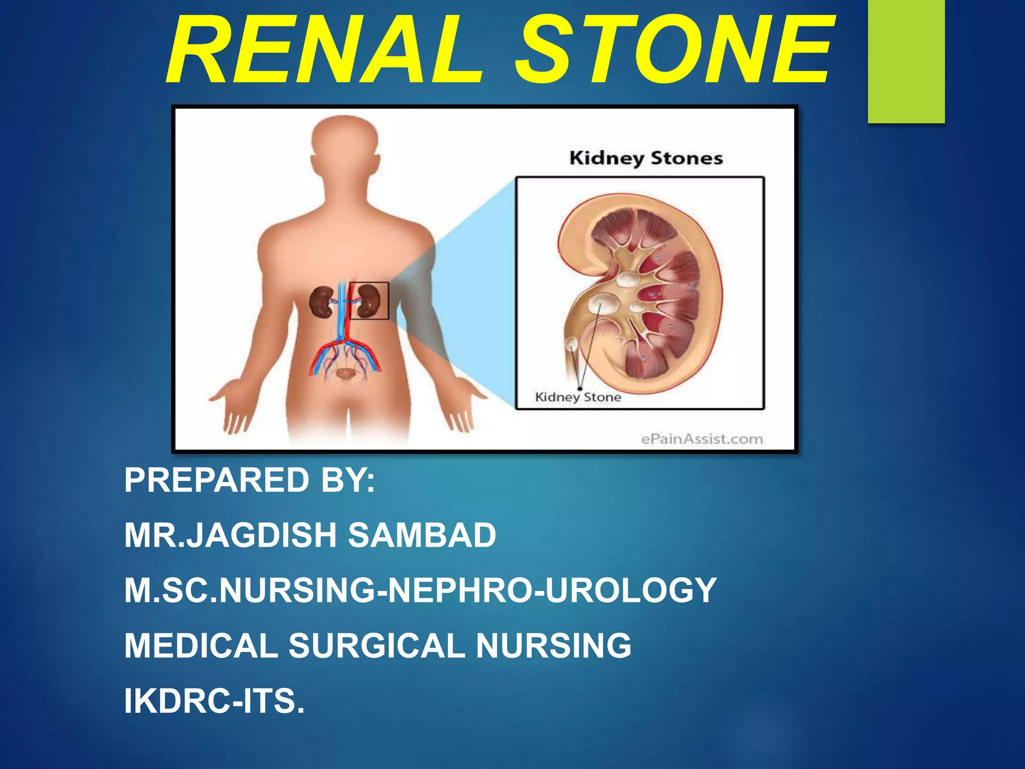
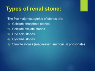
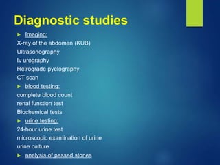
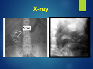
RENAL STONE
PREPARED BY:
MR.JAGDISH SAMBAD
M.SC.NURSING-NEPHRO-UROLOGY
MEDICAL SURGICAL NURSING
IKDRC-ITS.
Renal stone
Introduction
 Urolithiasis and nephrolithiasis refer to stones (calculi) in the urinary
tract and kidney, respectively.
 Kidney stones have many causes and can affect any part of urinary
tract- from kidney to bladder. Urinary stones account for more than
320,000 hospital admissions each year.
 The occurrence of urinary stones occurs predominantly in the third to
fifth decades of life and affects men more than women. About half of
patients with a single renal stone have another episode within 5
years. Stone formation occurs more often in the summer months,
thus supporting the role of dehydration in this process.
Risk factors
 Metabolic: abnormalities that result in increased urine levels of
calcium, oxaluric acid, uric acid, or citric acid.
 Climate: warm climates that cause increased fluid loss, low urine
volume, and increased solute concentration in urine.
 Diet:
 Large intake of dietary proteins that increases uric acid excretion.
 Excessive amounts of tea or fruit juices that elevate urinary oxalate
level.
 Large intake of calcium and oxalate.
 Low fluid intake that increases urinary concentration.
 Genetic factor: family history of stone formation, cystinuria, gout
and renal acidosis.
 Lifestyle: sedentary occupation, immobility, obesity.
Pathophysiology
Types of renal stone:
The five major categories of stones are:
1) Calcium phosphate stones
2) Calcium oxalate stones
3) Uric acid stones
4) Cysteine stones
5) Struvite stones (magnesium ammonium phosphate)
Clinical manifestation
 Severe pain in the side and back, below the ribs
 Pain that radiates to the lower abdomen and groin
 Pain that comes in waves and fluctuates in intensity
 Pain on urination
 Tenderness
 Hematuria
 Persistent need to urinate
 Cloudy or foul-smelling urine
 Nausea and vomiting
 Fever and chills if an infection is present
Diagnostic studies
 Imaging:
X-ray of the abdomen (KUB)
Ultrasonography
Iv urography
Retrograde pyelography
CT scan
 blood testing:
complete blood count
renal function test
Biochemical tests
 urine testing:
24-hour urine test
microscopic examination of urine
urine culture
 analysis of passed stones
X-ray
Management
Medical management:
 Opioid analgesics
 Non-steroidal anti-inflammatory drugs
(NSAIDS)
Surgical management
 Extracorporeal Shock-Wave Lithotripsy
 Percutaneous Nephrolithotomy
 Ureteroscopy
 Percutaneous Stone Dissolution
 Cystolithotomy
 Partial Total Nephrectomy
Extracorporeal Shock-
Wave Lithotripsy
 ESWL is used to remove stones slightly smaller than a half inch
that are located near the kidneys. This method uses ultrasonic
waves or shock waves to break up stones. Then, the stones
leave the body in the urine.
Percutaneous
Nephrolithotomy
 Percutaneous Nephrolithotomy or
nephrostomy is used for large
stones in or near the kidney, or
when kidneys or surrounding
areas are incorrectly formed. The
stone is removed with an
endoscope that is inserted into the
kidney through a small opening.
Ureteroscopy
 It may be used for stones in
the lower urinary tract. It
involves first visualizing the
stone and then destroying it.
Access to the stone is
accomplished by inserting an
ureteroscope into the ureter
and then inserting a laser
electrohydraulic lithotripter or
ultrasound device through the
ureteroscope to fragment and
remove stone.
Percutaneous Stone
Dissolution
 Stone dissolution using infusions of chemical
solutions (chemo lysis) such as alkylating
agents, acidifying agent for the purpose of
dissolving the stone.
Cystolithotomy
 Removal of bladder calculi through a suprapubic
incision is used only stones cannot be crushed
and removed transurethral.
Partial Total Nephrectomy
 Partial Total Nephrectomy is necessary because of extensive
kidney damage, overwhelming renal infection abnormal renal
parenchyma, which can be responsible for stone formation.
Nursing management:
Assessment:
 Subjective data
 Objective data
Nursing diagnosis:
 Acute pain related to effects of renal stone and inadequate pain
control or comfort measures as evidenced by complain of pain,
facial grimacing, restlessness.
 Impaired urinary elimination related to trauma or blockage of
ureters or urethra as evidenced by decreased urinary output,
hematuria.
 Ineffective therapeutic regimen management related to lack
of knowledge regarding disease process, prevention of
recurrence, diet and fluid requirements as evidenced by
questions about how to prevent future renal stones.
 Risk for deficient fluid volume related to nausea and vomiting
as evidenced by observe patient’s condition.
Nutritional management:
 Increase fluid intake is the mainstay
 calcium stones: Patient with calcium-based renal
stones were advised to restrict calcium in their diet.
 Uric acid stones: low-purine diet. foods high in purine
(shellfish, anchovies, asparagus, mushroom, and
organ meats)
 Cysteine stones: low-protein diet
 Oxalate stones: Intake of oxalate is limited. Theses
include spinach, strawberries, chocolate, tea, peanuts,
and wheat barn.
Complication
 Decrease or loss of kidney function
 Scarring, kidney damage
 Obstruction of the ureter
 Stones recurrence
 Urinary tract infection (UTI)
 Renal colic
Research study:
 Renal stone epidemiology: A 25-year study in Rochester,
Minnesota. There are no adequate studies of the incidence of
urolithiasis in the United States, in spite of earlier claims that a
“stone belt” exists in the southeastern section of the country. This
report is the first description of the incidence and recurrence rates
for symptomatic noninfected renal stones in a well-defined
population. A total of 798 patients were enrolled in the study group,
of whom 672 were incidence cases having had their first episode as
documented residents of Rochester, Minnesota, between 1950 and
the end of 1974. The annual age-adjusted incidence rate for
females was stable over the 25-year study period at 36.0 per
100,000 population. That for males increased significantly (P <
0.02) from 78.5 per 100,000 to 123.6 per 100,000. Recurrence
calculations showed a high rate for both sexes in the first year,
followed by lower but constant rates for all succeeding years.
Thank you

Renal stone

  • 1.
    RENAL STONE PREPARED BY: MR.JAGDISHSAMBAD M.SC.NURSING-NEPHRO-UROLOGY MEDICAL SURGICAL NURSING IKDRC-ITS.
  • 2.
  • 3.
    Introduction  Urolithiasis andnephrolithiasis refer to stones (calculi) in the urinary tract and kidney, respectively.  Kidney stones have many causes and can affect any part of urinary tract- from kidney to bladder. Urinary stones account for more than 320,000 hospital admissions each year.  The occurrence of urinary stones occurs predominantly in the third to fifth decades of life and affects men more than women. About half of patients with a single renal stone have another episode within 5 years. Stone formation occurs more often in the summer months, thus supporting the role of dehydration in this process.
  • 4.
    Risk factors  Metabolic:abnormalities that result in increased urine levels of calcium, oxaluric acid, uric acid, or citric acid.  Climate: warm climates that cause increased fluid loss, low urine volume, and increased solute concentration in urine.  Diet:  Large intake of dietary proteins that increases uric acid excretion.  Excessive amounts of tea or fruit juices that elevate urinary oxalate level.  Large intake of calcium and oxalate.  Low fluid intake that increases urinary concentration.  Genetic factor: family history of stone formation, cystinuria, gout and renal acidosis.  Lifestyle: sedentary occupation, immobility, obesity.
  • 5.
  • 6.
    Types of renalstone: The five major categories of stones are: 1) Calcium phosphate stones 2) Calcium oxalate stones 3) Uric acid stones 4) Cysteine stones 5) Struvite stones (magnesium ammonium phosphate)
  • 7.
    Clinical manifestation  Severepain in the side and back, below the ribs  Pain that radiates to the lower abdomen and groin  Pain that comes in waves and fluctuates in intensity  Pain on urination  Tenderness  Hematuria  Persistent need to urinate  Cloudy or foul-smelling urine  Nausea and vomiting  Fever and chills if an infection is present
  • 8.
    Diagnostic studies  Imaging: X-rayof the abdomen (KUB) Ultrasonography Iv urography Retrograde pyelography CT scan  blood testing: complete blood count renal function test Biochemical tests  urine testing: 24-hour urine test microscopic examination of urine urine culture  analysis of passed stones
  • 9.
  • 10.
    Management Medical management:  Opioidanalgesics  Non-steroidal anti-inflammatory drugs (NSAIDS)
  • 11.
    Surgical management  ExtracorporealShock-Wave Lithotripsy  Percutaneous Nephrolithotomy  Ureteroscopy  Percutaneous Stone Dissolution  Cystolithotomy  Partial Total Nephrectomy
  • 12.
    Extracorporeal Shock- Wave Lithotripsy ESWL is used to remove stones slightly smaller than a half inch that are located near the kidneys. This method uses ultrasonic waves or shock waves to break up stones. Then, the stones leave the body in the urine.
  • 13.
    Percutaneous Nephrolithotomy  Percutaneous Nephrolithotomyor nephrostomy is used for large stones in or near the kidney, or when kidneys or surrounding areas are incorrectly formed. The stone is removed with an endoscope that is inserted into the kidney through a small opening.
  • 14.
    Ureteroscopy  It maybe used for stones in the lower urinary tract. It involves first visualizing the stone and then destroying it. Access to the stone is accomplished by inserting an ureteroscope into the ureter and then inserting a laser electrohydraulic lithotripter or ultrasound device through the ureteroscope to fragment and remove stone.
  • 16.
    Percutaneous Stone Dissolution  Stonedissolution using infusions of chemical solutions (chemo lysis) such as alkylating agents, acidifying agent for the purpose of dissolving the stone.
  • 17.
    Cystolithotomy  Removal ofbladder calculi through a suprapubic incision is used only stones cannot be crushed and removed transurethral.
  • 18.
    Partial Total Nephrectomy Partial Total Nephrectomy is necessary because of extensive kidney damage, overwhelming renal infection abnormal renal parenchyma, which can be responsible for stone formation.
  • 19.
  • 20.
    Nursing diagnosis:  Acutepain related to effects of renal stone and inadequate pain control or comfort measures as evidenced by complain of pain, facial grimacing, restlessness.  Impaired urinary elimination related to trauma or blockage of ureters or urethra as evidenced by decreased urinary output, hematuria.  Ineffective therapeutic regimen management related to lack of knowledge regarding disease process, prevention of recurrence, diet and fluid requirements as evidenced by questions about how to prevent future renal stones.  Risk for deficient fluid volume related to nausea and vomiting as evidenced by observe patient’s condition.
  • 21.
    Nutritional management:  Increasefluid intake is the mainstay  calcium stones: Patient with calcium-based renal stones were advised to restrict calcium in their diet.  Uric acid stones: low-purine diet. foods high in purine (shellfish, anchovies, asparagus, mushroom, and organ meats)  Cysteine stones: low-protein diet  Oxalate stones: Intake of oxalate is limited. Theses include spinach, strawberries, chocolate, tea, peanuts, and wheat barn.
  • 22.
    Complication  Decrease orloss of kidney function  Scarring, kidney damage  Obstruction of the ureter  Stones recurrence  Urinary tract infection (UTI)  Renal colic
  • 23.
    Research study:  Renalstone epidemiology: A 25-year study in Rochester, Minnesota. There are no adequate studies of the incidence of urolithiasis in the United States, in spite of earlier claims that a “stone belt” exists in the southeastern section of the country. This report is the first description of the incidence and recurrence rates for symptomatic noninfected renal stones in a well-defined population. A total of 798 patients were enrolled in the study group, of whom 672 were incidence cases having had their first episode as documented residents of Rochester, Minnesota, between 1950 and the end of 1974. The annual age-adjusted incidence rate for females was stable over the 25-year study period at 36.0 per 100,000 population. That for males increased significantly (P < 0.02) from 78.5 per 100,000 to 123.6 per 100,000. Recurrence calculations showed a high rate for both sexes in the first year, followed by lower but constant rates for all succeeding years.
  • 24.