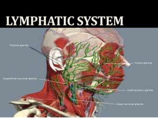
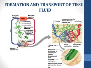
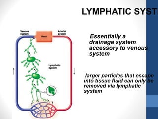
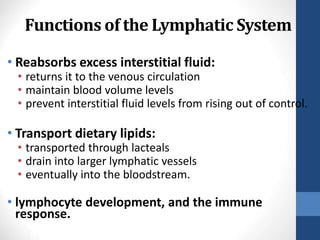
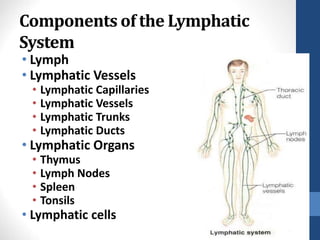
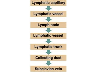
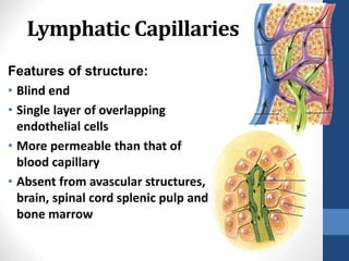
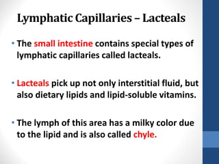
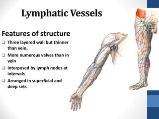
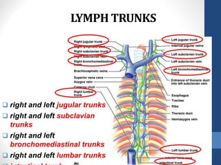
LYMPHATIC SYSTEM
By-
Dr Garima Sehgal
Lecturer
Department of Anatomy
KGMU
LYMPH
•What is a lymph ?
It is a Tissue fluid (interstitial fluid) that
enters the lymphatic vessels
FORMATION AND TRANSPORT OF TISSUE
FLUID
Essentially a
drainage system
accessory to venous
system
larger particles that escape
into tissue fluid can only be
removed via lymphatic
system
LYMPHATIC SYSTE
Functions of the Lymphatic System
• Reabsorbs excess interstitial fluid:
• returns it to the venous circulation
• maintain blood volume levels
• prevent interstitial fluid levels from rising out of control.
• Transport dietary lipids:
• transported through lacteals
• drain into larger lymphatic vessels
• eventually into the bloodstream.
• lymphocyte development, and the immune
response.
24-
5
Components of the Lymphatic
System
• Lymph
• Lymphatic Vessels
• Lymphatic Capillaries
• Lymphatic Vessels
• Lymphatic Trunks
• Lymphatic Ducts
• Lymphatic Organs
• Thymus
• Lymph Nodes
• Spleen
• Tonsils
• Lymphatic cells
24-
6
Lymph Vessels
• Lymphatic capillaries –
• Lymphatic collecting vessels
• Lymphatic trunks –
• Lymphatic ducts –
Lymphatic Capillaries
Features of structure:
• Blind end
• Single layer of overlapping
endothelial cells
• More permeable than that of
blood capillary
• Absent from avascular structures,
brain, spinal cord splenic pulp and
bone marrow
24-
9
Lymphatic Capillaries – Lacteals
• The small intestine contains special types of
lymphatic capillaries called lacteals.
• Lacteals pick up not only interstitial fluid, but
also dietary lipids and lipid-soluble vitamins.
• The lymph of this area has a milky color due
to the lipid and is also called chyle.
24-
10
Lymphatic Vessels
Features of structure
 Three layered wall but thinner
than vein,
 More numerous valves than in
vein
 Interposed by lymph nodes at
intervals
 Arranged in superficial and
deep sets
24-
11
LYMPH TRUNKS
 right and left jugular trunks
 right and left subclavian
trunks
 right and left
bronchomediastinal trunks
 right and left lumbar trunks
LYMPHATIC DUCTS
24-
13
Right lymphatic duct
 Formed by union of
right jugular,
subclavian, and
bronchomediastinal
trunks
 Ends by entering the
right venous angle
Thoracic duct
• Begins in front of L1 as a
dilated sac, the cisterna chyli,
• formed by left and right
lumbar trunks and intestinal
trunk
• Enter thoracic cavity &
ascends
• Travels upward, veering to
the left at the level of T5
LYMPHATIC
DUCTS
THORACIC DUCT…..
15
• At the root of the neck, it
turns laterally
• arches forwards and
descends to enter the left
venous angle
• before termination, it
receives the left jugular,
Subclavian and broncho-
mediastinal trunk
DRAINAGE PATTERN
THORACIC DUCT -
Drains lymph from lower
limbs, pelvic cavity,
abdominal cavity, left
side of thorax, and left
side of the head, neck
RIGHT LYMPHATIC DUCT
-Receives lymph from
right half of head, neck,
thorax and right upper
limb, right lung, right side
of heart, right surface of
liver
Lymphatic Cells
• Also called lymphoid cells.
• Located in both the lymphatic system and the
cardiovascular system.
• Work together to elicit an immune response.
• Types of lymphatic cells are:
• macrophages
• epithelial cells
• dendritic cells
• lymphocytes
24-
17
LYMPHATIC ORGANS
Primary organs
• Red bone marrow
• Thymus gland
Secondary organs
• Lymph nodes
• Lymph nodules
• Spleen
Lymph
Nodes
• Small, round or oval
• located along the
pathways of lymph vessels.
• length from 1 - 25
millimeters
• Typically found in clusters
• receive lymph from many
body regions.
• Lymph nodes are also
found individually
throughout the body
tissues.
24-
19
Lymph node
Features
 Bean-shaped bodies
 With afferent vessels
(entering at the periphery)
and efferent lymph
vessels(emerging at the
hilus)
 Arranged in groups, along
the blood vessels or the
flexural side of the joint
 Divided into superficial and
21
• Regional Lymph Node is the
lymph node where the lymph of the
organ or part of the body drainge to
firstly
Sentinel Lymph Node(in clinic)
Regional Lymph drainage
Spleen
22
Location
Left epigastric region
between 9th-11th rib
in line of 10th rib
• Largest lymphatic organ in the
body.
• Can vary considerably in size
and weight
Function
THYMUS
23
Features
Consists of two
elongated lobes
Is a large organ in the
fetus
Occupies the thoracic
cavity behind the
sternum
Secrete
lymphopoietin
Lymphatic Nodules
• Oval clusters of lymphatic cells with some extracellular
matrix that are not surrounded by a connective tissue
capsule.
• Filter and attack antigens.
• In some areas of the body, many lymphatic nodules group
together to form larger structures.
• mucosa-associated lymphatic tissue (MALT) or tonsils
• very prominent in the mucosa of the small intestine,
primarily in the ileum
• Peyer patches
• also present in the appendix
24-
24
Tonsils
• clusters of lymphatic cells and extracellular matrix
not completely surrounded by a connective tissue
capsule.
• Consist of multiple germinal centers and crypts
• Several groups of tonsils form a protective ring
around the pharynx.
• pharyngeal tonsils (or adenoids) in nasopharynx
• palatine tonsils in oral cavity
• lingual tonsils along posterior one-third of the
tongue
24-
26
MALT (Mucosa Associated
Lymphoid Tissue)
27
APPLIED
ANATOMY
LYMPHANGITIS
• Inflammation of the lymph
vessels
• Commonest cause bacteria
called streptococcus
pyogenes(most common).
• Lymph vessels appear as
red streaks through the
skin
FILARIASIS
LYMPHEDEMA
• Occurs due to
accumulation of
lymphatic fluid in the
interstitial tissue
• Sometimes can be
appreciated after
wearing tight clothing
or jewellary on
affected limb
LYMPHADENOPATHY
• Means a disease of
the lymph nodes
• Lymph nodes
become swollen/
enlarged and may be
painful to touch
LYMPHOMAS
• Cancers originating either
from the lymphocytes in the
lymph nodes or the
lymphatic tissue in organs
• Risk factors -- HIV,
HEPATITIS, EBV infections
TONSILLITIS
• Infection of the
pharyngeal tonsils
• Tonsils are swollen,
• Fever and pain during
swallowing usually
present
• Treatment – surgical
removal of tonsils
(TONSILLECTOMY)
SPLENOMEGALY
•Enlarged Spleen
•Various causes

lymphatic_system.ppt1111111111111111111111111111

  • 1.
    LYMPHATIC SYSTEM By- Dr GarimaSehgal Lecturer Department of Anatomy KGMU
  • 2.
    LYMPH •What is alymph ? It is a Tissue fluid (interstitial fluid) that enters the lymphatic vessels
  • 3.
    FORMATION AND TRANSPORTOF TISSUE FLUID
  • 4.
    Essentially a drainage system accessoryto venous system larger particles that escape into tissue fluid can only be removed via lymphatic system LYMPHATIC SYSTE
  • 5.
    Functions of theLymphatic System • Reabsorbs excess interstitial fluid: • returns it to the venous circulation • maintain blood volume levels • prevent interstitial fluid levels from rising out of control. • Transport dietary lipids: • transported through lacteals • drain into larger lymphatic vessels • eventually into the bloodstream. • lymphocyte development, and the immune response. 24- 5
  • 6.
    Components of theLymphatic System • Lymph • Lymphatic Vessels • Lymphatic Capillaries • Lymphatic Vessels • Lymphatic Trunks • Lymphatic Ducts • Lymphatic Organs • Thymus • Lymph Nodes • Spleen • Tonsils • Lymphatic cells 24- 6
  • 7.
    Lymph Vessels • Lymphaticcapillaries – • Lymphatic collecting vessels • Lymphatic trunks – • Lymphatic ducts –
  • 9.
    Lymphatic Capillaries Features ofstructure: • Blind end • Single layer of overlapping endothelial cells • More permeable than that of blood capillary • Absent from avascular structures, brain, spinal cord splenic pulp and bone marrow 24- 9
  • 10.
    Lymphatic Capillaries –Lacteals • The small intestine contains special types of lymphatic capillaries called lacteals. • Lacteals pick up not only interstitial fluid, but also dietary lipids and lipid-soluble vitamins. • The lymph of this area has a milky color due to the lipid and is also called chyle. 24- 10
  • 11.
    Lymphatic Vessels Features ofstructure  Three layered wall but thinner than vein,  More numerous valves than in vein  Interposed by lymph nodes at intervals  Arranged in superficial and deep sets 24- 11
  • 12.
    LYMPH TRUNKS  rightand left jugular trunks  right and left subclavian trunks  right and left bronchomediastinal trunks  right and left lumbar trunks
  • 13.
    LYMPHATIC DUCTS 24- 13 Right lymphaticduct  Formed by union of right jugular, subclavian, and bronchomediastinal trunks  Ends by entering the right venous angle
  • 14.
    Thoracic duct • Beginsin front of L1 as a dilated sac, the cisterna chyli, • formed by left and right lumbar trunks and intestinal trunk • Enter thoracic cavity & ascends • Travels upward, veering to the left at the level of T5 LYMPHATIC DUCTS
  • 15.
    THORACIC DUCT….. 15 • Atthe root of the neck, it turns laterally • arches forwards and descends to enter the left venous angle • before termination, it receives the left jugular, Subclavian and broncho- mediastinal trunk
  • 16.
    DRAINAGE PATTERN THORACIC DUCT- Drains lymph from lower limbs, pelvic cavity, abdominal cavity, left side of thorax, and left side of the head, neck RIGHT LYMPHATIC DUCT -Receives lymph from right half of head, neck, thorax and right upper limb, right lung, right side of heart, right surface of liver
  • 17.
    Lymphatic Cells • Alsocalled lymphoid cells. • Located in both the lymphatic system and the cardiovascular system. • Work together to elicit an immune response. • Types of lymphatic cells are: • macrophages • epithelial cells • dendritic cells • lymphocytes 24- 17
  • 18.
    LYMPHATIC ORGANS Primary organs •Red bone marrow • Thymus gland Secondary organs • Lymph nodes • Lymph nodules • Spleen
  • 19.
    Lymph Nodes • Small, roundor oval • located along the pathways of lymph vessels. • length from 1 - 25 millimeters • Typically found in clusters • receive lymph from many body regions. • Lymph nodes are also found individually throughout the body tissues. 24- 19
  • 20.
    Lymph node Features  Bean-shapedbodies  With afferent vessels (entering at the periphery) and efferent lymph vessels(emerging at the hilus)  Arranged in groups, along the blood vessels or the flexural side of the joint  Divided into superficial and
  • 21.
    21 • Regional LymphNode is the lymph node where the lymph of the organ or part of the body drainge to firstly Sentinel Lymph Node(in clinic) Regional Lymph drainage
  • 22.
    Spleen 22 Location Left epigastric region between9th-11th rib in line of 10th rib • Largest lymphatic organ in the body. • Can vary considerably in size and weight Function
  • 23.
    THYMUS 23 Features Consists of two elongatedlobes Is a large organ in the fetus Occupies the thoracic cavity behind the sternum Secrete lymphopoietin
  • 24.
    Lymphatic Nodules • Ovalclusters of lymphatic cells with some extracellular matrix that are not surrounded by a connective tissue capsule. • Filter and attack antigens. • In some areas of the body, many lymphatic nodules group together to form larger structures. • mucosa-associated lymphatic tissue (MALT) or tonsils • very prominent in the mucosa of the small intestine, primarily in the ileum • Peyer patches • also present in the appendix 24- 24
  • 26.
    Tonsils • clusters oflymphatic cells and extracellular matrix not completely surrounded by a connective tissue capsule. • Consist of multiple germinal centers and crypts • Several groups of tonsils form a protective ring around the pharynx. • pharyngeal tonsils (or adenoids) in nasopharynx • palatine tonsils in oral cavity • lingual tonsils along posterior one-third of the tongue 24- 26
  • 27.
  • 28.
  • 29.
    LYMPHANGITIS • Inflammation ofthe lymph vessels • Commonest cause bacteria called streptococcus pyogenes(most common). • Lymph vessels appear as red streaks through the skin
  • 30.
  • 31.
    LYMPHEDEMA • Occurs dueto accumulation of lymphatic fluid in the interstitial tissue • Sometimes can be appreciated after wearing tight clothing or jewellary on affected limb
  • 32.
    LYMPHADENOPATHY • Means adisease of the lymph nodes • Lymph nodes become swollen/ enlarged and may be painful to touch
  • 33.
    LYMPHOMAS • Cancers originatingeither from the lymphocytes in the lymph nodes or the lymphatic tissue in organs • Risk factors -- HIV, HEPATITIS, EBV infections
  • 34.
    TONSILLITIS • Infection ofthe pharyngeal tonsils • Tonsils are swollen, • Fever and pain during swallowing usually present • Treatment – surgical removal of tonsils (TONSILLECTOMY)
  • 35.