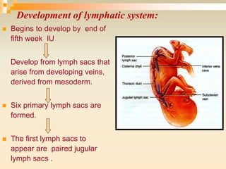
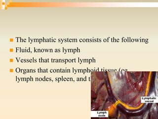
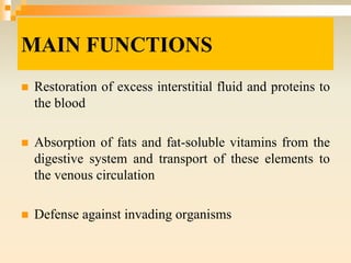
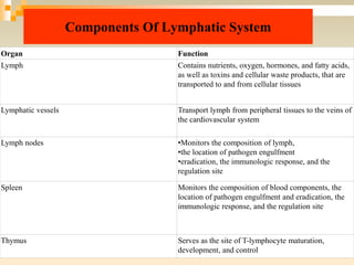
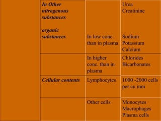
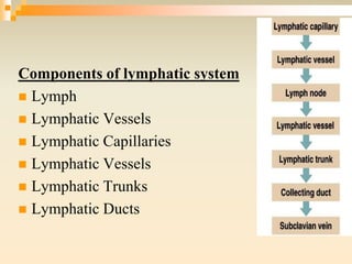
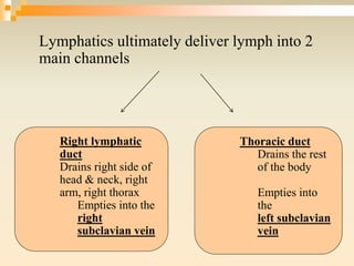
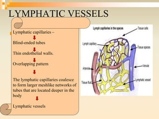
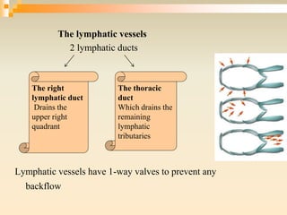
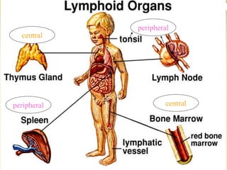
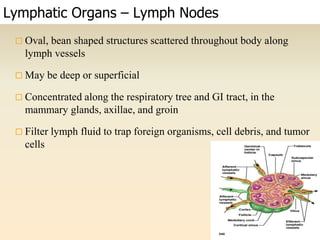
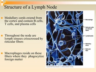
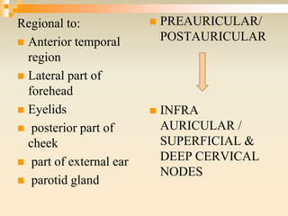
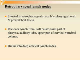
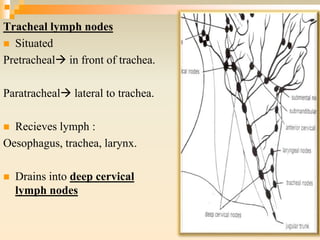
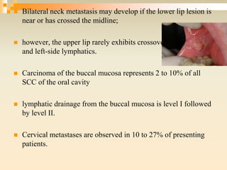
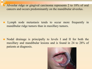
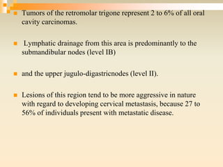
3. īą The lymphatic system represents an accessory route
through which fluid flows from the interstitial spaces
into blood
īą It is an essential part of bodyâs immune system.
Introduction
5. īŽ Begins to develop by end of
fifth week IU
Develop from lymph sacs that
arise from developing veins,
derived from mesoderm.
īŽ Six primary lymph sacs are
formed.
īŽ The first lymph sacs to
appear are paired jugular
lymph sacs .
Development of lymphatic system:
6. Capillary plexuses enlarge.
Form lymphatic vessels .
Each jugular lymph sac
retains at least one
connection with its jugular
vein.
Left one develops into the
superior portion of the
thoracic duct.
7. 8th wk of IU-Retroperitoneal lymph sacs forms.
9th wk of IU cisterna chili develops-lower part of the thoracic
duct develops from left jugular sac.
Later stages-lymph sacs are invaded by lymphocytes.
Transformed into group of lymph nodes
8. Development of Spleen & Thymus
īŽ The spleen develops from mesenchymal cells
between layers of the dorsal mesentery of the
stomach.
īŽ The thymus arises as an outgrowth of the third
pharyngeal pouch.
9. īŽ The lymph nodes develop in the early fetal
period through a septation of the lymph sacs by
mesenchymal cells.
The spaces thus delimited become the sinus of the adult
lymph nodes.
11. īŽ The lymphatic system consists of the following
īŽ Fluid, known as lymph
īŽ Vessels that transport lymph
īŽ Organs that contain lymphoid tissue (eg,
lymph nodes, spleen, and thymus)
12. MAIN FUNCTIONS
īŽ Restoration of excess interstitial fluid and proteins to
the blood
īŽ Absorption of fats and fat-soluble vitamins from the
digestive system and transport of these elements to
the venous circulation
īŽ Defense against invading organisms
13. Components Of Lymphatic System
Organ Function
Lymph Contains nutrients, oxygen, hormones, and fatty acids,
as well as toxins and cellular waste products, that are
transported to and from cellular tissues
Lymphatic vessels Transport lymph from peripheral tissues to the veins of
the cardiovascular system
Lymph nodes âĸMonitors the composition of lymph,
âĸthe location of pathogen engulfment
âĸeradication, the immunologic response, and the
regulation site
Spleen Monitors the composition of blood components, the
location of pathogen engulfment and eradication, the
immunologic response, and the regulation site
Thymus Serves as the site of T-lymphocyte maturation,
development, and control
14. LYMPH
īŽ Lymph blood plasma.
īŽ It is pushed out through the capillary wall by pressure exerted
by the heart or by osmotic pressure at the cellular level.
īŽ Lymph contains
As the lymph passes through the lymph nodes, lymphocytes and
monocytes enter it.
Nutrients
Oxygen
Hormones
Toxins
Cellular Waste
15. Water (96%)
Solids (4 %) Organic
substances
Proteins
(2 â 6 % of solids)
Lipids
(5â 15% of solids)
Carbohydrates
Amino acids
Albumin
Globulin
Fibrinogen
Prothrombin
Other clotting factors
Antibodies
Enzymes
Chylomicrons
Lipoproteins
Glucose(120 mg%)
Allaminoacidspresentsinplasma
Composition of lymph
16. In Other
nitrogenous
substances
organic
substances In low conc.
than in plasma
Urea
Creatinine
Sodium
Potassium
Calcium
In higher
conc. than in
plasma
Chlorides
Bicarbonates
Cellular contents Lymphocytes 1000 -2000 cells
per cu mm
Other cells Monocytes
Macrophages
Plasma cells
18. All tissues of body have special lymph channels to drain
excess fluid directly from interstitial spaces except :
ī superficial portion of skin,
ī CNS
ī endomysium of muscles,
ī bones
They have minute interstitial channels called prelymphatics .
Fluid eventually empties into lymphatic vessels , or in case of
brain into CSF & then directly back into blood.
19. Lymphatics ultimately deliver lymph into 2
main channels
Right lymphatic
duct
Drains right side of
head & neck, right
arm, right thorax
Empties into the
right
subclavian vein
Thoracic duct
Drains the rest
of the body
Empties into
the
left subclavian
vein
20. Only 2 areas in head and neck have no direct lymphatics:
īŽ a) orbit- is virtually devoid of lymphatics.
īŽ b) muscles- do not have lymphatics
Their lymph drains in fascial planes between muscles and around
the blood vessels that supply them.
īŽ LYMPH VESSELS ARE NOT PRESENT IN :
īŽ CNS
īŽ Bones
īŽ Alveoli of lungs
21.
22. LYMPHATIC VESSELS
īŽ .Lymphatic capillaries â
Blind-ended tubes
Thin endothelial walls.
Overlapping pattern
The lymphatic capillaries coalesce
to form larger meshlike networks of
tubes that are located deeper in the
body
Lymphatic vessels
23. The lymphatic vessels
2 lymphatic ducts
Lymphatic vessels have 1-way valves to prevent any
backflow
The right
lymphatic duct
Drains the
upper right
quadrant
The thoracic
duct
Which drains the
remaining
lymphatic
tributaries
24. RATE OF FLOW
īŽ About 120 ml lymph flows into blood per hour
īŽ 100 ml/hr â Thoracic duct
īŽ 20 ml/hr - Rt. Lymphatic duct
27. Thymus
īŽ In the thymus, t lymphocytes dont respond to pathogens and foreign
organisms.
īŽ After maturation
īŽ They enter the blood and go to other lymphatic organs where they
help provide defense.
Bilobed lymphoid organ
Superior mediastinum of the thorax, posterior to the
sternum
Function
īąProcessing and maturation of t lymphocytes.
īąProduces thymosin, a hormone that helps stimulate
maturation of t lymphocytes in other lymphatic organs
28. SPLEEN
īŽ It is surrounded by a connective tissue capsule that extends inward to
divide the organ into lobules
īŽ Red pulp venous sinuses filled with blood and cords of
lymphocytes and macrophages
īŽ White pulp lymphatic tissue consisting of lymphocytes around
the arteries.
īŽ Lymphocytes are densely packed within the cortex of the spleen.
âĸLargest lymphatic organ
âĸConvex lymphoid structure located Below
the diaphragm and behind The stomach.
âĸCells
âĸsmall blood vessels
âĸtissue known as red and white pulp.
29. FUNCTIONS
īŽ Reservoir of lymphocytes
īŽ It filters blood
īŽ It plays an important role in red blood cell and iron metabolism
through macrophage phagocytosis of old and damaged red blood cells
īŽ It recycles iron by sending it to the liver
īŽ It serves as a storage reservoir for blood
īŽ It contains T lymphocytes and B lymphocytes for immunologic
response
30. Mucosa Associated Lymphoid Tissue
MALT
Non encapsulated lymphoid tissue
īļ 2 major components of MALT:
ī BALT (Bronchial Associated Lymphoid Tissue)
ī GALT (Gut Associated Lymphoid Tissue) GALT
īŧ Peyerâs patches
īŧ Appendix â also known as belly tonsil / intestinal tonsil
īļ Minor components of MALT
ī Nose-associated lymphoid tissue (NALT)
ī Vulvovaginal-associated lymphoid tissue (VALT)
ī Skin associated lymphoid tissue (SALT) is not mucosal but has
the same characteristics of the MALT
31. Tonsils
īŽ Aggregates of lymph node tissue located under the epithelial lining of the
oral and pharyngeal areas.
īŽ The predominance of lymphocytes and macrophages in these tonsillar
tissues offers protection against harmful pathogens and substances that may
enter through the oral cavity or airway
âĸ The palatine tonsils (on the sides of the oropharynx)
âĸ The pharyngeal tonsils (on the roof of the nasopharynx; also known
as adenoids)
âĸLingual tonsils (on the base of the posterior surface of the tongue).
32. Type Epithelium Capsule Crypts Location
Adenoids (also
termed "pharyngeal
tonsils")
Ciliated
pseudostratified
columnar (respirator
y epithelium)
Incompletely
encapsulated
No Roof of pharynx
Tubal tonsils
Ciliated
pseudostratified
columnar
(respiratory
epithelium)
Partially
encapsulated
Roof of pharynx
Palatine tonsils
Non-keratinized
stratified squamous
Incompletely
encapsulated
Long, branched
Sides
of oropharynx betwe
en palatoglossal
and
palatopharyngeal
arches
Lingual tonsils
Non-keratinized
stratified squamous
Incompletely
encapsulated
Long, unbranched
Behind terminal
sulcus (tongue)
33. Lymphatic Organs â Lymph Nodes
ī¨ Oval, bean shaped structures scattered throughout body along
lymph vessels
ī¨ May be deep or superficial
ī¨ Concentrated along the respiratory tree and GI tract, in the
mammary glands, axillae, and groin
ī¨ Filter lymph fluid to trap foreign organisms, cell debris, and tumor
cells
34. Lymphatic Organs â Lymph Nodes
īŽ Covered by a fibrous connective tissue capsule
īŽ Trabeculae extend from cortex to medulla
īŽ Stroma â the internal supportive connective tissue network of reticular fibers
35. Structure of a Lymph Node
īŽ outer cortex - filled with lymph follicles
ī¨ outer edge of follicle contains more T cells
ī¨ inner germinal center is the site of B-cell
proliferation
īŽ inner medulla - medullary cords of
lymphocytes, macrophages, plasma cells
(activated B cells)
Cortex
Medulla
36. Structure of a Lymph Node
īŽ Medullary cords extend from
the cortex and contain B cells,
T cells, and plasma cells
īŽ Throughout the node are
lymph sinuses crisscrossed by
reticular fibers
īŽ Macrophages reside on these
fibers where they phagocytize
foreign matter
38. Circulation in the Lymph Nodes
īŽ Lymph enters via a number of afferent
lymphatic vessels
īŽ It then enters a large subcapsular sinus
and travels into a number of smaller
sinuses
īŽ It meanders through these sinuses and
exits the node at the hilus via efferent
vessels
īŽ The node acts as a âsettling tank,â
because there are fewer efferent vessels,
lymph stagnates somewhat in the node
īŽ This allows lymphocytes and
macrophages time to carry out their
protective functions
Only lymph nodes filter lymph!
39. īŽ Fluid enters cortex through
afferent vessels
ī¨ Filter and trap damaged cells,
microorganisms, foreign
substances, tumor cells by
reticular fibers
ī¨ Macrophages phagocytize
some, lymphocytes destroy
some by immune defenses
īŽ Exits medulla by efferent
vessels at hilus
Lymph Flow Through Lymph Nodes
40. Blood Flow Through Lymph Nodes
īŽ Blood vessels enter
and exit at the hilus
īŽ This blood provides
nutrition for the
nodeâs tissues
īŽ route for leukocytes to
enter into or exit from
the lymphatic tissue
of the node
42. īŽ Deep lymph nodes
1. Prelaryngeal and pretracheal
2. Paratracheal
3. Retropharyngeal
43. OCCIPITAL NODES
īŽ Situated at the apex of
posterior triangle of
neck
īŽ Recieves lymph from
back of scalp
īŽ Drains into deep
cervical lymph nodes
44. MASTOID /
RETROAURICULAR
LYMPH NODES
īŽ Situated over lateral surface of
mastoid process of temporal
bone
īŽ Recieves lymph from
a) Strip of scalp above auricle.
b) Posterior wall of external
auditory meatus
īŽ Drains into
deep cervical lymph nodes
45. PAROTID LYMPH
NODES
īŽ Situated on/ within
parotid gland.
īŽ Receives lymph from
a) Strip of scalp above
parotid salivary
gland.
B) lateral surface of
auricle.
C) anterior wall of
external auditory
meatus
D) lateral wall of
external auditory
meatus.
E)lateral wall of eyelid
Drains into deep cervical nodes
46. Regional to:
īŽ Anterior temporal
region
īŽ Lateral part of
forehead
īŽ Eyelids
īŽ posterior part of
cheek
īŽ part of external ear
īŽ parotid gland
īŽ PREAURICULAR/
POSTAURICULAR
īŽ INFRA
AURICULAR /
SUPERFICIAL &
DEEP CERVICAL
NODES
47. CLINICAL SIGNIFICANCE
īŽ The most common area that drains into
these nodes is skin, and thus the most
common tumors to metastasize to them are
melanoma and squamous cell carcinoma.
48. Buccal lymph nodes
īŽ Situated over
buccinator muscle
close to facial vein.
īŽ Recieves lymph from
Eyelids, cheek, mid
portion of face Rarely
gums & palate
Drains into submandibular
lymph nodes
49. Regional to:
īŽ Skin on the anterior
surface of face
Secondary to:
īŽ Deeper part of face
īŽ Mucous memberane
of lips & cheek.
īŽ Occasionally even
from upper/lower
teeth & adjacent
gingiva.
51. īŽ They are divided into:
īŽ Anterior group :submental vein close to chin.
īŽ Middle group : around facial vein& facial
artery above submandibular salivary gland.
īŽ Posterior group : behind facial vein.
52. Recieves lymph from:
īŽ Front of scalp.
īŽ Anterior part of nasal cavity, palate & adjacent cheek.
īŽ Upper & lower lip except central part.
īŽ Frontal, maxillary, ethmoidal air sinuses.
īŽ Upper& lower teeth except lower incisors.
īŽ Anterior 2/3rd of tongue.
īŽ Floor of mouth, vestibule.
Drains into deep cervical nodes.
53. Submental lymph nodes
īŽ Lies b/w chin & hyoid
bone
īŽ b/w anterior bellies of
digastric muscles in
submental triangles.
Recieves lymph from
A. Tip of tongue
B. floor beneath tongue
C. lower incisors
D. central part of lower lip
E. skin over chin Drains into submandibular &
deep cervical nodes
54. īŽ Regional to
īŧ Middle part of lower lip
īŧ Skin of chin
īŧ tip of tongue
īŧ lower incisors & gingiva
īŽ Secondary lymph nodes of this region are in part
submandibular & in part superior deep cervical
lymph nodes
55. Cervical lymph nodes
īŽ Distributed along the internal & external jugular veins.
īŽ Acc. To their relation to deep fascia of neck, they are
divided into superficial & deep groups
īŽ Superficial nodes restricted to upper region of neck&
found in angle b/w mandibular ramus & SCM muscle.
īŽ Receive lymph from
īŽ ear lobe
īŽ adjacent part of skin.
īŽ secondary to preauricular & postauricular lymph nodes.
56.
57. īŽ Deep cervical nodes divided into upper & lower group
īŽ The superior & inferior deep cervical nodes that are situated in
front of SCM muscle: c/a anterior/ medial deep cervical
nodes.
īŽ It follows the internal jugular vein so c/a JUGULAR CHAIN
īŽ Those situated in posterior triangles of neck behind SCM
muscle are c/a posterior/ lateral deep cervical nodes.
īŽ They are in close relation to accassory nerve, known as
ACCESSORY CHAIN
58. Primary to :
īŽ Base of tongue
īŽ Sublingual
region
īŽ Posterior part
of palate
They are secondary and tertiary
nodes into which the lymph of
auricular, submental,
submandibular & accessory nodes
of face empty.
They are also secondary to nuchal
nodes, deep lymph nodes of neck,
retropharyngeal, infrahyoid,
pretracheal, paratracheal lymph
nodes.
59. Jugulo digastric lymph nodes
īŽ Situated at the level of greator horn of hyoid bone.
īŽ Recieves lymph from tonsil and tongue.
Juglo-omohyoid nodes
īŽ Situatedī related to the intermediate tendon of
omohyoid muscle.
īŽ Recieves lymph from posterior 1/3rd of tongue.
īŽ In general deep cervical nodes receive lymph from
regional lymph nodes and drain into jugular
lymph trunk
60. SUPERIOR DEEP CERVICAL NODES
INFERIOR DEEP CERVICAL/ SUPRACLAVICULAR
NODES.
THORASIC DUCT(left side)
LYMPHATIC DUCT (RIGHT SIDE)
VENOUS ANGLE (on either side), where internal jugular &
subclavian veins unite.
Thus the lymph enters the system of superior vena cava
61. Retropharyngeal lymph nodes
īŽ Situated in retropharyngeal space b/w pharyngeal wall
& prevertebral fascia .
īŽ Recieves lymph from: soft palate,nasal part of
pharynx, auditory tube, upper part of cervical vertebral
column.
īŽ Drains into deep cervical lymph nodes.
62. Laryngeal lymph nodes
īŽ Situated in front of larynx
on cricothyroid ligament.
īŽ Recieves lymph from
larynx, trachea, isthmus of
thyroid.
īŽ Drains into deep cervical
lymph nodes.
63. Tracheal lymph nodes
īŽ Situated
Pretrachealī in front of trachea.
Paratrachealī lateral to trachea.
īŽ Recieves lymph :
Oesophagus, trachea, larynx.
īŽ Drains into deep cervical
lymph nodes
64. WALDEYER RING
īŽ Waldeyer's tonsillar ring (or pharyngeal lymphoid ring) is an
anatomical term describing the Lymphoid tissue ring located in
the pharynx and to the back of the oral cavity.
īŽ Heinrich Wilhelm Gottfried von Waldeyer-Hartz.
66. Dr. owais pg Ist yr ENT
SMHS
Grading the Size of Tonsils
Grading system:
A. 0 â tonsils in fossa
B. +1 â tonsils less than 25%
C. +2 â tonsils less than 50%
D. +3 â tonsils less than 75%
E. +4 â tonsils greater than 75%
68. Anatomy
Blood supply â Adenoids
īŽ Ascending palatine branch of facial a.
īŽ Ascending pharyngeal a.
īŽ Pharyngeal branch of IMAX.
īŽ Ascending cervical branch of thyrocervical trunk.
69. LYMPHATIC DRAINAGE OF TONGUE
ī§ Rich network of lymphtics
Enormous swelling
Carcinma of tongue:
Affected side is removed Surgically .With deep cervical node
Carcinoma of posterior one- third is more dangerous due to bilateral
lymphatic spread
70. īŽ Tip of tongue drains bilaterally ī
sub-mental nodes
īŽ Right & left halves of remaining
halves of anterior 2/3rd drain
unilaterally ī submandibular
nodes.
īŽ Posterior 1/3rd drains bilaterally
ī juglo-digastric nodes.
71. APPLIED ANATOMY
The latest classification has been created by the
American Joint Committee on Cancer and
the American Academy of Otolaryngology -
Head and Neck Surgery.
72. Staging
īŽ The TNM system devised by the AJCC is
designed to stratify cancer patients into
different stages based on the characteristics of
the primary tumor (T), regional lymph node
metastasis (N), and distant metastasis (M).
73. Regional Lymph Nodes (N)
Node Description
īŽ NX Regional lymph nodes cannot be assessed
īŽ N0 No regional lymph node metastasis
īŽ N1 Metastasis in a single ipsilateral lymph node, 3 cm or less
in greatest dimension
īŽ N2 Metastasis in a single ipsilateral lymph node, more than 3
cm but not more than 6 cm in greatest dimension; or in
multiple ipsilateral lymph nodes, none more than 6 cm in
greatest dimension; or in bilateral or contralateral lymph
nodes, none more than 6 cm in greatest dimension
74. īŽ N2a Metastasis in a single ipsilateral lymph node more than 3
cm but not more than 6 cm in greatest dimension
īŽ N2b Metastasis in multiple ipsilateral lymph nodes, none more
than 6 cm in greatest dimension
īŽ N2c Metastasis in bilateral or contralateral lymph nodes, none
more than 6 cm in greatest dimension
īŽ N3 Metastasis in a lymph node more than 6 cm in greatest
dimension
75. īŽ Level I - all nodes above hyoid
bone, below mylohyoid muscle,
and anterior to posterior edge of
submandibular gland
Level IA - all nodes between
medial margins of anterior
digastric muscles, above hyoid
bone, below mylohyoid muscle
Level IB - all nodes below
mylohyoid muscle, above hyoid
bone, posterior and lateral to
medial anterior digastric
muscle and anterior to
submandibular gland
76. īŽ Level II - all nodes below skull
base at jugular fossa to hyoid
bone, anterior to posterior edge of
sternocleidomastoid muscle and
posterior to submandibular gland
īŽ Level IIA - all nodes that lie
posterior to internal jugular
vein and are inseperable from
the vein or lie anterior, lateral or
medial to the vein
īŽ Level IIB - all nodes that lie
posterior to internal jugular
vein and have a fat plane
separating the nodes and the
vein
77. īŽ Level III - all nodes between
hyoid bone and cricoid
cartilage arch and anterior to
posterior
sternoclediomastoid muscle,
and lateral to the internal
carotid artery
Level IV - all nodes between
cricoid cartilage arch and
clavicle, anterior to posterior
sternocleidomastoid
muscleand posterolateral to
anterior scalene muscle and
lateral to common carotid
artery
78. īŽ Level V - all nodes from skull
base posterior down to
posterior border of
sternocleidomastoid muscle to
level of clavicle, anterior to
trapezius muscle
īŽ Level VA - all nodes between
skull base and cricoid cartilage
arch, behind posterior edge of
sternocleidomastoid muscle
īŽ
Level VB - all nodes between
cricoid cartilage arch and
clavicle, behind
sternoclediomastoid muscle
79. īŽ Level VI - all nodes
inferior to hyoid bone and
above top of manubrium,
between medial margins of
bilateral common carotid
and internal carotid
arteries
Level VII - all nodes
behind the manubrium
between medial margins of
common carotid arteries
bilaterally, extending
inferiorly to level of
innominate vein
80. Face and Scalp Anterior Facial, Ib
Lateral Parotid
Posterior Occipital, V
Eyelids Medial Ib
Lateral Parotid, II
Chin Ia, Ib, II
External Ear Anterior Parotid, II
Posterior Post auricular, II, V
Middle Ear Parotid, II
Floor of mouth Anterior Ia, Ib, IIa > IIb
Lower incisors Ia, Ib, IIa > IIb
Lateral Ib, IIa > IIb, III
Teeth except incisors Ib, IIa > IIb, III
Nasal Cavity Anterior Ib
Posterior Retropharyngeal, II, V
Common Nodal Drainage Patterns
81. Nasal Cavity Posterior Retropharyngeal, II, V
Nasopharynx Retropharyngeal, II, III, V
Oropharynx IIb > IIa, III, IV, V
Larynx Supraglottic IIa > IIb, III, IV
Subglottic VI, IV
Cervical
esophagus IV, VI
Thyroid VI, IV, V, Mediastinal
Tongue Tip Ia, Ib, IIa > IIb, III, IV
Lateral Ib, IIa > IIb, III, IV
Common Nodal Drainage Patterns
82. METASTASIS
īŽ Spread of tumor in such a way by invasion that
discontinuous secondary tumor masses are formed at the
site of lodgement.
īŽ Routes of metasis:
1 Lymphatic spread
2 Haematogenous spread
3 Spread along body cavities and natural passages
( transcoelomic fluid, CSF)
83. īŽ carcinomas metastatise by lymphatic route
īŽ sarcomas by haematogenous route.
īŽ The wall of lymphatics is readily invaded by cancer
cells & forms a continuous growth in lymphatic
channels c/a lymphatic permeation, or may detach to
form tumor emboli to be carried along to the next
lymph node.
īŽ Tumor emboli enter the lymph node at itâs convex
surface & are lodged in subcapsular sinus.
84.
85. SPREAD OF ORAL CANCER VIA LYMPH
NODES
īŽ Mucosal lip cancers represent approximately 2 to 42% of oral cavity
cancers.
īŽ 10% of lower lip cancers and 20% of cancers in the upper lip and
commissure are found to metastasize to the nodes.
īŽ Metastasis from the lower lip is to the submental, submandibular,
and perifacial nodes (level I more commonlythan level II).
īŽ Preauricular, periparotid,and submandibular nodes drain cancers
of the upper lip and commissure (level II more commonly than level
I).
86. īŽ Bilateral neck metastasis may develop if the lower lip lesion is
near or has crossed the midline;
īŽ however, the upper lip rarely exhibits crossover between right-
and left-side lymphatics.
īŽ Carcinoma of the buccal mucosa represents 2 to 10% of all
SCC of the oral cavity
īŽ lymphatic drainage from the buccal mucosa is level I followed
by level II.
īŽ Cervical metastases are observed in 10 to 27% of presenting
patients.
87. īŽ Alveolar ridge or gingival carcinoma represents 2 to 18% of oral
cancers and occurs predominantly on the mandibular alveolus.
īŽ Lymph node metastasis tends to occur more frequently in
mandibular ridge tumors than in maxillary tumors.
īŽ Nodal drainage is principally to levels I and II for both the
maxillary and mandibular lesions and is found in 24 to 28% of
patients at diagnosis.
88. īŽ Tumors of the retromolar trigone represent 2 to 6% of all oral
cavity carcinomas.
īŽ Lymphatic drainage from this area is predominantly to the
submandibular nodes (level IB)
īŽ and the upper jugulo-digastricnodes (level II).
īŽ Lesions of this region tend to be more aggressive in nature
with regard to developing cervical metastasis, because 27 to
56% of individuals present with metastatic disease.
89. īŽ There is a paucity of lymphatics to the hard palate.
īŽ Approximately 10 to 25% of individuals present with
evidence of metastasis, generally to levels I and II.
īŽ Hard palate lesions may also metastasize to retropharyngeal
nodes
īŽ or nodes that are not palpable on a clinical examination or
readily removable with a traditional neck dissection.
90. īŽ Lymphatic drainage of the oral tongue is principally to level II,
followed by levels III
īŽ Carcinoma of the lateral border generally metastasizes
ipsilaterally
īŽ but SCC of the tip or body of the tongue may exhibit bilateral
metastases.
īŽ Approximately 40% of patients have evidence of clinical node
metastasis at the time of diagnosis.
91. Sentinel Lymph Node History
īŽ 1955 ī First echelon node
īŽ 1960 ī âSentinel nodeâ
īŽ 1977 ī Demonstrated in penile cancer
īŽ 1992 ī Morton reintroduced concept in N0
melanoma
īŽ Currently widely used in melanoma and breast cancer
therapy.
92. Sentinel lymph node concept
īŽ Tumor spreads via lymphatics to a primary node.
īŽ Examination of primary echelon nodes for tumor direct the
need for surgical management of the nodal basins.
93. Sentinel lymph node concept
īŽ Difficulties of lymphatic mapping in head and neck
(OâBrien).
1. It is difficult to visualize lymphatic channels using
lymphoscintigraphy because of proximity to the injection site.
2. The radiotracer travels fast in the lymphatic vessels.
3. If more than one node is visible, it can be difficult to
distinguish first echelon nodes from second-echelon nodes.
4. The SLN may be small and not easily accessible (eg, in the
parotid gland).
94. īŽ see if cancer has spread from the primary tumour to the lymph
nodes
ī¨ This information is used to determine the stage (the extent
of cancer in the body).
īŽ help plan treatment
īŽ reduce the chance of lymphedema (buildup of lymph fluid)
developing
ī¨ SLNB reduces, but does not completely eliminate, the risk
of lymphedema.
95. Senital node biopsy
īŽ The surgeon injects a radioactive substance (radiotracer), a blue dye or both into the
tissue around the tumour or into the area from where the tumour was removed.
ī¨ The radiotracer is injected anywhere from 1â16 hours before the surgical
procedure.
ī¨ It takes about 5 minutes for the blue dye to reach the sentinel nodes, so the dye
is often injected in the operating room just before the surgery.
īŽ The dye or radioactive substance is taken up by the lymph vessels. It travels along
the lymph vessels draining the area around the cancer to the sentinel lymph node(s).
96. īŽ A special scanning device detects the radioactivity in the sentinel lymph
node(s), or the surgeon looks for the lymph node(s) stained blue.
ī¨ Sometimes, the sentinel lymph node cannot be identified.
ī¨ If the sentinel lymph node is positive or if it cannot be identified, then more
lymph nodes will need to be removed.
īŽ The surgeon makes a small cut (incision) over the node(s).
īŽ The radioactive or blue lymph node(s) is removed and sent to the laboratory to
be examined under a microscope by a pathologist (a doctor who specializes in
the causes and nature of disease).
97. EXAMINATION OF LYMPHATI C SYSTEM
īŽ LOCAL EXAMINATION
īŽ Inspection
īŽ Swelling
1. Number
2. Position
3. Size
4. Shape
5. Surface
īŽ Skin over the swelling
98. Palpation
1. Rise in local temperature
2. Tenderness
3. Situation and extent
4. Size and shape
5. Surface
6. Margin
7. Consistency (Soft, elastic and rubbery, firm, hard and stony hard)
8. Nodes separate or matted together- periadenitis
9. Fixity to surrounding structures(skin, muscle,nerve,vessel,bone or
any viscus)
99. īŽ Look for the primary focus in the drainage area
īŽ Examine the lymph vessels
100. īŽ Acute lymphangitis- lymph vessels show reddened, tender, indurated
streaks ascending to the regional lymph nodes from the point of
infection
īŽ Carcinoma- multiple hard subcutaneous nodules in path b/w primary
focus and lymph nodes
īŽ Lymphedema-stasis of lymph(lymphatic obstruction)
swelling of affected limb
Early- pitting is seen
Late â fibrosis, prolonged pressure to pit
Finally extreme fibrosis- no pitting
101. EXAMINATION OF LYMPH NODES
1. Lymph nodes should be examined from patientâs behind.
2. Examination is done by asking patient to flex his neck
slightly to reduce tension of muscles
3. To palpate, use the pads of all four fingertips.
4. Examine both sides of head simultaneously while applying
steady gentle pressure.
102. ANTERIOR/POSTERIOR CERVICAL LYMPH
NODES
īŽ They lie anterior & posterior to sternomastoid muscle.
īŽ Tip of fingers are used to palpate anterior nodes, medial to
sternomastoid muscle and posterior nodes behind the muscle
while patient,s head tipped slightly forwards.
108. Supraclavicular lymph
nodes
īŽ While patientâs head is tipped
forward, the index finger of
the examiner is placed in the
triangle and the area is
palpated with a rotary motion.
110. Laboratory Studies
īŽ Directed by the history and physical examination, overall clinical assessment
īŽ CBC count, peripheral blood smear.
īŽ Evaluation of hepatic and renal function, urine underlying systemic
disorders
īŽ Skin testing for tuberculosis is usually indicated.
īŽ Specific regional adenopathy, lymph node aspirate for culture may be
important if lymphadenitis is clinically suspected.
īŽ Titers for specific microorganisms-generalized adenopathy is present.
īŽ These may include epstein-barr virus, cytomegalovirus (cmv), b henselae,
toxoplasma species, and human immunodeficiency virus (hiv).
111. Imaging Studies
īŽ Chest radiography -primary screening tool
īŽ Elucidating mediastinal adenopathy and underlying diseases affecting
the lungs
īŽ .
īŽ Supraclavicular adenopathy,-CT scanning of the chest, abdomen, or
both.
īŽ Positron-emission tomography (PET) scanning is not helpful as a
screening tool as benign and malignant conditions may cause intense
uptake
âĸTuberculosis,
âĸ Coccidioidomycosis,
âĸLymphomas,
âĸNeuroblastoma,
âĸHistiocytoses,
âĸGaucher disease
112. īŽ PET scanning is helpful in the evaluation of lymphomas once a
clinical or tissue-based diagnosis is made.
īŽ scanning is helpful in the evaluation of lymphomas.
īŽ Ultrasonography -evaluating the changes in the lymph nodes and
in evaluating the extent of lymph node involvement in patients
with lymphadenopathy
113. īŽ Patients with matted nodes were more likely to
develop distant metastases, whereas patients
with normal nodes were more likely to develop
a local recurrence
114. Sensitivity %
(range)
Specificity %
(range)
Palpation 35 (30-40) 35 (27-42)
CT 45 (17-86) 11 (3-21)
US 46 (42-50) 21 (11-33)
MRI 42 (20-70) 14 (5-26)
Accuracy of diagnostic methods in detecting occult cervical metastases.
A new approach to pre-treatment assessment of the N0 neck in oral squamous cell carcinoma: the role of
sentinel node biopsy and positron emission tomography
115. BIOPSY
īŽ If the size, location, or character of the lymphadenopathy suggests
malignancy and laboratory testing is inconclusive, a lymph node biopsy is
immediately indicated.
īŽ Best performed on regional lymph nodes suggestive of metastasis using a
fine-bore needle to aspirate cells for cytologic examination.
īŽ Ultrasound-guided fine-needle aspiration cytology is now favored.
īŽ Fine needle aspiration -small samples with limited ability to perform flow
cytometry and chromosomal analysis
īŽ So some prefer excisional biopsy.
124. Characters of L.N. Enlargement in Some Diseases
1- Streptococcal infection of tonsils:
2- Scarlet Fever
Sore throat.
3-Diphtheria
Uni or Bilateral * Tender & unmatted
*Usually submandibular but may extend to lower
cervical group.
marked enlargement of submandibular L.N.
*Other cervical L.N. (bilateral, tender, discrete,
suppuration is common
Enlarged submandibular L.N. usually bilateral,
tender, not matted.
125. 4-German Measle:
âĸOccipitaI L.N. enlargement are nearly always present, closely
resembles that of infectious mononucleosis.
5-Infectious Mononucleosis:
* Sore throat, Fever, sometimes headache, myalgia.
* Palatal petechiae often, are present
* Mild splenomegally in 50% of cases
*Lymphocytosis in 75% of cases with some atypical lymphocytes.
Bilateral L.N. enlargement, firm, discrete, mobile.
* Appear first in posterior cervical area, adjacent to
cervical spines, few days later , submandibular L.N. will
be enlarged
126. 6- T.B.:
* The chiefly affected group is upper cervical group, generalized
L.N. enlargement is exceptional.
* Unilateral or Bilateral.
* Often firm, matted, painful, may become adherent to skin or deep
structures.
* Cystic areas may occur due to caseation and later on cold abscess
formation.
* Overlying skin may break down giving T.B. ulcers or sinuses.
127. Syphilis:
Primary
L.N draining a chancre
-Rocky hard, uni Or bilateral, not tender.
Secondary
-Generalized L.N. enlargement especially posterior
triangle of the neck or epitrochlear gp
(slightly enlarged, shotty, discrete, painless).
128. 8- LYMPHOMATOUS L. N:
âĸMay be associated with constitutional symptoms.(anorexia, fever,
weight loss, sweating, âĻ.. etc).
âĸPel Ebstein fever: may be observed in H.D., it is a period of fever
lasting for few days or weeks alternating with longer or shorter
apyrexial periods .
âĸ L.N. usually discrete at start & not tender (but may become tender
during febrile periods).
âĸL.N. may increase in size during pyrexial periods and decrease in size
during apyrexial periods
129. a-H.D.:
* may be confined to one group at first esp. lower cervical group then
later on generalized L.N. enlargement.
âĸGlands are:
a- moderately enlarged, not tender.
b- Firm, rubbery in consistency.
c- Discrete, mobile however as a result of later extension
outside the capsule glands become matted or fixed
b-N.H .L:
*Also the cervical group is firstly affected
*Rapid rate of growth results in large number of variable sized nodes
which are hard in consistency, tend to become fused and fixed to deep
structures & may give pressure manifestations.
130. 9- LEUKAEMIC L. N:
*May be associated with general manifestations (fever, malaise,
anorexia, headache, Hemorhagic tendency)
a- Acute Leukaemia:
*Late, slightly or moderately enlarged
*Soft, discrete esp. cervical L.N. due to oral sepsis
*May be tender bone.
b-C.L.L:
* May affect cervica1 L.N. but mostly all superficial L.N. are
enlarged.
*The glands usually are (firm, not tender, not matted, usually
moderately enlarged, but in advanced stages may be markedly
enlarged)
c-C.M.L.:
*Rare to be manifested by L.N. enlargement.
131. 10- CARCINOMATOUS L.N.:
*Firm, but some times hard.
*A stoney hard nodes fixed to underlying tissues are nearly
always neoplastic in nature, however the reverse is not true.
*Carcinomatous L.N. may be freely mobile
132. lymphangioma
īŽ Lymphangioma is a benign hamartomatous tumor of lymphatic
channels, with a marked predilection for the head and neck
region, at submandibular and parotid area .
133.
134. CONCLUSION
īŽ Lymphatic system is a closed system of lymph
channels through which lymph flows.
īŽ It is an one way system.
īŽ The entire lymph from the head and neck drains
ultimately into deep cervical nodes either directly
or through peripheral nodes.
īŽ In CNS- lymph is replaced by CSF
īŽ It is essential to have appropriate knowledge of
tumor metastases for most appropriate treatment.