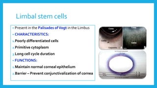
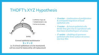
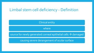
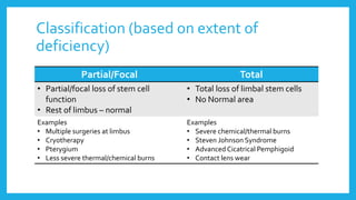
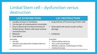
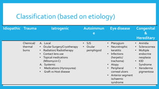
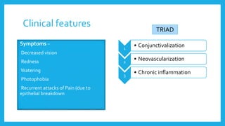
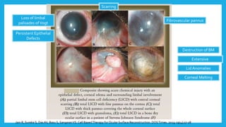
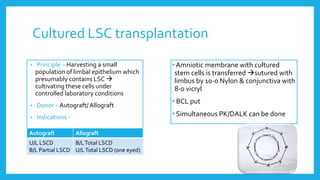
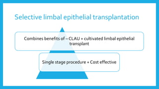
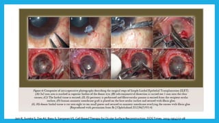
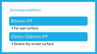
LIMBAL STEM CELL
DEFICIENCY & ITS
MANAGEMENT
Dr. Karan Bhatia
DOMS, DNB
Department of Cornea & Refractive Surgery
M. M. Joshi Eye Institute, Hubli, Karnataka
Introduction – Concept of stem cell
Specialized
Undifferentiated
Self-renewing
Capable of indefinite proliferation
Responsible for cellular
replacement & regeneration
Limbal stem cells
• Present in the Palisades ofVogt in the Limbus
• CHARACTERISTICS:
1.Poorly differentiated cells
2.Primitive cytoplasm
3.Long cell cycle duration
• FUNCTIONS:
1.Maintain normal corneal epithelium
2.Barrier – Prevent conjunctivalization of cornea
Tseng, S. C. G. and Sun,T.T. (2000) In: Corneal Surgery:Theory,Technique, andTissue. (F. S. Brightbill, ed.), Mosby-Year Book, Inc., St. Louis, Missouri, pp. 12.
SC (Parent)
SC
(Daughter
)
SC
SC SC TAC
PMC
TDC
SC – Stem Cell
TAC –Transiently Amplifying Cells
PMC – Post-mitotic Cells
TDC –Terminally Differentiating
Cells
Result of this migration & differentiation  Corneal epithelium renewed every 7–10 days
THOFT’s XYZ Hypothesis
• X vector – combination of proliferation
& centripetal migration of basal
epithelial cells
• Y vector – As basal epithelial cells
divide they give rise to suprabasal cells
that form stratified layers of cornea
• Z vector – shedding of squamous
epithelial cells from surface of cornea
into tear film
Corneal epithelial maintenance
X + Y = Z
i.e. if corneal epithelium is to be maintained,
cell loss must be balanced by cell replacement
Limbal stem cell deficiency - Definition
Clinical entity
where
source for newly generated corneal epithelial cells  damaged
causing severe derangement of ocular surface
Classification (based on extent of
deficiency)
Partial/Focal Total
• Partial/focal loss of stem cell
function
• Rest of limbus – normal
• Total loss of limbal stem cells
• No Normal area
Examples
• Multiple surgeries at limbus
• Cryotherapy
• Pterygium
• Less severe thermal/chemical burns
Examples
• Severe chemical/thermal burns
• Steven Johnson Syndrome
• Advanced Cicatrical Pemphigoid
• Contact lens wear
Limbal Stem cell – dysfunction versus
destruction
LSC DYSFUNCTION LSC DESTRUCTION
• Usually, primary or hereditary
• Can be part of ocular/systemic anomalies
• Due to abnormal microenvironment of
limbal tissues stem cells never achieve
normal function
• Bilateral
• Less severe
• Acquired loss of functioning of stem cells
• Associated with severe ocular surface
damage
• Unilateral
Examples
• Aniridia
• Keratitis associated with multiple endocrine
deficiencies
Examples
• Chemical/thermal burns
• SJS, ocular pemphigoid
• Multiple surgeries, cryotherapy to limbus
• Contact lens wear
Classification (based on etiology)
Idiopathic Trauma Iatrogenic Autoimmun
e
Eye disease Congenital
&
Hereditary
Chemical/
thermal
burns
A. Local
• Ocular Surgery/Cryotherapy
• Radiation/ Radiotherapy
• Contact lens use
• Topical medications
(Mitomycin C)
A. Systemic
• Medications (Hyroxyurea)
• Graft vs Host disease
• SJS
• Ocular
pemphigoid
• Pterygium
• Neurotrophic
keratitis
• Infections
(herpetic/
trachoma)
• Atopy
• Peripheral
corneal ulcers
• Anterior segment
ischaemic
syndrome
• Aniridia
• Sclerocornea
• Multiple
endocrine
neoplasia
• KID
Syndrome
• Xeroderma
pigmentosa
Clinical features
1
• Conjunctivalization
2
• Neovascularization
3
• Chronic inflammation
Symptoms –
• Decreased vision
• Redness
• Watering
• Photophobia
• Recurrent attacks of Pain (due to
epithelial breakdown
TRIAD
Jain R, Sureka S, DasAV, Basu S, SangwanVS. Cell BasedTherapy for Ocular Surface Reconstruction. DOSTimes. 2013; 19(4);17-28
Fibrovascular pannus
Persistent Epithelial
Defects
Loss of limbal
palisades ofVogt
Scarring
Extensive
symblepharon
Destruction of BM
Corneal Melting
Lid Anomalies
Diagnosis
 History & Clinical signs
 Impression Cytology
• Can detect presence of goblet cells on cornea
• Can be used to perform immunohistochemistry of for
different markers
• Corneal epithelium specific K3/K12
• Conjunctiva K-19
 Histopathology of pannus
 Flurophotometry
Management – step wise approach
Corneal
transplantation
Limbal stem
cell
transplantation
Dry eye
management
Associated
Adnexal
conditions
Acute phase of
inflammation
Topical Steroids
 Lubricants
 Amniotic
membrane
Transplantation
ProKera
 Lid abnormalities
 Symblepharon
 Ankyloblepharon
Algorithm for Management of LSCD
Acute
Stage
Chronic Stage
Unilateral Bilateral
Partial Diffuse Partial Diffuse
Topical Steroids
AMT
Prevent
Perforation (PK,
tenonplasty)
Conservative
Mx
Scleral CL
SSCE
AMT
CLAU
CLET
SLET
KLAL (one
eyed)
Allo-CLET
CLET
Allo-CLET
KLAL
CLAL-lr/lnr
KLAL
Allo-CLET
KLAL
Keratoprosthesis
PK = Penetrating Keratoplasty, SSCE = Sequential Sector Conjunctival Epitheliectomy,AMT = Amniotic Membrane
Transplantation, CLAU = Conjunctival Limbal autograft, SLET = Selective Limbal EpithelialTransplant, CLET =
Cultivated Limbal EpithelialTransplantation, Allo-CLET = AllogenicCLET, KLAL = Keratolimbal allograft, CLAL-
lr/lnr = Conjunctival Limbal Allograft-live related/nonrelated
Lal I, Gupta N, Purushotam J, SangwanVS. Limbal stem cell deficiency:Current Management. Journal of Clinical Ophthalmology and Research. 2016 (4)
Surgery steps
• Release symblepharon
• Insert speculum
• Conjunctival peritomy (4-5 mm from limbus)
• Remove conjunctival pannus
• Haemostasis (cautery/adrenaline)
Conjunctival limbal autograft (CLAU)
1st described by Kenyon andTseng in
1989
Indication Unilateral partial/total
LSCD  chemical injury, multiple
surgeries or contact lens use
• 2 strips of limbal conjunctival grafts 6-7 limbal arc
lengths removed
• Lenticule  1 mm clear cornea + 1 mm limbus + 3
mm bulbar conjunctiva = 5 mm
• Lenticules sutured with 10-
0 nylon & 8-0 vicry sutures
 AMT patch can be used to cover lesion site
 Recommendation (to prevent donor site LSCD) –
 <6 clock hours
 Partial thickness limbal graft (40-60µ)
Conjunctival limbal allograft
• Indication – Bilateral LSCD, Unilateral LSCD (one eyed)
• Donor – Living related person/ HLA matched cadaver
Living related allograft Cadaver allograft
Chance of LSCD Yes No
360˚ coverage Not possible with one
donor
Possible
Simultaneous PK tissue Not available Available
Chance of rejection Less More
Viability of stem cells More Doubtful
Excision same as autograft
Donor Lenticules
Suturing of 4
lenticules from 2
donors inTotal LSCD
Keratolimbal allograft (KLAL)
• Indication – Partial/Total Bilateral LSCD
Unilateral LSCD (one eyed)
• Donor – Cadaveric tissue within 72 hours of death (50 years)
Conjunctival limbal gaft Keratolimbal allograft
Chances of LSCD in donor More Less
Suturing Easy, less mismatch Difficult, more mismatch
Simultaneous PK tissue Not available Available
Chances of rejection More Less
Viability of stem cells More Doubtful
• Corneo-scleral Rim
• Excised with 7.5 trephine
• Excess peripheral rim excised (leaving 1 mm
sclera peripheral to limbus
• Posterior 1/2 to 2/3 of ring removed (lamellar
dissection)
• Ring shaped limbal tissue sutured to recipient
limbal area
• + Penetrating/Lamellar Keratoplasty
Cultured LSC transplantation
• Principle – Harvesting a small
population of limbal epithelium which
presumably contains LSC 
cultivating these cells under
controlled laboratory conditions
• Donor – Autograft/ Allograft
• Indications -
Autograft Allograft
U/L LSCD
B/L Partial LSCD
B/LTotal LSCD
U/LTotal LSCD (one eyed)
• Amniotic membrane with cultured
stem cells is transferred sutured with
limbus by 10-0 Nylon & conjunctiva with
8-0 vicryl
• BCL put
• Simultaneous PK/DALK can be done
Selective limbal epithelial transplantation
Combines benefits of – CLAU + cultivated limbal epithelial
transplant
Single stage procedure + Cost effective
Jain R, Sureka S, DasAV, Basu S, SangwanVS. Cell BasedTherapy for Ocular Surface Reconstruction. DOSTimes. 2013; 19(4);17-28
Keratoprosthesis
Boston KP
•For wet surface
Osteo-Odonto KP
• Severe dry ocular surface
Post-operative care
• Topical antibiotics - till defects heal
• Topical corticosteroids
• Tear substitutes (preservative free)
• Topical CyclosporinA
• Oral Immunosuprresants – For atleast a year (sometimes lifelong)
• Follow up – Close till 6-8 weeks, thereafter monthly
Future directions
Alternatives to
AMT
Human
embryonic
stem cells
Skin epidermal
stem cells
Hair follicle cells
Bone marrow-
derived
mesenchymal
stem cells
Immature
dental pulp
stem cells
Thankyou

Limbal Stem Cell Deficiency & its management

  • 1.
    LIMBAL STEM CELL DEFICIENCY& ITS MANAGEMENT Dr. Karan Bhatia DOMS, DNB Department of Cornea & Refractive Surgery M. M. Joshi Eye Institute, Hubli, Karnataka
  • 2.
    Introduction – Conceptof stem cell Specialized Undifferentiated Self-renewing Capable of indefinite proliferation Responsible for cellular replacement & regeneration
  • 3.
    Limbal stem cells •Present in the Palisades ofVogt in the Limbus • CHARACTERISTICS: 1.Poorly differentiated cells 2.Primitive cytoplasm 3.Long cell cycle duration • FUNCTIONS: 1.Maintain normal corneal epithelium 2.Barrier – Prevent conjunctivalization of cornea
  • 4.
    Tseng, S. C.G. and Sun,T.T. (2000) In: Corneal Surgery:Theory,Technique, andTissue. (F. S. Brightbill, ed.), Mosby-Year Book, Inc., St. Louis, Missouri, pp. 12. SC (Parent) SC (Daughter ) SC SC SC TAC PMC TDC SC – Stem Cell TAC –Transiently Amplifying Cells PMC – Post-mitotic Cells TDC –Terminally Differentiating Cells Result of this migration & differentiation  Corneal epithelium renewed every 7–10 days
  • 5.
    THOFT’s XYZ Hypothesis •X vector – combination of proliferation & centripetal migration of basal epithelial cells • Y vector – As basal epithelial cells divide they give rise to suprabasal cells that form stratified layers of cornea • Z vector – shedding of squamous epithelial cells from surface of cornea into tear film Corneal epithelial maintenance X + Y = Z i.e. if corneal epithelium is to be maintained, cell loss must be balanced by cell replacement
  • 6.
    Limbal stem celldeficiency - Definition Clinical entity where source for newly generated corneal epithelial cells  damaged causing severe derangement of ocular surface
  • 7.
    Classification (based onextent of deficiency) Partial/Focal Total • Partial/focal loss of stem cell function • Rest of limbus – normal • Total loss of limbal stem cells • No Normal area Examples • Multiple surgeries at limbus • Cryotherapy • Pterygium • Less severe thermal/chemical burns Examples • Severe chemical/thermal burns • Steven Johnson Syndrome • Advanced Cicatrical Pemphigoid • Contact lens wear
  • 8.
    Limbal Stem cell– dysfunction versus destruction LSC DYSFUNCTION LSC DESTRUCTION • Usually, primary or hereditary • Can be part of ocular/systemic anomalies • Due to abnormal microenvironment of limbal tissues stem cells never achieve normal function • Bilateral • Less severe • Acquired loss of functioning of stem cells • Associated with severe ocular surface damage • Unilateral Examples • Aniridia • Keratitis associated with multiple endocrine deficiencies Examples • Chemical/thermal burns • SJS, ocular pemphigoid • Multiple surgeries, cryotherapy to limbus • Contact lens wear
  • 9.
    Classification (based onetiology) Idiopathic Trauma Iatrogenic Autoimmun e Eye disease Congenital & Hereditary Chemical/ thermal burns A. Local • Ocular Surgery/Cryotherapy • Radiation/ Radiotherapy • Contact lens use • Topical medications (Mitomycin C) A. Systemic • Medications (Hyroxyurea) • Graft vs Host disease • SJS • Ocular pemphigoid • Pterygium • Neurotrophic keratitis • Infections (herpetic/ trachoma) • Atopy • Peripheral corneal ulcers • Anterior segment ischaemic syndrome • Aniridia • Sclerocornea • Multiple endocrine neoplasia • KID Syndrome • Xeroderma pigmentosa
  • 10.
    Clinical features 1 • Conjunctivalization 2 •Neovascularization 3 • Chronic inflammation Symptoms – • Decreased vision • Redness • Watering • Photophobia • Recurrent attacks of Pain (due to epithelial breakdown TRIAD
  • 11.
    Jain R, SurekaS, DasAV, Basu S, SangwanVS. Cell BasedTherapy for Ocular Surface Reconstruction. DOSTimes. 2013; 19(4);17-28 Fibrovascular pannus Persistent Epithelial Defects Loss of limbal palisades ofVogt Scarring Extensive symblepharon Destruction of BM Corneal Melting Lid Anomalies
  • 12.
    Diagnosis  History &Clinical signs  Impression Cytology • Can detect presence of goblet cells on cornea • Can be used to perform immunohistochemistry of for different markers • Corneal epithelium specific K3/K12 • Conjunctiva K-19  Histopathology of pannus  Flurophotometry
  • 13.
    Management – stepwise approach Corneal transplantation Limbal stem cell transplantation Dry eye management Associated Adnexal conditions Acute phase of inflammation Topical Steroids  Lubricants  Amniotic membrane Transplantation ProKera  Lid abnormalities  Symblepharon  Ankyloblepharon
  • 14.
    Algorithm for Managementof LSCD Acute Stage Chronic Stage Unilateral Bilateral Partial Diffuse Partial Diffuse Topical Steroids AMT Prevent Perforation (PK, tenonplasty) Conservative Mx Scleral CL SSCE AMT CLAU CLET SLET KLAL (one eyed) Allo-CLET CLET Allo-CLET KLAL CLAL-lr/lnr KLAL Allo-CLET KLAL Keratoprosthesis PK = Penetrating Keratoplasty, SSCE = Sequential Sector Conjunctival Epitheliectomy,AMT = Amniotic Membrane Transplantation, CLAU = Conjunctival Limbal autograft, SLET = Selective Limbal EpithelialTransplant, CLET = Cultivated Limbal EpithelialTransplantation, Allo-CLET = AllogenicCLET, KLAL = Keratolimbal allograft, CLAL- lr/lnr = Conjunctival Limbal Allograft-live related/nonrelated Lal I, Gupta N, Purushotam J, SangwanVS. Limbal stem cell deficiency:Current Management. Journal of Clinical Ophthalmology and Research. 2016 (4)
  • 15.
    Surgery steps • Releasesymblepharon • Insert speculum • Conjunctival peritomy (4-5 mm from limbus) • Remove conjunctival pannus • Haemostasis (cautery/adrenaline)
  • 16.
    Conjunctival limbal autograft(CLAU) 1st described by Kenyon andTseng in 1989 Indication Unilateral partial/total LSCD  chemical injury, multiple surgeries or contact lens use
  • 17.
    • 2 stripsof limbal conjunctival grafts 6-7 limbal arc lengths removed • Lenticule  1 mm clear cornea + 1 mm limbus + 3 mm bulbar conjunctiva = 5 mm • Lenticules sutured with 10- 0 nylon & 8-0 vicry sutures  AMT patch can be used to cover lesion site  Recommendation (to prevent donor site LSCD) –  <6 clock hours  Partial thickness limbal graft (40-60µ)
  • 18.
    Conjunctival limbal allograft •Indication – Bilateral LSCD, Unilateral LSCD (one eyed) • Donor – Living related person/ HLA matched cadaver Living related allograft Cadaver allograft Chance of LSCD Yes No 360˚ coverage Not possible with one donor Possible Simultaneous PK tissue Not available Available Chance of rejection Less More Viability of stem cells More Doubtful
  • 19.
    Excision same asautograft Donor Lenticules Suturing of 4 lenticules from 2 donors inTotal LSCD
  • 20.
    Keratolimbal allograft (KLAL) •Indication – Partial/Total Bilateral LSCD Unilateral LSCD (one eyed) • Donor – Cadaveric tissue within 72 hours of death (50 years) Conjunctival limbal gaft Keratolimbal allograft Chances of LSCD in donor More Less Suturing Easy, less mismatch Difficult, more mismatch Simultaneous PK tissue Not available Available Chances of rejection More Less Viability of stem cells More Doubtful
  • 21.
    • Corneo-scleral Rim •Excised with 7.5 trephine • Excess peripheral rim excised (leaving 1 mm sclera peripheral to limbus • Posterior 1/2 to 2/3 of ring removed (lamellar dissection) • Ring shaped limbal tissue sutured to recipient limbal area • + Penetrating/Lamellar Keratoplasty
  • 22.
    Cultured LSC transplantation •Principle – Harvesting a small population of limbal epithelium which presumably contains LSC  cultivating these cells under controlled laboratory conditions • Donor – Autograft/ Allograft • Indications - Autograft Allograft U/L LSCD B/L Partial LSCD B/LTotal LSCD U/LTotal LSCD (one eyed) • Amniotic membrane with cultured stem cells is transferred sutured with limbus by 10-0 Nylon & conjunctiva with 8-0 vicryl • BCL put • Simultaneous PK/DALK can be done
  • 23.
    Selective limbal epithelialtransplantation Combines benefits of – CLAU + cultivated limbal epithelial transplant Single stage procedure + Cost effective
  • 24.
    Jain R, SurekaS, DasAV, Basu S, SangwanVS. Cell BasedTherapy for Ocular Surface Reconstruction. DOSTimes. 2013; 19(4);17-28
  • 25.
    Keratoprosthesis Boston KP •For wetsurface Osteo-Odonto KP • Severe dry ocular surface
  • 26.
    Post-operative care • Topicalantibiotics - till defects heal • Topical corticosteroids • Tear substitutes (preservative free) • Topical CyclosporinA • Oral Immunosuprresants – For atleast a year (sometimes lifelong) • Follow up – Close till 6-8 weeks, thereafter monthly
  • 27.
    Future directions Alternatives to AMT Human embryonic stemcells Skin epidermal stem cells Hair follicle cells Bone marrow- derived mesenchymal stem cells Immature dental pulp stem cells
  • 28.

Editor's Notes

  • #5 Ocular surface epithelia covering the cornea, limbus, and conjunctiva. The conjunctival epithelium differs from the limbal corneal epithelium in that it has mucin-expressing goblet cells. Some of the limbal basal epithelial cells are considered to be stem cells (SCs) for the corneal epithelium. By means of centripetal movement (open arrow), SCs generate corneal transient amplifying cells (TACs) located in the corneal epithelial basal layer. Both SCs and TACs are regarded as progenitor cells in the proliferative compartment, and they give rise to postmitotic cells of the suprabasal layers, as well as terminally differentiated cells of the super fi cial layers. The latter two cell types belong to the differentiative compartment. Suprabasal cell movement at the limbus creates a barrier to separate the conjunctival epithelium from the corneal epithelium (solid arrow).
  • #14 1. Any surgical treatment in an inflamed eye  NOT give desired results Active inflammation  detrimental to transplanted stem cells. Inflammation causes greater Stem cell damage than primary injury itself Supportive therapy  to prevent further complications AMT application in acute stage of insult serves as a biological bandage Prabasawat et al reported that AMT within 5 days of grade II-III chemical burns resulted in faster epithelial healing as well as decreased corneal haze and LSCD. A newer approach involves the implantation of ProKera—an FDA-approved class II medical device consisting of a sheet of cryopreserved amniotic membrane clipped into a dual symblepharon ring system — during the acute stage of chemical injury or SJS. It allows for an early intervention, at bedside, promoting the beneficial effect of reduction of inflammation and early healing of epithelial defect of the corneaand conjunctiva.
  • #15 Conservative Mx- lubricants, anti-inflammatory agents, BCL Gas permeable Scleral CL fluid interphase appears to aid re-epithelialization through oxygenation, moisture & protection. Definitive treatment of Partial LSCD – surgical SSCE – removal of conjunctival epithelium that is covering a sector of cornea and limbus or adjacent bulbar conjunctiva, so that corneal epithelial cells may regrow over denuded surface.