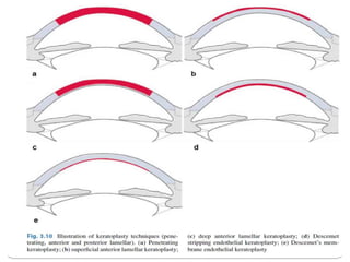
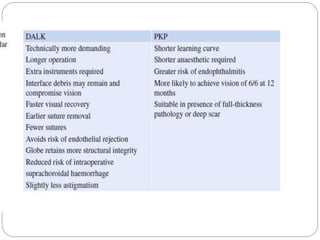
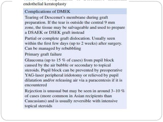
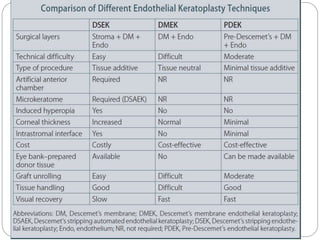
-Dr. Pravda Chaturvedi
Keratoplasty
DEFINITION
 The replacement of diseased cornea with
autologus or heterologus cornea is called as
keratoplasty. In most of the cases the donor
cornea is taken from a deceased person.
 Either full thickness of the cornea or a part of it
may be transplanted.
INDICATIONS
 Optical
 Therpeutic
 Tectonic
 Cosmetic
Optic
 A healthy, clear donor cornea is used to replace an
opaque, cloudy, or distorted cornea in an attempt to
improve vision and hence quality of life
 Scar due to ulcer, trauma
 Degenerations and dystrophies
 Chemical injuries
 Pseudophakic bullous keratopathy
 Keratoconus
 Regraft secondary to rejection
 Keratoglobus
 Aphakic bullous keratopathy
 Congenital opacities
Tectonic
Keratoplasty that is performed to restore
structural integrity of the cornea is named as
tectonic KP. No emphasis is made here for
optical outcome. Tissue addition is made in
areas of corneal thinning and restoration to
near normal surface contour of the cornea is
carried out.
 Descemetocele.
 Corneal perforation.
 Corneal stromal thinning
Therapeutic
 Keratoplasty that is performed to remove
infected corneal tissue that has failed medical
treatment.
 Rarely,KP is performed to remove corneal
tumor that has not invaded the full-thickness
of the cornea.
 Therapeutic KP may also be utilized in
corneal inflammatory processes, and in
selected cases of viral, bacterial, and fungal
keratitis.
Cosmetic
 Corneal transplantation is performed to
improve the appearance of the patient and
has no bearing on the visual outcome. This
could also be done in a nonseeing eye. An
opaque cornea with a white or blue-gray hue
may be disturbing to the patient, who may
request PK.
Types:
A. Based on Location
1. Central
2. Peripheral—Circular, oval, crescentic, annular,
semilunar, rectangular or strip graft
3. Total—central and peripheral
4. Corneoscleral
B. Based on Stem-cell Transplantation
1. Non-stem cell KP
2. KP with stem cell transplantation (SCT)
Contraindictions
 Dry eye, blepharitis, ectropion, entropion
 Recurrent occular infection,
 Melting cornea due to connective tissue disorder,
 Herpetic infection,
 Uveitis,
 Uncontrolled glaucoma.
HISTORY
The history of keratoplasty can be divided into four
great periods as follows:
 1. The period of the precursors and the first trials,
before 1900.
 2. The developmental period, between 1900 and
1945.
 3. The period of constant improvement and
changes, from 1945 until the middle of the
nineties.
 4. The current period, namely, the modern era.
 1824 Reisinger – First animal graft and coined
the term “keratoplasty”
 1831 Dieffenbach proposes partial-thickness
keratoplasty (LKP, extraocular procedure)
 1846 First use of general anesthesia – ether, at
the Massachusetts General Hospital, Boston, MA
 1878 Arthur von Hippel, invented circular cutting
trephine, blades of different diameters, a key for
winding up the watch mechanism
 1888 Arthur von Hippel, first successful LKP in
man
 1908 Plange – First human lamellar autograft
(clear cornea from blind eye to opposite, scarred
eye of the same patient). Graft remained clear for
5 years.
 1912 Magitot – Use of human cornea previously
preserved in an antiseptic fluid for corneal
transplantation.
Types of keratoplasty
Based on the thickness of the cornea transplanted,
keratoplasty can be divided as:
 Penetrating keraoplasty- involved full thickness of
the cornea.
 Lamellar keratoplasty- involves atransplantation
of a part.
Anterior lamellar : SALK, MALK, DALK, TALK
Posterior lamellar : DLEK, DMEK, DSAEK
Donor tissue
 Removed as early as possible (6-12 hours of
death).
 Corneas from infants (3 years and under) are
rarely used -surgical, refractive and rejection
problems.
 It should be stored under sterile conditions.
 Evaluation –medical history review and donor
blood screening to exclude contraindications, and
microscopic examination of the cornea including
endothelial cell count determination
Corneal preservation
Short-term storage (up to 2 days) -The whole globe is
preserved at 40C in a moist chamber.
Intermediate storage (up to 2 weeks) -McCarey-
Kaufman (MK) medium and various chondroitin sulfate
enriched media such as optisol medium used.
Long-term storage (up to 35 days) -It is done by
organ culture method.
Contraindications for donation of
corneal tissue
•Death of unknown cause.
•Certain systemic infections such as HIV, viral
hepatitis, syphilis, congenital rubella,
tuberculosis, septicaemia and active malaria.
•Prior high-risk behavior for HIV and hepatitis.
infectious diseases of the CNS.
•Receipt of a transplanted organ.
•Most hematological malignancies.
•Ocular disease such as inflammation and
malignancies (e.g. retinoblastoma) and
corneal refractive surgery.
PENETRATING
KERATOPLASTY
Pre op evaluation:
 Evaluation of visual potential.
 Ocular surface disease – must be recognized
and treated prior to PK.e.g. rosacea, dry eyes,
blepharitis, trichiasis, exposure keratopathy,
ectropion, and entropion.
 IOP must be controlled prior to surgery.
 Ocular inflammation – must be recognized
and treated.
 Prior corneal diseases and vascularisation.
 decompression of globe is ensured.
Intravenous mannitol or mechanical ocular
decompression should be considered.
 Miotics are placed preoperatively to protect
the lens during surgery.
 Graft size determination is based on three
main factors: the size of the recipient cornea,
the targeted disease, and the known
increased risk of rejection with increasing graft
size.
- An ideal size is 7.5 mm.
- grafts smaller than this may give rise to high
astigmatism.
- Grafts of diameter 8.5 mm or more are prone to
postoperative anterior synechiae formation,
vascularization and increased intraocular
pressure.
 Excision of donor corneal button -The donor
corneal button should be trephined 0.25 mm
larger than the recipient, taking care not to
damage the endothelium.
- to facilitate watertight closure, minimize
postoperative flattening and reduce the possibility
of postoperative glaucoma.
 Excision of donor corneal button -The donor
corneal button should be trephined 0.25 mm
larger than the recipient, taking care not to
damage the endothelium.
- to facilitate watertight closure, minimize
postoperative flattening and reduce the possibility
of postoperative glaucoma.
 Four
cardinal
sutures in
place
12 interrupted sutures
Continuous
running sutures
Knots burried
 Four categories of suturing techniques
employed in keratoplasty will be addressed in
subsequent sections:
 1 Interrupted sutures (IS)
 2 Combined continuous and interrupted
sutures (CCIS)
 3 Single continuous suture (SCS)
 4 Double continuous suture (DCS)
Cardinal sutures
The second suture
is 180 degrees
from the first suture
so that the donor
tissue and wound
are divided equally
into nasal and
temporal halves .
The third and the
fourth sutures also
divide the donor
INTRA OP COMPLICATIONS
 Poor centration of graft
 Excessive bleeding
 Damage to host iris, lens or graft endothelium
 Expulsive hemorrhage
POST OP COMPLICATIONS
EARLY
 Wound leak
 Flat AC with raised IOP. Causes: pupillary block, ant
rotation of iris lens diaphragm, choroidal effusion,
malignant glaucoma.
 Endophthalmitis
 Persistent epithelial defect
 Loose or broken sutures
LATE
• astigmatism, recurrence of initial disease process,
late wound separation, retro-corneal membrane
formation, glaucoma and cystoid macular oedema.
Postoperative management:
 Topical steroids are used to decrease the risk of
immunological graft rejection.
 Other immunosuppressants –azathioprine,
ciclosporin may be rarely used in high-risk for
prevention of rejection.
 Mydriatics - if uveitis persists.
 Monitoring of IOP is performed during the early
postoperative period.
 Removal of sutures when the graft-host junction
has healed. This is usually after 12–18 months.
 Rigid contact lenses -to optimize visual acuity in
eyes with astigmatism.
Graft failure and Graft rejection
 Persistent edema in
donor tissue in non
inflammed eye
 Poor donor
endothelial function or
damage to it while
surgery.
 Observation
 Regraft if necessary
 A most common
cause of graft failure
 Decreased vision,
photophobia, pain,
redness.
 Topical corticosteroid:
hrly
 Subconj corticosteroid
 Systemic may be
given for serious
cases
Graft rejection
 Epithelial rejection: an epithelial line
 Sub epithelial rejection: multiple sub epithelial
infiltrates limited to the graft
 Endothelial rejection: KP, iritis, corneal oedema.
Khodadoust line is the advancing front of the
host immunological and inflammatory cells
against donor endothelium.
LAMELLAR KERATOPLASTY
 Selective removal of diseased tissue and
replacement with donor cornea.
 TYPES : anterior and posterior.
 Anterior lamellar keratoplasty:
 Transplanted tissue doesn’t include corneal
endothelium. A closed globe procedure.
Avoids the high risk of endothelial rejection.
Also the dnor tissue can be retrieved from old
cornea.
 Indications-
 Opacification of the superficial one-third of the corneal
stroma.
 Disease involving the anterior 95% of corneal
thickness with a normal endothelium and absence
of breaks or scars in Descemet membrane
 Marginal corneal thinning or infiltration as in recurrent
pterygium, marginal degeneration.
 Localized thinning or descemetocele formation.
 Chronic inflammatory disease such as atopic
keratoconjunctivitis which carries an increased risk
of graft rejection.
Posterior lamellar keratoplasty:
 Diseased corneal endothelium is replaced. Maintains
structural integrity. Decreases surgically induced
astigmatism.
 It involves removal only of diseased endothelium
along with Descemet’s membrane, through a
corneoscleral or corneal incision.
 Folded donor tissue is introduced through the same
small (about 5 mm) incision.
Indications:
 include endothelial disease such as pseudophakic
bullous keratopathy.
DALK
 DALK has become an alternative to PK for a
variety of indications . Studies have
demonstrated equivalent best corrected visual
outcomes and superior preservation of
endothelial cell density with DALK compared
with PK. Interest in DALK has been piqued by
its advantages, including enhanced structural
integrity, decreased recovery time, and lower
incidence of rejection and infection.
DLEK
 In contrast to PK, EK causes little to no
change in corneal topography, resulting in far
less change in spherical equivalent or
cylinder.
 PK induces 4–5 D of refractive cylinder on
average and may exceed 8 D, DSEK and
DMEK cause no significant increase in mean
refractive cylinder.
 DSEK generally induces 0.5–2.0 D of
hyperopia, while DMEK causes a smaller
mean hyperopic shift of about 0.25–0.50 D.
The expected hyperopic shift should be
factored into intraocular lens calculations
Lack of adherence or partial adherence post op:
 complete removal of fluid from donor/ host
interface,
 achieving a firm air tamponade,
 cautioning patient not to rub eye in the early
postoperative period.
Keratoplasty
Keratoplasty

Keratoplasty

  • 1.
  • 2.
    DEFINITION  The replacementof diseased cornea with autologus or heterologus cornea is called as keratoplasty. In most of the cases the donor cornea is taken from a deceased person.  Either full thickness of the cornea or a part of it may be transplanted.
  • 3.
  • 4.
    Optic  A healthy,clear donor cornea is used to replace an opaque, cloudy, or distorted cornea in an attempt to improve vision and hence quality of life  Scar due to ulcer, trauma  Degenerations and dystrophies  Chemical injuries  Pseudophakic bullous keratopathy  Keratoconus  Regraft secondary to rejection  Keratoglobus  Aphakic bullous keratopathy  Congenital opacities
  • 5.
    Tectonic Keratoplasty that isperformed to restore structural integrity of the cornea is named as tectonic KP. No emphasis is made here for optical outcome. Tissue addition is made in areas of corneal thinning and restoration to near normal surface contour of the cornea is carried out.  Descemetocele.  Corneal perforation.  Corneal stromal thinning
  • 6.
    Therapeutic  Keratoplasty thatis performed to remove infected corneal tissue that has failed medical treatment.  Rarely,KP is performed to remove corneal tumor that has not invaded the full-thickness of the cornea.  Therapeutic KP may also be utilized in corneal inflammatory processes, and in selected cases of viral, bacterial, and fungal keratitis.
  • 7.
    Cosmetic  Corneal transplantationis performed to improve the appearance of the patient and has no bearing on the visual outcome. This could also be done in a nonseeing eye. An opaque cornea with a white or blue-gray hue may be disturbing to the patient, who may request PK.
  • 8.
    Types: A. Based onLocation 1. Central 2. Peripheral—Circular, oval, crescentic, annular, semilunar, rectangular or strip graft 3. Total—central and peripheral 4. Corneoscleral B. Based on Stem-cell Transplantation 1. Non-stem cell KP 2. KP with stem cell transplantation (SCT)
  • 9.
    Contraindictions  Dry eye,blepharitis, ectropion, entropion  Recurrent occular infection,  Melting cornea due to connective tissue disorder,  Herpetic infection,  Uveitis,  Uncontrolled glaucoma.
  • 10.
    HISTORY The history ofkeratoplasty can be divided into four great periods as follows:  1. The period of the precursors and the first trials, before 1900.  2. The developmental period, between 1900 and 1945.  3. The period of constant improvement and changes, from 1945 until the middle of the nineties.  4. The current period, namely, the modern era.
  • 11.
     1824 Reisinger– First animal graft and coined the term “keratoplasty”  1831 Dieffenbach proposes partial-thickness keratoplasty (LKP, extraocular procedure)  1846 First use of general anesthesia – ether, at the Massachusetts General Hospital, Boston, MA  1878 Arthur von Hippel, invented circular cutting trephine, blades of different diameters, a key for winding up the watch mechanism  1888 Arthur von Hippel, first successful LKP in man
  • 12.
     1908 Plange– First human lamellar autograft (clear cornea from blind eye to opposite, scarred eye of the same patient). Graft remained clear for 5 years.  1912 Magitot – Use of human cornea previously preserved in an antiseptic fluid for corneal transplantation.
  • 13.
    Types of keratoplasty Basedon the thickness of the cornea transplanted, keratoplasty can be divided as:  Penetrating keraoplasty- involved full thickness of the cornea.  Lamellar keratoplasty- involves atransplantation of a part. Anterior lamellar : SALK, MALK, DALK, TALK Posterior lamellar : DLEK, DMEK, DSAEK
  • 14.
    Donor tissue  Removedas early as possible (6-12 hours of death).  Corneas from infants (3 years and under) are rarely used -surgical, refractive and rejection problems.  It should be stored under sterile conditions.  Evaluation –medical history review and donor blood screening to exclude contraindications, and microscopic examination of the cornea including endothelial cell count determination
  • 17.
    Corneal preservation Short-term storage(up to 2 days) -The whole globe is preserved at 40C in a moist chamber. Intermediate storage (up to 2 weeks) -McCarey- Kaufman (MK) medium and various chondroitin sulfate enriched media such as optisol medium used. Long-term storage (up to 35 days) -It is done by organ culture method.
  • 18.
    Contraindications for donationof corneal tissue •Death of unknown cause. •Certain systemic infections such as HIV, viral hepatitis, syphilis, congenital rubella, tuberculosis, septicaemia and active malaria. •Prior high-risk behavior for HIV and hepatitis. infectious diseases of the CNS. •Receipt of a transplanted organ. •Most hematological malignancies. •Ocular disease such as inflammation and malignancies (e.g. retinoblastoma) and corneal refractive surgery.
  • 20.
    PENETRATING KERATOPLASTY Pre op evaluation: Evaluation of visual potential.  Ocular surface disease – must be recognized and treated prior to PK.e.g. rosacea, dry eyes, blepharitis, trichiasis, exposure keratopathy, ectropion, and entropion.  IOP must be controlled prior to surgery.  Ocular inflammation – must be recognized and treated.  Prior corneal diseases and vascularisation.
  • 21.
     decompression ofglobe is ensured. Intravenous mannitol or mechanical ocular decompression should be considered.  Miotics are placed preoperatively to protect the lens during surgery.  Graft size determination is based on three main factors: the size of the recipient cornea, the targeted disease, and the known increased risk of rejection with increasing graft size.
  • 22.
    - An idealsize is 7.5 mm. - grafts smaller than this may give rise to high astigmatism. - Grafts of diameter 8.5 mm or more are prone to postoperative anterior synechiae formation, vascularization and increased intraocular pressure.  Excision of donor corneal button -The donor corneal button should be trephined 0.25 mm larger than the recipient, taking care not to damage the endothelium. - to facilitate watertight closure, minimize postoperative flattening and reduce the possibility of postoperative glaucoma.
  • 23.
     Excision ofdonor corneal button -The donor corneal button should be trephined 0.25 mm larger than the recipient, taking care not to damage the endothelium. - to facilitate watertight closure, minimize postoperative flattening and reduce the possibility of postoperative glaucoma.
  • 28.
  • 29.
  • 30.
  • 31.
  • 33.
     Four categoriesof suturing techniques employed in keratoplasty will be addressed in subsequent sections:  1 Interrupted sutures (IS)  2 Combined continuous and interrupted sutures (CCIS)  3 Single continuous suture (SCS)  4 Double continuous suture (DCS)
  • 34.
    Cardinal sutures The secondsuture is 180 degrees from the first suture so that the donor tissue and wound are divided equally into nasal and temporal halves . The third and the fourth sutures also divide the donor
  • 39.
    INTRA OP COMPLICATIONS Poor centration of graft  Excessive bleeding  Damage to host iris, lens or graft endothelium  Expulsive hemorrhage
  • 40.
    POST OP COMPLICATIONS EARLY Wound leak  Flat AC with raised IOP. Causes: pupillary block, ant rotation of iris lens diaphragm, choroidal effusion, malignant glaucoma.  Endophthalmitis  Persistent epithelial defect  Loose or broken sutures LATE • astigmatism, recurrence of initial disease process, late wound separation, retro-corneal membrane formation, glaucoma and cystoid macular oedema.
  • 41.
    Postoperative management:  Topicalsteroids are used to decrease the risk of immunological graft rejection.  Other immunosuppressants –azathioprine, ciclosporin may be rarely used in high-risk for prevention of rejection.  Mydriatics - if uveitis persists.  Monitoring of IOP is performed during the early postoperative period.  Removal of sutures when the graft-host junction has healed. This is usually after 12–18 months.  Rigid contact lenses -to optimize visual acuity in eyes with astigmatism.
  • 42.
    Graft failure andGraft rejection  Persistent edema in donor tissue in non inflammed eye  Poor donor endothelial function or damage to it while surgery.  Observation  Regraft if necessary  A most common cause of graft failure  Decreased vision, photophobia, pain, redness.  Topical corticosteroid: hrly  Subconj corticosteroid  Systemic may be given for serious cases
  • 43.
    Graft rejection  Epithelialrejection: an epithelial line  Sub epithelial rejection: multiple sub epithelial infiltrates limited to the graft  Endothelial rejection: KP, iritis, corneal oedema. Khodadoust line is the advancing front of the host immunological and inflammatory cells against donor endothelium.
  • 44.
    LAMELLAR KERATOPLASTY  Selectiveremoval of diseased tissue and replacement with donor cornea.  TYPES : anterior and posterior.  Anterior lamellar keratoplasty:  Transplanted tissue doesn’t include corneal endothelium. A closed globe procedure. Avoids the high risk of endothelial rejection. Also the dnor tissue can be retrieved from old cornea.
  • 45.
     Indications-  Opacificationof the superficial one-third of the corneal stroma.  Disease involving the anterior 95% of corneal thickness with a normal endothelium and absence of breaks or scars in Descemet membrane  Marginal corneal thinning or infiltration as in recurrent pterygium, marginal degeneration.  Localized thinning or descemetocele formation.  Chronic inflammatory disease such as atopic keratoconjunctivitis which carries an increased risk of graft rejection.
  • 46.
    Posterior lamellar keratoplasty: Diseased corneal endothelium is replaced. Maintains structural integrity. Decreases surgically induced astigmatism.  It involves removal only of diseased endothelium along with Descemet’s membrane, through a corneoscleral or corneal incision.  Folded donor tissue is introduced through the same small (about 5 mm) incision. Indications:  include endothelial disease such as pseudophakic bullous keratopathy.
  • 47.
    DALK  DALK hasbecome an alternative to PK for a variety of indications . Studies have demonstrated equivalent best corrected visual outcomes and superior preservation of endothelial cell density with DALK compared with PK. Interest in DALK has been piqued by its advantages, including enhanced structural integrity, decreased recovery time, and lower incidence of rejection and infection.
  • 51.
  • 52.
     In contrastto PK, EK causes little to no change in corneal topography, resulting in far less change in spherical equivalent or cylinder.  PK induces 4–5 D of refractive cylinder on average and may exceed 8 D, DSEK and DMEK cause no significant increase in mean refractive cylinder.  DSEK generally induces 0.5–2.0 D of hyperopia, while DMEK causes a smaller mean hyperopic shift of about 0.25–0.50 D. The expected hyperopic shift should be factored into intraocular lens calculations
  • 53.
    Lack of adherenceor partial adherence post op:  complete removal of fluid from donor/ host interface,  achieving a firm air tamponade,  cautioning patient not to rub eye in the early postoperative period.