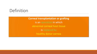
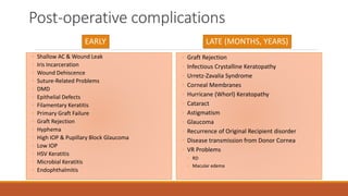
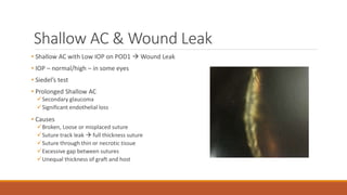
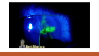
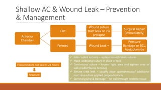
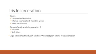
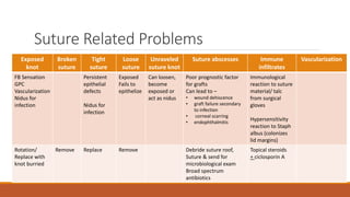
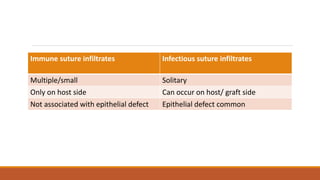
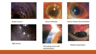
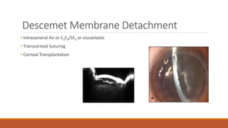
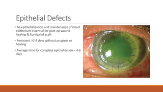
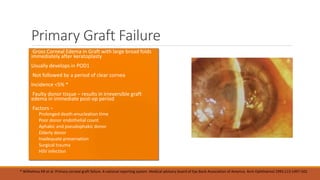
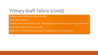
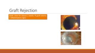
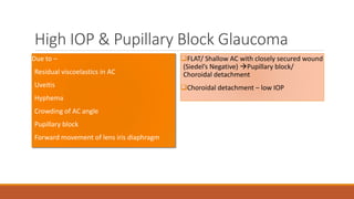
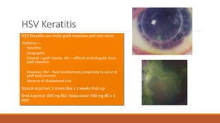
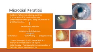
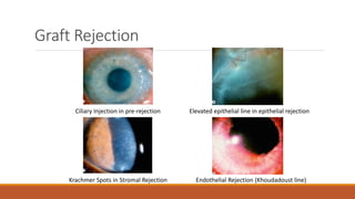
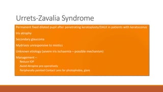
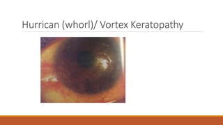
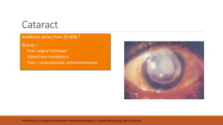
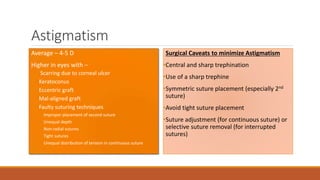
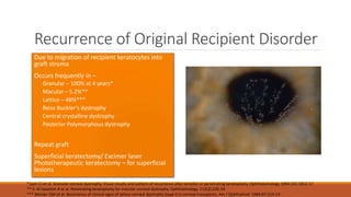
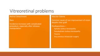
Keratoplasty associated
complications
DR. KARAN BHATIA
FELLOW CORNEA, PHACOEMULSIFICATION AND REFRACTIVE SURGERY
MM JOSHI EYE INSTITUTE, HUBLI, KARNATAKA, INDIA
Introduction
The history of "tissue transplantation" starts from Adam and Eve in
Eden. Throughout history, however, the eye, as the avenue to the Sun
God, has symbolized virtue and wisdom, with blindness as a penalty for
impiety and the stigma of sexual shame. Blind people were generally
regarded as social outcasts, for whom treatment of any sort
represented a tampering with God's proper judgment. In myths and
folklore, although occasionally the damaged eye was replaced by the
fresh one, the eye is more usually replaced by its symbolic equivalent of
wisdom or second sight.
Definition
Corneal transplantation or grafting
is an operation in which
abnormal corneal host tissue
is replaced by
healthy donor cornea
Post-operative complications
EARLY
◦ Shallow AC & Wound Leak
◦ Iris Incarceration
◦ Wound Dehiscence
◦ Suture-Related Problems
◦ DMD
◦ Epithelial Defects
◦ Filamentary Keratitis
◦ Primary Graft Failure
◦ Graft Rejection
◦ Hyphema
◦ High IOP & Pupillary Block Glaucoma
◦ Low IOP
◦ HSV Keratitis
◦ Microbial Keratitis
◦ Endophthalmitis
LATE (MONTHS, YEARS)
◦ Graft Rejection
◦ Infectious Crystalline Keratopathy
◦ Urretz-Zavalia Syndrome
◦ Corneal Membranes
◦ Hurricane (Whorl) Keratopathy
◦ Cataract
◦ Astigmatism
◦ Glaucoma
◦ Recurrence of Original Recipient disorder
◦ Disease transmission from Donor Cornea
◦ VR Problems
◦ RD
◦ Macular edema
Early Post Operative
Complications
VARY FROM MINOR TO TRUE OPHTHALMIC EMERGENCIES  LOSS OF EYE
METICULOUS FOLLOW UP, EARLY DIAGNOSIS, TIMELY INTERVENTION  MANDATORY
Shallow AC & Wound Leak
• Shallow AC with Low IOP on POD1  Wound Leak
• IOP – normal/high – in some eyes
• Siedel’s test
• Prolonged Shallow AC
Secondary glaucoma
Significant endothelial loss
• Causes
Broken, Loose or misplaced suture
Suture track leak  full thickness suture
Suture through thin or necrotic tissue
Excessive gap between sutures
Unequal thickness of graft and host
Shallow AC & Wound Leak – Prevention
& Management
Anterior
Chamber
Flat
Wound suture
tract leak or iris
prolapse
Surgical Repair
(immediately)
Formed Wound Leak +
Pressure
Bandage or BCL,
Acetzolamide
If wound does not seal in 24 hours
Resuture
 Interrupted sutures – replace loose/broken sutures
 Place additional suture in place of leak
 Continuous suture – loosen tight area and tighten area of
leak (redistributes tension)
 Suture tract leak – usually close spontaneously/ additional
mattress suture applied perpendicularly
 Corneal gluing & Bandage – for leak through necrotic tissue
Iris Incarceration
• Causes
Collapse of AC/wound leak
Inflamed eyes/ Swollen & Flaccid Iris (preop)
Poorly placed sutures
• Closes AC angle at site incarceration 
Glaucoma
Graft failure
• Large adhesions at host-graft junction localized graft edema  vascularization
Wound Dehiscence
• Can occur immediately/several years later
• Causes
Trauma
Infectious Keratitis
Suture Failure
Spontaneous wound separation
• Resuture immediately
Suture Related Problems
Exposed
knot
Broken
suture
Tight
suture
Loose
suture
Unraveled
suture knot
Suture abscesses Immune
infiltrates
Vascularization
FB Sensation
GPC
Vascularization
Nidus for
infection
Persistent
epithelial
defects
Nidus for
infection
Exposed
Fails to
epithelize
Can loosen,
become
exposed or
act as nidus
Poor prognostic factor
for grafts
Can lead to –
• wound dehiscence
• graft failure secondary
to infection
• corneal scarring
• endophthalmitis
Immunological
reaction to suture
material/ talc
from surgical
gloves
Hypersensitivity
reaction to Staph
albus (colonizes
lid margins)
Rotation/
Replace with
knot burried
Remove Replace Remove Debride suture roof,
Suture & send for
microbiological exam
Broad spectrum
antibiotics
Topical steroids
+ ciclosporin A
Immune suture infiltrates Infectious suture infiltrates
Multiple/small Solitary
Only on host side Can occur on host/ graft side
Not associated with epithelial defect Epithelial defect common
Broken Suture
Tight Suture
Suture Infiltrates Suture induces Vascularization
Protruding suture with
vascularization
Papillary hyperplasia
Descemet Membrane Detachment
• Intracameral Air or C3F8/SF6 or viscoelastic
• Transcorneal Suturing
• Corneal Transplantation
Epithelial Defects
• Re-epithelialization and maintenance of intact
epithelium essential for post-op wound
healing & Survival of graft
• Persistent >2-4 days without progress or
healing
• Average time for complete epithelization – 4-6
days
Epithelial defects (contd) – Risk Factors
Ocular Surface Disorders
• Lid abnormalities – ectropion, entropion,
lagophthalmos
• Infection & Inflammation – HSV
• Iatrogenic – tight sutures, dryness, poor
apposition of graft-host junction
• Epitheliotoxic drugs – gentamicin, timolol,
ciprofloxacin, prednisolone, dorzolamide,
NSAIDs
•Damaged donor epithelium
•Basement membrane disorders
•Intrinsic epithelial disorder – Stem cell
deficiency secondary to thermal/ chemical
burns, SJS, Ocular cicatrical pemphigoid
•Trauma
•Poor nutrition – Vitamin A deficiency, PEM
•Metalbolic diseases – DM (both in donor and
host)
Epithelial defects (contd) – Management
• Prevent and Treat Risk Factors
• Adequate lid closure
• Prevent Corneal Exposure
• BCL
•Temporary Tarsorrhaphy
• Choramphenicol/ Panthogel
• Autologous Serum
• AMT
• HSV – Oral Acyclovir 300 mg BD
Filamentary Keratitis
Reported Incidence of 27% in one case series*
*Rotkis WM et al. Filamentary Keratitis following penetrating keratoplasty. Ophthalmology. 1982;89:946-9.
Primary Graft Failure
• Gross Corneal Edema in Graft with large broad folds
immediately after keratoplasty
•Usually develops in POD1
• Not followed by a period of clear cornea
•Incidence <5% *
• Faulty donor tissue – results in irreversible graft
edema in immediate post-op period
• Factors –
 Prolonged death-enucleation time
 Poor donor endothelial count
 Aphakic and pseudophakic donor
 Elderly donor
 Inadequate preservation
 Surgical trauma
 HSV infection
* Wilhelmus KR et al. Primary corneal graft failure. A national reporting system. Medical advisory board of Eye Bank Association of America. Arch Ophthalmol 1995;113:1497-502
Primary Graft Failure (contd)
• Unresponsive to hypertonic saline/ steroids
•Proper donor selection
•Prolonged death to enucleation time – MK media can preserve donor tissue only up to 2 hours
•Early surgery & Minimal surgical trauma
•Observe for 3-4 weeks. No improvement  Repeat Penetrating Keratoplasty
Graft Rejection
•Graft clear for atleast 2 weeks  graft edema
+ inflammatory signs
Hyphema
•Incidence increases with intraoperative manipulations like extensive synechiolysis, iridoplasty or
iridotomy
•Clears spontaneously without treatment
•IOP high – then treat aggressively
•Β-Blockers + Briminodine/Acetazolamide
•Prolonged persistence – Clot irrigation and aspiration
High IOP & Pupillary Block Glaucoma
Due to –
•Residual viscoelastics in AC
•Uveitis
•Hyphema
•Crowding of AC angle
•Pupillary block
•Forward movement of lens iris diaphragm
FLAT/ Shallow AC with closely secured wound
(Siedel’s Negative) Pupillary block/
Choroidal detachment
Choroidal detachment – low IOP
Low IOP
Causes –
•Wound Leak
•Iridocyclitis: Ciliary shock
•Cyclodialysis
•Choroidal detachment
•Retinal Detachment
HSV Keratitis
•HSV Keratitis can incite graft rejection and vice versa
• Patterns –
Dendritic
Geographic
Stromal – graft edema, KPs – difficult to distinguish from
graft rejection
However, HSV – focal involvement, propensity to occur at
graft host junction
Absence of Khadadoust Line
•Topical Acyclovir 5 times/day x 2 weeks Post-op
•Oral Acyclovir 400 mg BD/ Valacyclovir 500 mg BD x 1
year
Microbial Keratitis
• Incidence higher in developing countries
• ½ occur within 1st 6 months of surgery
• Either infection within graft/ along suture tracts at
graft host junction
Inflammatory Reaction
Initiation of Graft Rejection
Graft Failure Graft Melting Endophthalmitis
• Corneal scrapings – Gram’s stain/KOH/C & S
• Therapy modified based on lab report
• Initial therapy – Fluoroquinolone or combination of
Cefazolin 5% and Tobramycin 1.3%
Endophthalmitis
• Vitreous Tap
•Intensive topical, intravitreal and systemic
antibiotics
Late Post-operative
MONTHS, YEARS
Graft Rejection
Ciliary Injection in pre-rejection Elevated epithelial line in epithelial rejection
Krachmer Spots in Stromal Rejection Endothelial Rejection (Khoudadoust line)
Infectious Crystalline Keratopathy
•Chronic, progressive corneal infection
•Anterior lamella of graft involved
•No clinically evident stromal inflammation
•Crystalline branching opacities in anterior &
mid stroma
Urrets-Zavalia Syndrome
•Permanent fixed dilated pupil after penetrating keratoplasty/DALK in patients with keratoconus
•Iris atrophy
•Secondary glaucoma
•Mydriasis unresponsive to miotics
•Unknown etiology (severe iris ischaemia – possible mechanism)
•Management –
• Reduce IOP
• Avoid Atropine pre-operatively
• Peripherally painted Contact Lens for photophobia, glare
Corneal Membranes
Epithelial ingrowth (conjunctival/corneal) – through gap at host-graft junction
Fibrous ingrowth (retrocorneal membrane) – gray/white fibrous membranes between DM and
endothelium
Hurrican (whorl)/ Vortex Keratopathy
Cataract
•Incidence varies from 25-80% *
•Due to –
Poor surgical technique
Altered lens metabolism
Toxic – corticosteroids, anticholinesterase
*Rathi VM et al. Cataract formation after Penetraing keratoplasty. J Cataract Refract Surg. 1997;23:562-64
Astigmatism
•Average – 4-5 D
•Higher in eyes with –
 Scarring due to corneal ulcer
Keratoconus
Eccentric graft
Mal-aligned graft
Faulty suturing techniques
 Improper placement of second suture
 Unequal depth
 Non-radial sutures
 Tight sutures
 Unequal distribution of tension in continuous suture
Surgical Caveats to minimize Astigmatism
•Central and sharp trephination
•Use of a sharp trephine
•Symmetric suture placement (especially 2nd
suture)
•Avoid tight suture placement
•Suture adjustment (for continuous suture) or
selective suture removal (for interrupted
sutures)
Glaucoma
•Due to PAS and epithelial downgrowth
•2 unique mechanisms –
Collapse of trabecular meshwork
Compression of AC angle
•Larger Donor Grafts – associated with deeper AC  lower incidence of post-op progressive
angle closure and lower post-op IOPs
•Avoid Dorzolamide
•Laser Trabeculoplasty
•Trabeculectomy with MMC  GDD Surgery
Recurrence of Original Recipient Disorder
•Due to migration of recipient keratocytes into
graft stroma
•Occurs frequently in –
Granular – 100% at 4 years*
Macular – 5.2%**
Lattice – 48%***
Reiss Buckler’s dystrophy
Central crystalline dystrophy
Posterior Polymorphous dystrophy
•Repeat graft
•Superficial keratectomy/ Excimer laser
Phototherapeutic keratectomy – for superficial
lesions
*Lyon CJ et al. Granular corneal dystrophy. Visual results and pattern of recurrence after lamellar or penetrating keratoplasty. Ophthalomology 1994;101:1812-17
** S. Al-Swailem A et al. Penetrating keratoplasty for macular corneal dystrophy. Ophthalmology. 112(2):220-24
*** Meisler DM et al. Recurrence of clinical signs of lattice corneal dystrophy (type I) in corneal transplants. Am J Ophthalmol. 1984;97:210-14
Vitreoretinal problems
Retinal Detachment
•Rare
•Incidence increases with complicated
procedure, especially after vitreous
manipulation
Macular Edema
•Common cause of non improvement of vision
despite clear graft
•Predispositions –
Aphakic bullous keratopathy
Pseudophakic bullous keratopathy
Trauma
Any previous intraocular surgery
Thankyou

Keratoplasty associated complications

  • 1.
    Keratoplasty associated complications DR. KARANBHATIA FELLOW CORNEA, PHACOEMULSIFICATION AND REFRACTIVE SURGERY MM JOSHI EYE INSTITUTE, HUBLI, KARNATAKA, INDIA
  • 2.
    Introduction The history of"tissue transplantation" starts from Adam and Eve in Eden. Throughout history, however, the eye, as the avenue to the Sun God, has symbolized virtue and wisdom, with blindness as a penalty for impiety and the stigma of sexual shame. Blind people were generally regarded as social outcasts, for whom treatment of any sort represented a tampering with God's proper judgment. In myths and folklore, although occasionally the damaged eye was replaced by the fresh one, the eye is more usually replaced by its symbolic equivalent of wisdom or second sight.
  • 3.
    Definition Corneal transplantation orgrafting is an operation in which abnormal corneal host tissue is replaced by healthy donor cornea
  • 4.
    Post-operative complications EARLY ◦ ShallowAC & Wound Leak ◦ Iris Incarceration ◦ Wound Dehiscence ◦ Suture-Related Problems ◦ DMD ◦ Epithelial Defects ◦ Filamentary Keratitis ◦ Primary Graft Failure ◦ Graft Rejection ◦ Hyphema ◦ High IOP & Pupillary Block Glaucoma ◦ Low IOP ◦ HSV Keratitis ◦ Microbial Keratitis ◦ Endophthalmitis LATE (MONTHS, YEARS) ◦ Graft Rejection ◦ Infectious Crystalline Keratopathy ◦ Urretz-Zavalia Syndrome ◦ Corneal Membranes ◦ Hurricane (Whorl) Keratopathy ◦ Cataract ◦ Astigmatism ◦ Glaucoma ◦ Recurrence of Original Recipient disorder ◦ Disease transmission from Donor Cornea ◦ VR Problems ◦ RD ◦ Macular edema
  • 5.
    Early Post Operative Complications VARYFROM MINOR TO TRUE OPHTHALMIC EMERGENCIES  LOSS OF EYE METICULOUS FOLLOW UP, EARLY DIAGNOSIS, TIMELY INTERVENTION  MANDATORY
  • 6.
    Shallow AC &Wound Leak • Shallow AC with Low IOP on POD1  Wound Leak • IOP – normal/high – in some eyes • Siedel’s test • Prolonged Shallow AC Secondary glaucoma Significant endothelial loss • Causes Broken, Loose or misplaced suture Suture track leak  full thickness suture Suture through thin or necrotic tissue Excessive gap between sutures Unequal thickness of graft and host
  • 8.
    Shallow AC &Wound Leak – Prevention & Management Anterior Chamber Flat Wound suture tract leak or iris prolapse Surgical Repair (immediately) Formed Wound Leak + Pressure Bandage or BCL, Acetzolamide If wound does not seal in 24 hours Resuture  Interrupted sutures – replace loose/broken sutures  Place additional suture in place of leak  Continuous suture – loosen tight area and tighten area of leak (redistributes tension)  Suture tract leak – usually close spontaneously/ additional mattress suture applied perpendicularly  Corneal gluing & Bandage – for leak through necrotic tissue
  • 9.
    Iris Incarceration • Causes Collapseof AC/wound leak Inflamed eyes/ Swollen & Flaccid Iris (preop) Poorly placed sutures • Closes AC angle at site incarceration  Glaucoma Graft failure • Large adhesions at host-graft junction localized graft edema  vascularization
  • 10.
    Wound Dehiscence • Canoccur immediately/several years later • Causes Trauma Infectious Keratitis Suture Failure Spontaneous wound separation • Resuture immediately
  • 11.
    Suture Related Problems Exposed knot Broken suture Tight suture Loose suture Unraveled sutureknot Suture abscesses Immune infiltrates Vascularization FB Sensation GPC Vascularization Nidus for infection Persistent epithelial defects Nidus for infection Exposed Fails to epithelize Can loosen, become exposed or act as nidus Poor prognostic factor for grafts Can lead to – • wound dehiscence • graft failure secondary to infection • corneal scarring • endophthalmitis Immunological reaction to suture material/ talc from surgical gloves Hypersensitivity reaction to Staph albus (colonizes lid margins) Rotation/ Replace with knot burried Remove Replace Remove Debride suture roof, Suture & send for microbiological exam Broad spectrum antibiotics Topical steroids + ciclosporin A
  • 12.
    Immune suture infiltratesInfectious suture infiltrates Multiple/small Solitary Only on host side Can occur on host/ graft side Not associated with epithelial defect Epithelial defect common
  • 13.
    Broken Suture Tight Suture SutureInfiltrates Suture induces Vascularization Protruding suture with vascularization Papillary hyperplasia
  • 14.
    Descemet Membrane Detachment •Intracameral Air or C3F8/SF6 or viscoelastic • Transcorneal Suturing • Corneal Transplantation
  • 15.
    Epithelial Defects • Re-epithelializationand maintenance of intact epithelium essential for post-op wound healing & Survival of graft • Persistent >2-4 days without progress or healing • Average time for complete epithelization – 4-6 days
  • 16.
    Epithelial defects (contd)– Risk Factors Ocular Surface Disorders • Lid abnormalities – ectropion, entropion, lagophthalmos • Infection & Inflammation – HSV • Iatrogenic – tight sutures, dryness, poor apposition of graft-host junction • Epitheliotoxic drugs – gentamicin, timolol, ciprofloxacin, prednisolone, dorzolamide, NSAIDs •Damaged donor epithelium •Basement membrane disorders •Intrinsic epithelial disorder – Stem cell deficiency secondary to thermal/ chemical burns, SJS, Ocular cicatrical pemphigoid •Trauma •Poor nutrition – Vitamin A deficiency, PEM •Metalbolic diseases – DM (both in donor and host)
  • 17.
    Epithelial defects (contd)– Management • Prevent and Treat Risk Factors • Adequate lid closure • Prevent Corneal Exposure • BCL •Temporary Tarsorrhaphy • Choramphenicol/ Panthogel • Autologous Serum • AMT • HSV – Oral Acyclovir 300 mg BD
  • 18.
    Filamentary Keratitis Reported Incidenceof 27% in one case series* *Rotkis WM et al. Filamentary Keratitis following penetrating keratoplasty. Ophthalmology. 1982;89:946-9.
  • 19.
    Primary Graft Failure •Gross Corneal Edema in Graft with large broad folds immediately after keratoplasty •Usually develops in POD1 • Not followed by a period of clear cornea •Incidence <5% * • Faulty donor tissue – results in irreversible graft edema in immediate post-op period • Factors –  Prolonged death-enucleation time  Poor donor endothelial count  Aphakic and pseudophakic donor  Elderly donor  Inadequate preservation  Surgical trauma  HSV infection * Wilhelmus KR et al. Primary corneal graft failure. A national reporting system. Medical advisory board of Eye Bank Association of America. Arch Ophthalmol 1995;113:1497-502
  • 20.
    Primary Graft Failure(contd) • Unresponsive to hypertonic saline/ steroids •Proper donor selection •Prolonged death to enucleation time – MK media can preserve donor tissue only up to 2 hours •Early surgery & Minimal surgical trauma •Observe for 3-4 weeks. No improvement  Repeat Penetrating Keratoplasty
  • 21.
    Graft Rejection •Graft clearfor atleast 2 weeks  graft edema + inflammatory signs
  • 22.
    Hyphema •Incidence increases withintraoperative manipulations like extensive synechiolysis, iridoplasty or iridotomy •Clears spontaneously without treatment •IOP high – then treat aggressively •Β-Blockers + Briminodine/Acetazolamide •Prolonged persistence – Clot irrigation and aspiration
  • 23.
    High IOP &Pupillary Block Glaucoma Due to – •Residual viscoelastics in AC •Uveitis •Hyphema •Crowding of AC angle •Pupillary block •Forward movement of lens iris diaphragm FLAT/ Shallow AC with closely secured wound (Siedel’s Negative) Pupillary block/ Choroidal detachment Choroidal detachment – low IOP
  • 24.
    Low IOP Causes – •WoundLeak •Iridocyclitis: Ciliary shock •Cyclodialysis •Choroidal detachment •Retinal Detachment
  • 25.
    HSV Keratitis •HSV Keratitiscan incite graft rejection and vice versa • Patterns – Dendritic Geographic Stromal – graft edema, KPs – difficult to distinguish from graft rejection However, HSV – focal involvement, propensity to occur at graft host junction Absence of Khadadoust Line •Topical Acyclovir 5 times/day x 2 weeks Post-op •Oral Acyclovir 400 mg BD/ Valacyclovir 500 mg BD x 1 year
  • 26.
    Microbial Keratitis • Incidencehigher in developing countries • ½ occur within 1st 6 months of surgery • Either infection within graft/ along suture tracts at graft host junction Inflammatory Reaction Initiation of Graft Rejection Graft Failure Graft Melting Endophthalmitis • Corneal scrapings – Gram’s stain/KOH/C & S • Therapy modified based on lab report • Initial therapy – Fluoroquinolone or combination of Cefazolin 5% and Tobramycin 1.3%
  • 27.
    Endophthalmitis • Vitreous Tap •Intensivetopical, intravitreal and systemic antibiotics
  • 28.
  • 29.
    Graft Rejection Ciliary Injectionin pre-rejection Elevated epithelial line in epithelial rejection Krachmer Spots in Stromal Rejection Endothelial Rejection (Khoudadoust line)
  • 30.
    Infectious Crystalline Keratopathy •Chronic,progressive corneal infection •Anterior lamella of graft involved •No clinically evident stromal inflammation •Crystalline branching opacities in anterior & mid stroma
  • 31.
    Urrets-Zavalia Syndrome •Permanent fixeddilated pupil after penetrating keratoplasty/DALK in patients with keratoconus •Iris atrophy •Secondary glaucoma •Mydriasis unresponsive to miotics •Unknown etiology (severe iris ischaemia – possible mechanism) •Management – • Reduce IOP • Avoid Atropine pre-operatively • Peripherally painted Contact Lens for photophobia, glare
  • 32.
    Corneal Membranes Epithelial ingrowth(conjunctival/corneal) – through gap at host-graft junction Fibrous ingrowth (retrocorneal membrane) – gray/white fibrous membranes between DM and endothelium
  • 33.
  • 34.
    Cataract •Incidence varies from25-80% * •Due to – Poor surgical technique Altered lens metabolism Toxic – corticosteroids, anticholinesterase *Rathi VM et al. Cataract formation after Penetraing keratoplasty. J Cataract Refract Surg. 1997;23:562-64
  • 35.
    Astigmatism •Average – 4-5D •Higher in eyes with –  Scarring due to corneal ulcer Keratoconus Eccentric graft Mal-aligned graft Faulty suturing techniques  Improper placement of second suture  Unequal depth  Non-radial sutures  Tight sutures  Unequal distribution of tension in continuous suture Surgical Caveats to minimize Astigmatism •Central and sharp trephination •Use of a sharp trephine •Symmetric suture placement (especially 2nd suture) •Avoid tight suture placement •Suture adjustment (for continuous suture) or selective suture removal (for interrupted sutures)
  • 37.
    Glaucoma •Due to PASand epithelial downgrowth •2 unique mechanisms – Collapse of trabecular meshwork Compression of AC angle •Larger Donor Grafts – associated with deeper AC  lower incidence of post-op progressive angle closure and lower post-op IOPs •Avoid Dorzolamide •Laser Trabeculoplasty •Trabeculectomy with MMC  GDD Surgery
  • 38.
    Recurrence of OriginalRecipient Disorder •Due to migration of recipient keratocytes into graft stroma •Occurs frequently in – Granular – 100% at 4 years* Macular – 5.2%** Lattice – 48%*** Reiss Buckler’s dystrophy Central crystalline dystrophy Posterior Polymorphous dystrophy •Repeat graft •Superficial keratectomy/ Excimer laser Phototherapeutic keratectomy – for superficial lesions *Lyon CJ et al. Granular corneal dystrophy. Visual results and pattern of recurrence after lamellar or penetrating keratoplasty. Ophthalomology 1994;101:1812-17 ** S. Al-Swailem A et al. Penetrating keratoplasty for macular corneal dystrophy. Ophthalmology. 112(2):220-24 *** Meisler DM et al. Recurrence of clinical signs of lattice corneal dystrophy (type I) in corneal transplants. Am J Ophthalmol. 1984;97:210-14
  • 39.
    Vitreoretinal problems Retinal Detachment •Rare •Incidenceincreases with complicated procedure, especially after vitreous manipulation Macular Edema •Common cause of non improvement of vision despite clear graft •Predispositions – Aphakic bullous keratopathy Pseudophakic bullous keratopathy Trauma Any previous intraocular surgery
  • 40.