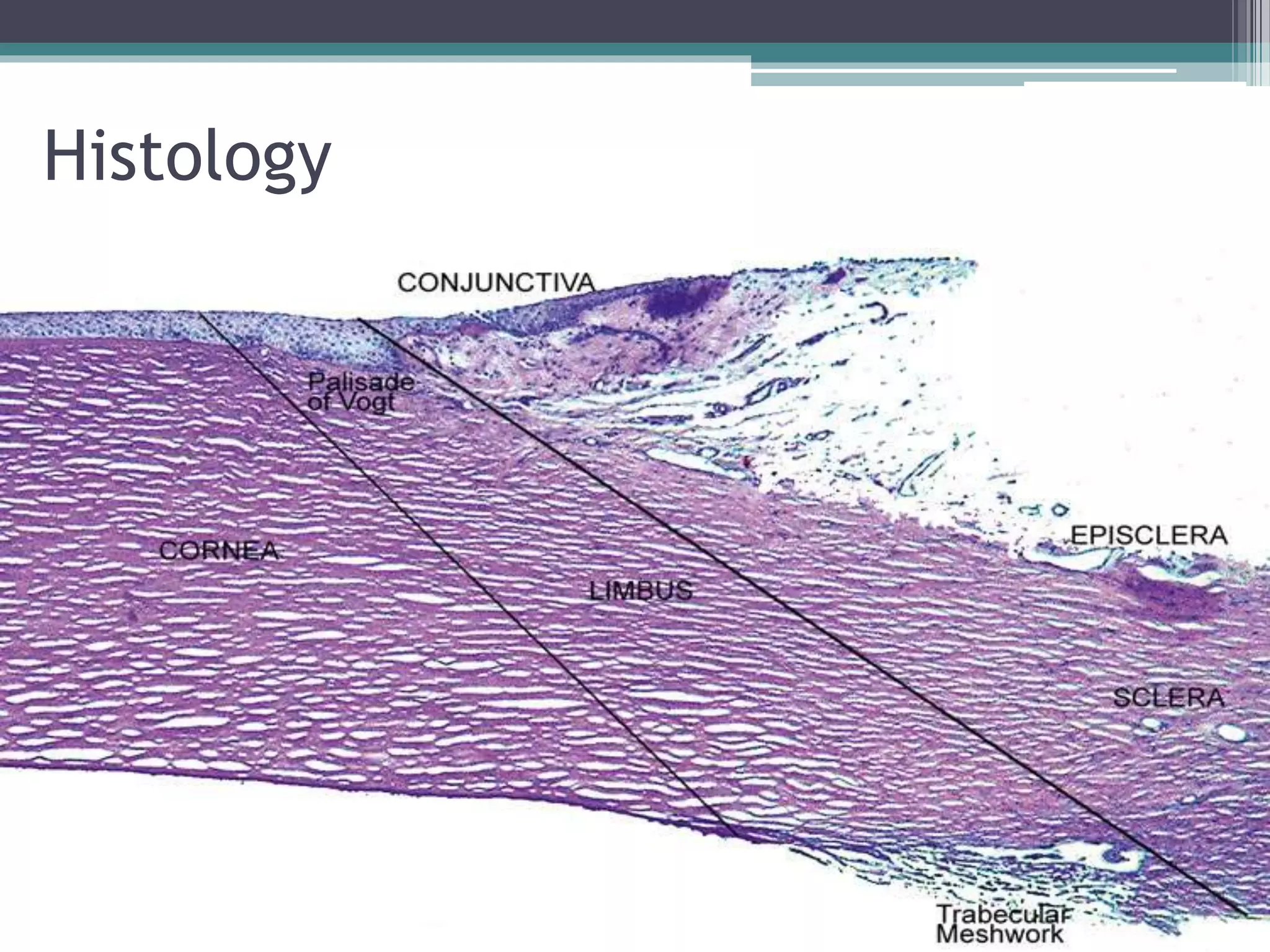
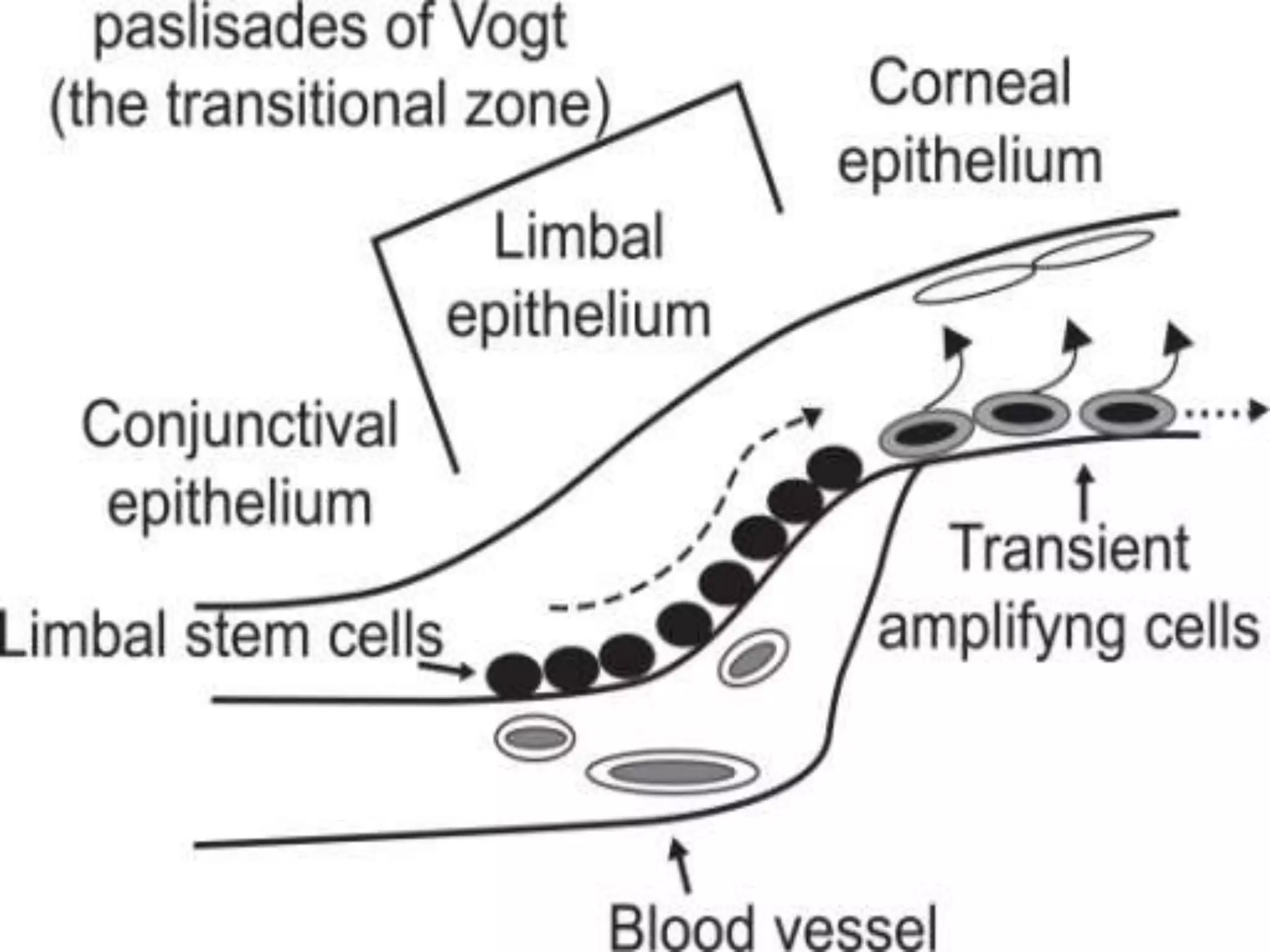
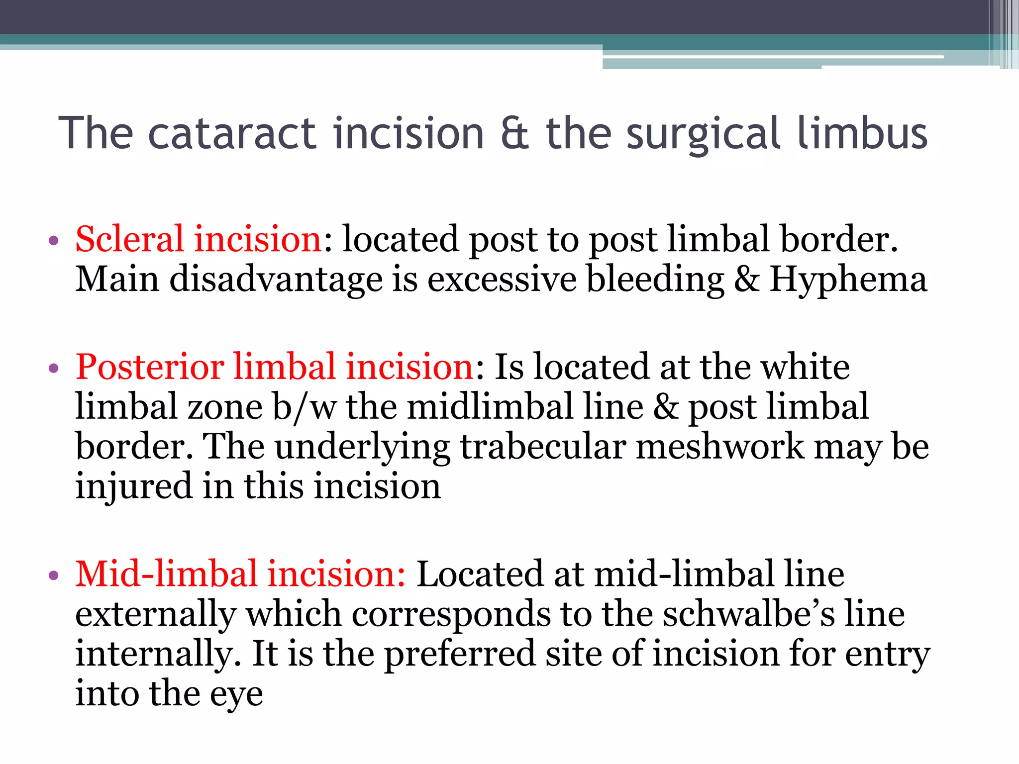
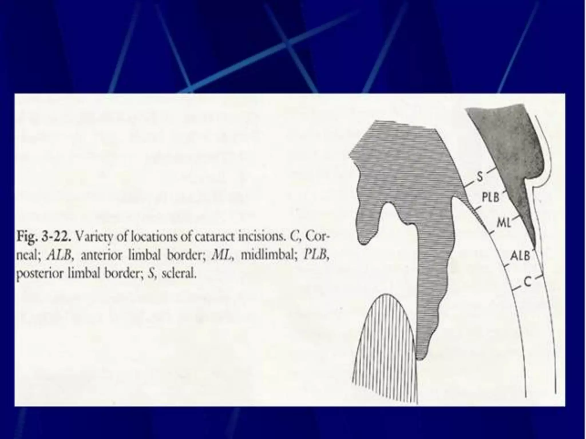
The Limbus
Anatomy and Surgical application
Dr.Mohammad Dmour- PGY1
Supervised by : Dr.Mohammad Bilal khalil
Ophthalmology Department
Islamic Hospital
LIMBUS, INC., a shadowy employment agency
that operates at the edge of the normal world.
• Eye (2007) Developmental Biology Unit, University College London Institute of
Child Health and Great Ormond Street Hospital for Children NHS Trust,
London, UK
Histology
Histology
Anatomical Limbus
• Anatomically, the limbus refers to a circumcorneal
transitional zone of the conjunctivocorneal and
the corneoscleral junction.
Anatomical Limbus
• Conjunctivocorneal junction:
1. At this point, the bulbar conjunctiva is firmly
adherent to the underlying structures.
2. The Substantia propria of the conjunctiva stops here
but its epithelium continues with that of the cornea.
3. At the transition zone, the epithelium becomes
several layers thick (10-12) and arranged irregularly
at the limbus.
• Sclerocorneal junction:
At this point, Transparent corneal lamellae become
continuous With the oblique, circular and opaque
fibres of sclera.
At Limbus…….
1. Corneal epithelium becomes bulbar
conjunctival epithelium
2. Bowman's membrane becomes continuous with
the lamina propria of the conjunctiva and
tenon's capsule.
3. Stroma becomes sclera
4. Descemet's membrane becomes schwalbe's line.
5. Endothelium lines the trabecular meshwork
and becomes continuous with the anterior
surface of the epithelium
Surgical Limbus
• Is a 2mm wide circumcorneal transitional
zone between clear cornea on one side and
the opaque sclera on the other side.
Surgical Limbus
• Anterior limbal border:
1. It is the ant boundary of surgical limbus seen
from exterior.
2. Marked by prominent ridge created by insertion
of the conjunctiva and tenons capsule into the
cornea.
3. It overlies the termination of the bowman’s
membranes.
Surgical Limbus
• Blue limbal zone:
1. is bluish translucent zone seen post to ant
limbal border.
2. Post to this bluish zone is the white sclera
3. Extent of blue limbal zone varies in different
quadrants(due to variable insertion of conj
&tenon’s capsule into the cornea)- 1mm in
supquad, 0.8mm in inf quad, 0.4mm in nasal
&temp quad.
Surgical Limbus
• Mid limbal line:
1. junction of blue zone with white area.
2. Overlies termination of DM(schwalbe’s line)
3. It is the most useful external landmark.
Surgical Limbus
• Posterior Limbal Border:
1. Lies 1mm post to mid-limbal line
2. Overlies scleral spur
3. Seen with sclerotic scatter illumination
4. Forms post border of surgical limbus
Surgical Limbus
• White limbal zone:
1. 1mm wide whitish area which lies b/w mid-
limbal line and the post limbal border
2. overlies the trabecular meshwork
Surgical Limbus
• Ant border….. overlies the termination of the
bowman’s membranes.
• Midlimbal ….. Overlies termination of
DM(schwalbe’s line)
• Post border….. Overlies scleral spur
• White zone……overlies the trabecular meshwork.
The cataract incision & the surgical limbus
• Scleral incision: located post to post limbal border.
Main disadvantage is excessive bleeding & Hyphema
• Posterior limbal incision: Is located at the white
limbal zone b/w the midlimbal line & post limbal
border. The underlying trabecular meshwork may be
injured in this incision
• Mid-limbal incision: Located at mid-limbal line
externally which corresponds to the schwalbe’s line
internally. It is the preferred site of incision for entry
into the eye
The cataract incision & the surgical limbus
• Anterior limbal incision: is located in blue
limbal zone and transverses the descemet’s
membrane & may cause stripping
• Clear corneal incision:
Located in front of the ant limbal border & is
associated with a high induced astigmatism
Greater chances of descemet’s stripping
Take Home Massage….
• The limbus forms the border between the
transparent cornea and opaque sclera, contains
the pathways of aqueous humour outflow, and is
the site of surgical incisions for cataract and
glaucoma.
• Limbal epithelium is thought to contain the
source of the stem cells (SC) that serve as the
source of corneal epithelial cell renewal.

The limbus

  • 1.
    The Limbus Anatomy andSurgical application Dr.Mohammad Dmour- PGY1 Supervised by : Dr.Mohammad Bilal khalil Ophthalmology Department Islamic Hospital
  • 3.
    LIMBUS, INC., ashadowy employment agency that operates at the edge of the normal world.
  • 5.
    • Eye (2007)Developmental Biology Unit, University College London Institute of Child Health and Great Ormond Street Hospital for Children NHS Trust, London, UK
  • 7.
  • 8.
  • 13.
    Anatomical Limbus • Anatomically,the limbus refers to a circumcorneal transitional zone of the conjunctivocorneal and the corneoscleral junction.
  • 14.
    Anatomical Limbus • Conjunctivocornealjunction: 1. At this point, the bulbar conjunctiva is firmly adherent to the underlying structures. 2. The Substantia propria of the conjunctiva stops here but its epithelium continues with that of the cornea. 3. At the transition zone, the epithelium becomes several layers thick (10-12) and arranged irregularly at the limbus. • Sclerocorneal junction: At this point, Transparent corneal lamellae become continuous With the oblique, circular and opaque fibres of sclera.
  • 18.
    At Limbus……. 1. Cornealepithelium becomes bulbar conjunctival epithelium 2. Bowman's membrane becomes continuous with the lamina propria of the conjunctiva and tenon's capsule. 3. Stroma becomes sclera 4. Descemet's membrane becomes schwalbe's line. 5. Endothelium lines the trabecular meshwork and becomes continuous with the anterior surface of the epithelium
  • 24.
    Surgical Limbus • Isa 2mm wide circumcorneal transitional zone between clear cornea on one side and the opaque sclera on the other side.
  • 26.
    Surgical Limbus • Anteriorlimbal border: 1. It is the ant boundary of surgical limbus seen from exterior. 2. Marked by prominent ridge created by insertion of the conjunctiva and tenons capsule into the cornea. 3. It overlies the termination of the bowman’s membranes.
  • 27.
    Surgical Limbus • Bluelimbal zone: 1. is bluish translucent zone seen post to ant limbal border. 2. Post to this bluish zone is the white sclera 3. Extent of blue limbal zone varies in different quadrants(due to variable insertion of conj &tenon’s capsule into the cornea)- 1mm in supquad, 0.8mm in inf quad, 0.4mm in nasal &temp quad.
  • 28.
    Surgical Limbus • Midlimbal line: 1. junction of blue zone with white area. 2. Overlies termination of DM(schwalbe’s line) 3. It is the most useful external landmark.
  • 29.
    Surgical Limbus • PosteriorLimbal Border: 1. Lies 1mm post to mid-limbal line 2. Overlies scleral spur 3. Seen with sclerotic scatter illumination 4. Forms post border of surgical limbus
  • 30.
    Surgical Limbus • Whitelimbal zone: 1. 1mm wide whitish area which lies b/w mid- limbal line and the post limbal border 2. overlies the trabecular meshwork
  • 31.
    Surgical Limbus • Antborder….. overlies the termination of the bowman’s membranes. • Midlimbal ….. Overlies termination of DM(schwalbe’s line) • Post border….. Overlies scleral spur • White zone……overlies the trabecular meshwork.
  • 36.
    The cataract incision& the surgical limbus • Scleral incision: located post to post limbal border. Main disadvantage is excessive bleeding & Hyphema • Posterior limbal incision: Is located at the white limbal zone b/w the midlimbal line & post limbal border. The underlying trabecular meshwork may be injured in this incision • Mid-limbal incision: Located at mid-limbal line externally which corresponds to the schwalbe’s line internally. It is the preferred site of incision for entry into the eye
  • 37.
    The cataract incision& the surgical limbus • Anterior limbal incision: is located in blue limbal zone and transverses the descemet’s membrane & may cause stripping • Clear corneal incision: Located in front of the ant limbal border & is associated with a high induced astigmatism Greater chances of descemet’s stripping
  • 42.
    Take Home Massage…. •The limbus forms the border between the transparent cornea and opaque sclera, contains the pathways of aqueous humour outflow, and is the site of surgical incisions for cataract and glaucoma. • Limbal epithelium is thought to contain the source of the stem cells (SC) that serve as the source of corneal epithelial cell renewal.