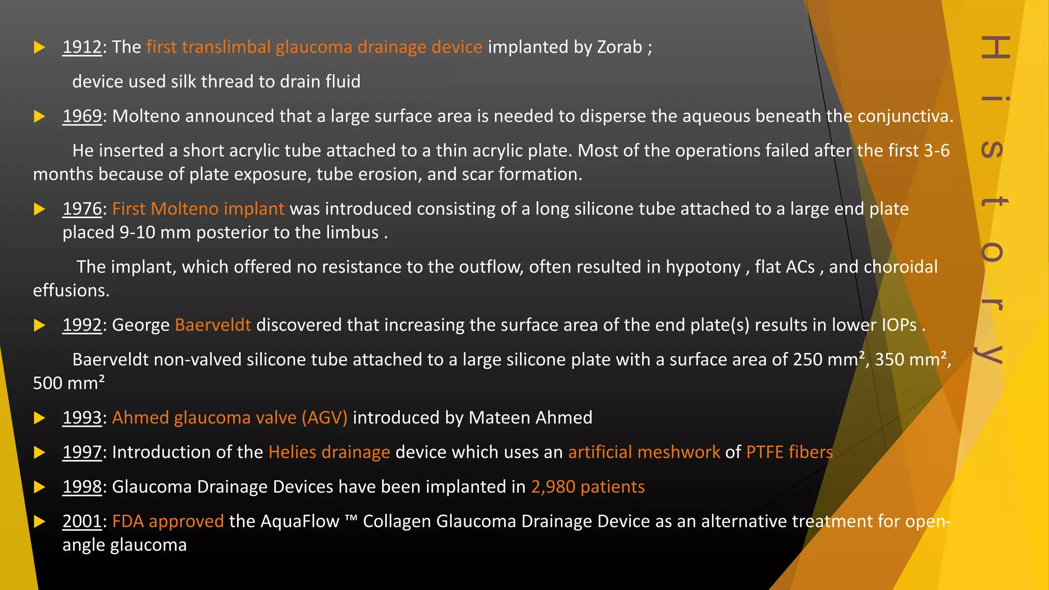
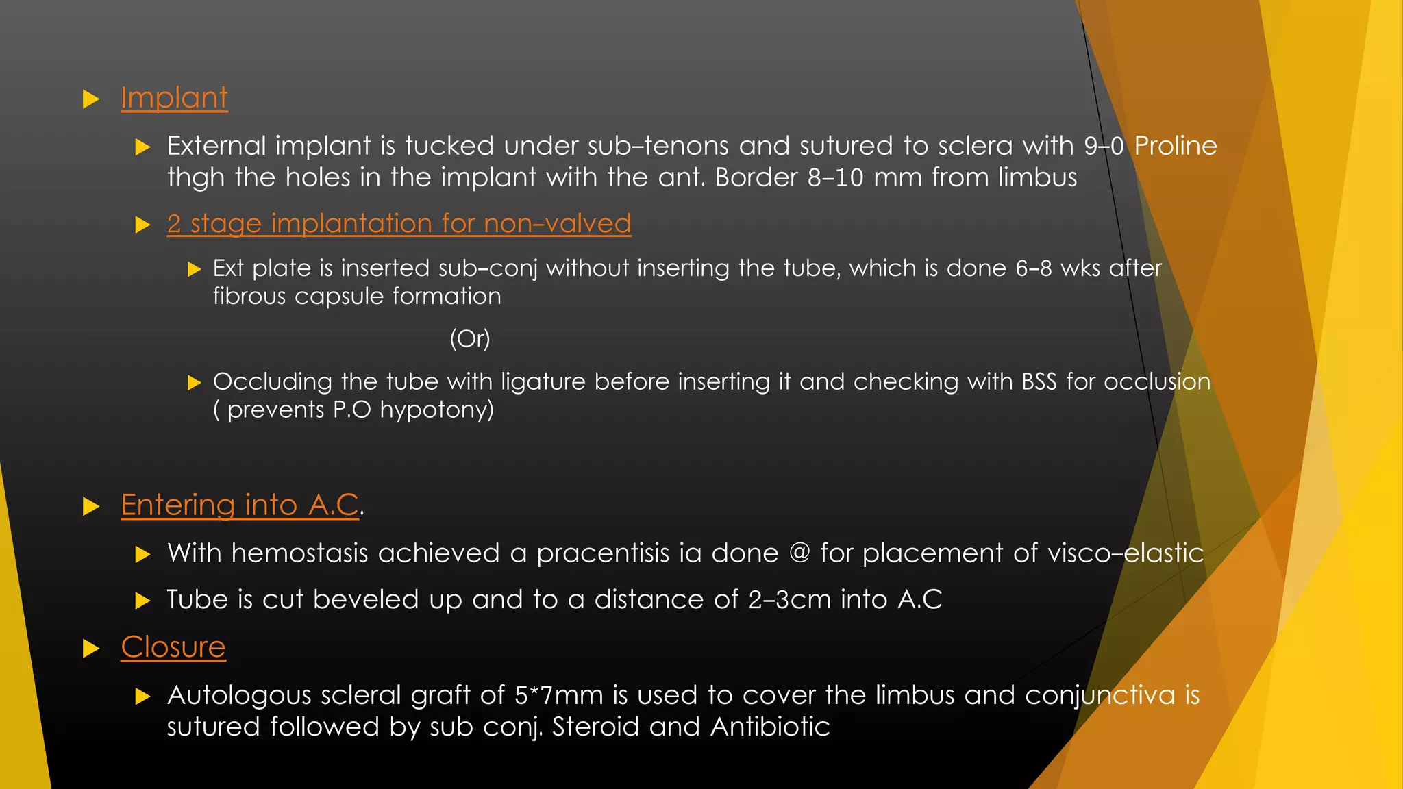
Glaucoma Drainage 
Devices 
Dr. Meenank 
M. S. Ophthalmology ( Post- Graduate) 
ASRAM
Introduction 
 Glaucoma – Multifactorial progressive optic neuropathy 
associated with visual field loss and characteristic structural 
changes, including thinning of the retinal nerve fibre layer and 
excavation of the optic nerve head 
 Elevated IOP is clearly the most frequent causative risk factor. 
 Choice of treatment is mainly 
 Medical Therapy 
 LASER Treatment 
 Filtering Surgeries 
 Choice of treatment in 
Refractory Glaucoma's is 
Glaucoma Drainage Devices 
(GDD)
Glaucoma Drainage Devices 
 Glaucoma drainage devices create an alternate aqueous pathway from the 
anterior chamber to the sub-conjunctival bleb or the supra-choroidal space 
by channeling aqueous out through a tube 
 Drainage Devices 
 Setons (Bristle) - Refers to solid shaft prevents wound apposition 
 Stunt (Tubular structure) - Fluid passes passively 
Valve - Tubular structure for unidirectional flow 
 Setons may be thread , wire , hair place in the wound to drain aq. Along side 
the inserted material 
 Setons were unsuccessful as they were unable to maintain fistula patency
Indications 
 GDD were designed mainly to enhance trabeculectomy surgeries 
 Indication 
 Trab. With adjuvant therapy failed 
 Young patients 
 NVG, uveitic glaucoma, refractive pediatric glaucoma, glaucoma in 
Aphakia, PPK, VRS, PRP 
 Sever conjunctival scarring
H i s t o r y 
 1912: The first translimbal glaucoma drainage device implanted by Zorab ; 
device used silk thread to drain fluid 
 1969: Molteno announced that a large surface area is needed to disperse the aqueous beneath the conjunctiva. 
He inserted a short acrylic tube attached to a thin acrylic plate. Most of the operations failed after the first 3-6 
months because of plate exposure, tube erosion, and scar formation. 
 1976: First Molteno implant was introduced consisting of a long silicone tube attached to a large end plate 
placed 9-10 mm posterior to the limbus . 
The implant, which offered no resistance to the outflow, often resulted in hypotony , flat ACs , and choroidal 
effusions. 
 1992: George Baerveldt discovered that increasing the surface area of the end plate(s) results in lower IOPs . 
Baerveldt non-valved silicone tube attached to a large silicone plate with a surface area of 250 mm², 350 mm², 
500 mm² 
 1993: Ahmed glaucoma valve (AGV) introduced by Mateen Ahmed 
 1997: Introduction of the Helies drainage device which uses an artificial meshwork of PTFE fibers 
 1998: Glaucoma Drainage Devices have been implanted in 2,980 patients 
 2001: FDA approved the AquaFlow ™ Collagen Glaucoma Drainage Device as an alternative treatment for open-angle 
glaucoma
Physiology 
 Basic design of drainage implant device shows 
 Fibrous capsule around effects the formation of bleb 
 Granulation leads to collagen capsule @ 1month and stable at 
6mnth all over 
 Show microcystic spaces for aq. Drainage 
Silicon tube from A.C 
to disc/plate 
Ridge- where tube is 
inserted. 
Dec. risk of obs. due 
to fibrous capsule 
Large surface area for 
formation of bleb
Implants 
 Implants can be studied under 
1. Type of plate 
2. Type of material 
3. Type of opening 
 Type of plate – of the available rigid and flexible plates the later 
causes less inflammation
Type of material 
 Most commonly used materials are 
 Silicon 
 Baerveldt, Krupin, Ahmed 
 Polypropylene 
 Ahmed, Molteno 
 Hydroxylapatite 
 Expandable polytetrafluoroethylene
 Medical grade silicone 
 Biocompatibility 
 Low tissue response to implantation 
 Odourless 
 Tasteless 
 Resistant to bacterial growth 
 Does not stain or corrode other materials 
 High tensile strength (1500psi), good elongation (to 1250%) 
and flexibility 
 Temperature Resistant 
 Stable at a temperature range of 75-500oC 
 Chemical Resistant 
 Resists water, oxidizing chemicals, ammonia, and isopropyl 
alcohol
 Polypropylene 
 High flexibility and dimensional stability 
 Low thrombogenicity 
 Poor abrasion resistance 
 Non-toxic 
 High tensile and compression strength 
 Chemically resistant to most alkalis and acids, organic 
solvents, degreasing agents, and electrolytic attack 
 Degrades in presence of UV light
 Polytetrafluoroethylene or PTFE 
 Chemically inert to most chemicals including nitric, sulfuric, 
and phosphoric acids 
 Hydrophobic 
 Highly crystalline and stable 
 Low friction 
 Low wear resistance 
 Inflammation caused by PTFE wear particles
Type of opening 
 Based on the type of opening implants can be classified into 
1. Non- valved 
 Rely on resistance formed by fibrous capsule or bleb 
 Bleb grows around the implant and creates a space for fluid 
to drain and be absorbed by surrounding tissue 
 Eg – Molteno, Baerveldt, Schocket 
1. Valved 
 Only drains fluid at a certain IOP 
 Valve opens and fluid is drained into a reservoir where it is 
absorbed by surrounding tissues 
 Eg – krupin, Ahmed
Non- valved devices 
 Baerveldt Implant 
 Plate features 
 barium impregnated 
 Large surface area – 250 mm², 350 mm²(less complication), 
500mm²(not available) 
 Location 
 Superior- temporal quadrant under the sup. rectus muscle 
 inserted thgh one quadrant conjunctival insertion 
 Fenestration seen on the plate to allow fibrous capsule 
formation and dec.ht. of bleb and diplopia 
 Indication – failed Trabeculectomy, cataract Sx 
 Side effects – restricted ocular movements @ 7yrs 
 Uncontrolled, complicated glaucoma
 Molteno (1969) 
 Original design – 
 Single acrylic thin plate ( 13 mm diameter & 135 mm² S.A) 
 Silicon tube (outer-0.62 mm , inner-0.30 mm) connected to 
upper end of plate 
 Thickened rim facilitating suturing to sclera adjacent to limbus
 With subsequent modification came 
 Single plate 
 Moved to a position a few millimetres away from limbus to allow for better 
drainage. 
 Aphakia, PPK, failed filters, NVG, < 3yr – 25% to 40% 
 Double plate 
 Inc. S.A. 270 mm, 
 Better control but inc. complication – hypotony 
 Dual chamber 
 In view to combat hypotony 
 Single implant ‘V’ shaped pressure ridge on upper surface of plate – inc. S.A. – 
10.5mm 
 Molteno 3 ( 175mm² & 230 mm²) 
 Shows a bowel shaped structure on the implant plate at the tube opening 
 Acts a biological valve limiting aq. Flow during low aq. production
 Schocket Tube 
 Anterior Chamber Tube Shunt To Encircling Band ( ACTSEB) 
 Contains a silastic tube used in nasolacrimal intubation encircling at 
the equator under the rectus band 
 Show max IOP effect when coupled with krapin
Flow-Restricted Drainage Devices / Valved 
 Ahmed Glaucoma Valve (AGV) 
 Most commonly used valved implant 
 Implant design – silicon tube connected to silicon sheathed 
valve held by a polypropylene body 
Material Surface Area Type 
Silicon 
FP7 1.9mm² Single valve plate 
FP8 96 mm² Small valve plate 
FX1 180mm² 2 plates 
Polypropylene 
S2 189mm² Single valve plate 
S3 96 mm² Small valve plate 
B1 180mm² 2 plates
 Valve related obstruction may cause hypertensive phase @ 4th – 8th P.O weeks 
 Hypertensive Phase : elevated IOP in the weeks to months after implantation as 
a result of capsule formation around the implant plate. This is frequently termed 
the hypertensive phase 
 Hypertensive phase is a result of holding the implant at no touch zone 
 No touch zone : area on the implant covering the silicon leaflet which when 
grasped with forceps separated the valve from implant – definitive closure – 
early P.O hypotony, fibro-vascular ingrowth b/w leaflet and implant failure
 Krupin Implants (1976) 
 Krupin and associates developed a concept of a valve that opens at a 
predetermined IOP to dec. early P.O. complication 
 Krupin Denver valve ( Original) 
 Internal supramid tube cemented to external silastic tube 
 Valve effect – by making a slit at closed ext. end of silastic which open opens at 9 – 10 
mmHg IOP 
 Failure – no ext plate, and fibrosis eventually closed the valve 
 Krupin eye valve with disc 
 Longer version of Denver and attached to Schocket 180 implant which open at 10 – 12 
mmHg IOP and valve @ in side the rim of plate
Other Drainage Devices 
 Ex-PRESS 
 Miniature non-valved device without an external plate 
 Made of rigid 316LVM stainless steel – same as cardiac stents 
 < 3mm long 
 Internal lumen size – 50μm/200μm 
 Biocompatible 
 Allows for the development of a delimited potential space and the 
formation of a fibrous capsule to create the resistance to outflow 
 Implanted under a traditional trabeculectomy flap
 The Solx Gold Shunt 
 Made from 24-karat gold and works to connect the anterior chamber and 
suprachoroidal space 
 Pressure in the suprachoroidal space serves as a natural counter pressure to prevent 
severe postoperative hypotony 
 Implanted by using an ab external approach so, no bleb 
 iStent trabecular microbypass stent 
 Stainless-steel stent with a lumen that is implanted from an ab interno approach 
 Traverses the trabecular meshwork and drains aqueous from the anterior chamber 
into the Schlemms canal, enhancing aqueous drainage
SURGICAL TECHNIQUES 
 Basic Principal 
1) Proper Traction – achieved by 6-0 vicryl / silk through superficial 
cornea at superior limbus 
2) Exposure Of Scleral Bed – a fornix based conjunctival flap is created 
and elevated by a blunt dissection b/w tenons and episcleral with 
blunt Westcott scissors exposing the scleral bed which is supported 
by relaxing incision for surgical exposure 
 In case of large / double plate avoid superior-nasal ? 
Can cause strabismus & close to optic nerve 
3) Isolation Of Recti – muscle hooks are used to isolate the 2 recti on 
either side of the surgical site
 Implant 
 External implant is tucked under sub-tenons and sutured to sclera with 9-0 Proline 
thgh the holes in the implant with the ant. Border 8-10 mm from limbus 
 2 stage implantation for non-valved 
 Ext plate is inserted sub-conj without inserting the tube, which is done 6-8 wks after 
fibrous capsule formation 
(Or) 
 Occluding the tube with ligature before inserting it and checking with BSS for occlusion 
( prevents P.O hypotony) 
 Entering into A.C. 
 With hemostasis achieved a pracentisis ia done @ for placement of visco-elastic 
 Tube is cut beveled up and to a distance of 2-3cm into A.C 
 Closure 
 Autologous scleral graft of 5*7mm is used to cover the limbus and conjunctiva is 
sutured followed by sub conj. Steroid and Antibiotic
COMPLICATIONS 
 Hypotony 
 m.c complication with non- valved in early P.O days until fibrous capsule 
formation 
 Rx – temporary obs of lumen 
 Basic tech – suture ligation of tube 
 Valve implants 
 Hypotony + flat A.C – Inject visco elastic – observe for 24hrs – no 
change – reposition device to prevent corneal decompensation 
 Late hypotony – Rx permenet occlusion of proximal tube and replacement 
of implant
 Elevated IOP ( early / late ) 
 Early – Before the ligature around the suture dissolves – transient ↑ IOP 
 Rx – medically / combine trab. Without MMC to control IOP 
 htn phase (7 – 8 days ) –↓ IOP conj & corneal oedema, cong⁺ around implant 
 HTN phase – IOP ass with fibrous capsule formation and ↓ oedema – bleb formation 
 1 – 4 wks – bleb cong⁺ ↑IOP - cong⁺ ↓IOP – stabiles 
 ↑IOP due to tube obs – fibrin, blood, iris, vitreous membranes, or silicone oil 
 Rx – reopening tube with – Iridectomy / NdYAG membrenctomy 
 Distal tube occlusion – scleral buckling 
 Rx – BSS irrigation / intra-cameral Tissue Plasminogen Activator to dissolve fibrin clot
 Late ↑IOP 
 Seen with patent tube in thickened fibrous capsule 
 Rx – needle revision ; 
 complication – endophthalmitis
 Migration, Extrusion, and Erosion 
 If not adequately secured 
 Migrate post. In to A.C – Rx)- reposition and re suture with 9-0 Proline 
 Ant Migration is due to dislocation 
 Extrusion – m.c in pediatric cases due to the development of eye ball 
 Can cause erosion of cornea 
 Avulsion – causes corneal melting and requires explantation of the implant 
and possibly corneal grafting 
 Endophthalmitis 
 commonly seen after needling procedure 
 Cause – Propionibacterium acnes endophthalmitis 
 Rx – vancomycin (poor response) 
Explantation with new devise instillation is a must
 Corneal Decompensation 
 Related to the retrograde flow from the encapsulated reservoir to the 
anterior chamber & Tube-cornea touch 
 Diplopia and Ocular Motility 
 devices with larger plates, implanted in the superonasal quadrant, can 
interrupt extraocular muscle function and cause strabismus and diplopia 
(exotropia, hypertropia, or limitation of ocular rotations ) 
 Rx)- repositioning the device to superior-nasal
Other complication 
 Epidermal down growth 
 With tube inserted at limbus – 
implant failure, corneal 
decompensation 
 Sterile hypopyon 
 After removal of suture stents in 
eyes with silicon oil 
 Irregular pupil 
 Root of iris adherent to tube 
 Globe penetration during 
suturing 
 Cause – RD, Vit Hx 
 Retinal Complications 
 RD, choroidal effusion, 
suprachoroidal Hx ( old age, 
HTN, C. effusion, atherosclerosis) 
 Vision loss 
 Hypotony, shallow A.C., RD, Vit 
Hx, CME

Glaucoma drainage devices

  • 1.
    Glaucoma Drainage Devices Dr. Meenank M. S. Ophthalmology ( Post- Graduate) ASRAM
  • 2.
    Introduction  Glaucoma– Multifactorial progressive optic neuropathy associated with visual field loss and characteristic structural changes, including thinning of the retinal nerve fibre layer and excavation of the optic nerve head  Elevated IOP is clearly the most frequent causative risk factor.  Choice of treatment is mainly  Medical Therapy  LASER Treatment  Filtering Surgeries  Choice of treatment in Refractory Glaucoma's is Glaucoma Drainage Devices (GDD)
  • 3.
    Glaucoma Drainage Devices  Glaucoma drainage devices create an alternate aqueous pathway from the anterior chamber to the sub-conjunctival bleb or the supra-choroidal space by channeling aqueous out through a tube  Drainage Devices  Setons (Bristle) - Refers to solid shaft prevents wound apposition  Stunt (Tubular structure) - Fluid passes passively Valve - Tubular structure for unidirectional flow  Setons may be thread , wire , hair place in the wound to drain aq. Along side the inserted material  Setons were unsuccessful as they were unable to maintain fistula patency
  • 4.
    Indications  GDDwere designed mainly to enhance trabeculectomy surgeries  Indication  Trab. With adjuvant therapy failed  Young patients  NVG, uveitic glaucoma, refractive pediatric glaucoma, glaucoma in Aphakia, PPK, VRS, PRP  Sever conjunctival scarring
  • 5.
    H i st o r y  1912: The first translimbal glaucoma drainage device implanted by Zorab ; device used silk thread to drain fluid  1969: Molteno announced that a large surface area is needed to disperse the aqueous beneath the conjunctiva. He inserted a short acrylic tube attached to a thin acrylic plate. Most of the operations failed after the first 3-6 months because of plate exposure, tube erosion, and scar formation.  1976: First Molteno implant was introduced consisting of a long silicone tube attached to a large end plate placed 9-10 mm posterior to the limbus . The implant, which offered no resistance to the outflow, often resulted in hypotony , flat ACs , and choroidal effusions.  1992: George Baerveldt discovered that increasing the surface area of the end plate(s) results in lower IOPs . Baerveldt non-valved silicone tube attached to a large silicone plate with a surface area of 250 mm², 350 mm², 500 mm²  1993: Ahmed glaucoma valve (AGV) introduced by Mateen Ahmed  1997: Introduction of the Helies drainage device which uses an artificial meshwork of PTFE fibers  1998: Glaucoma Drainage Devices have been implanted in 2,980 patients  2001: FDA approved the AquaFlow ™ Collagen Glaucoma Drainage Device as an alternative treatment for open-angle glaucoma
  • 6.
    Physiology  Basicdesign of drainage implant device shows  Fibrous capsule around effects the formation of bleb  Granulation leads to collagen capsule @ 1month and stable at 6mnth all over  Show microcystic spaces for aq. Drainage Silicon tube from A.C to disc/plate Ridge- where tube is inserted. Dec. risk of obs. due to fibrous capsule Large surface area for formation of bleb
  • 7.
    Implants  Implantscan be studied under 1. Type of plate 2. Type of material 3. Type of opening  Type of plate – of the available rigid and flexible plates the later causes less inflammation
  • 8.
    Type of material  Most commonly used materials are  Silicon  Baerveldt, Krupin, Ahmed  Polypropylene  Ahmed, Molteno  Hydroxylapatite  Expandable polytetrafluoroethylene
  • 9.
     Medical gradesilicone  Biocompatibility  Low tissue response to implantation  Odourless  Tasteless  Resistant to bacterial growth  Does not stain or corrode other materials  High tensile strength (1500psi), good elongation (to 1250%) and flexibility  Temperature Resistant  Stable at a temperature range of 75-500oC  Chemical Resistant  Resists water, oxidizing chemicals, ammonia, and isopropyl alcohol
  • 10.
     Polypropylene High flexibility and dimensional stability  Low thrombogenicity  Poor abrasion resistance  Non-toxic  High tensile and compression strength  Chemically resistant to most alkalis and acids, organic solvents, degreasing agents, and electrolytic attack  Degrades in presence of UV light
  • 11.
     Polytetrafluoroethylene orPTFE  Chemically inert to most chemicals including nitric, sulfuric, and phosphoric acids  Hydrophobic  Highly crystalline and stable  Low friction  Low wear resistance  Inflammation caused by PTFE wear particles
  • 12.
    Type of opening  Based on the type of opening implants can be classified into 1. Non- valved  Rely on resistance formed by fibrous capsule or bleb  Bleb grows around the implant and creates a space for fluid to drain and be absorbed by surrounding tissue  Eg – Molteno, Baerveldt, Schocket 1. Valved  Only drains fluid at a certain IOP  Valve opens and fluid is drained into a reservoir where it is absorbed by surrounding tissues  Eg – krupin, Ahmed
  • 13.
    Non- valved devices  Baerveldt Implant  Plate features  barium impregnated  Large surface area – 250 mm², 350 mm²(less complication), 500mm²(not available)  Location  Superior- temporal quadrant under the sup. rectus muscle  inserted thgh one quadrant conjunctival insertion  Fenestration seen on the plate to allow fibrous capsule formation and dec.ht. of bleb and diplopia  Indication – failed Trabeculectomy, cataract Sx  Side effects – restricted ocular movements @ 7yrs  Uncontrolled, complicated glaucoma
  • 14.
     Molteno (1969)  Original design –  Single acrylic thin plate ( 13 mm diameter & 135 mm² S.A)  Silicon tube (outer-0.62 mm , inner-0.30 mm) connected to upper end of plate  Thickened rim facilitating suturing to sclera adjacent to limbus
  • 15.
     With subsequentmodification came  Single plate  Moved to a position a few millimetres away from limbus to allow for better drainage.  Aphakia, PPK, failed filters, NVG, < 3yr – 25% to 40%  Double plate  Inc. S.A. 270 mm,  Better control but inc. complication – hypotony  Dual chamber  In view to combat hypotony  Single implant ‘V’ shaped pressure ridge on upper surface of plate – inc. S.A. – 10.5mm  Molteno 3 ( 175mm² & 230 mm²)  Shows a bowel shaped structure on the implant plate at the tube opening  Acts a biological valve limiting aq. Flow during low aq. production
  • 16.
     Schocket Tube  Anterior Chamber Tube Shunt To Encircling Band ( ACTSEB)  Contains a silastic tube used in nasolacrimal intubation encircling at the equator under the rectus band  Show max IOP effect when coupled with krapin
  • 17.
    Flow-Restricted Drainage Devices/ Valved  Ahmed Glaucoma Valve (AGV)  Most commonly used valved implant  Implant design – silicon tube connected to silicon sheathed valve held by a polypropylene body Material Surface Area Type Silicon FP7 1.9mm² Single valve plate FP8 96 mm² Small valve plate FX1 180mm² 2 plates Polypropylene S2 189mm² Single valve plate S3 96 mm² Small valve plate B1 180mm² 2 plates
  • 18.
     Valve relatedobstruction may cause hypertensive phase @ 4th – 8th P.O weeks  Hypertensive Phase : elevated IOP in the weeks to months after implantation as a result of capsule formation around the implant plate. This is frequently termed the hypertensive phase  Hypertensive phase is a result of holding the implant at no touch zone  No touch zone : area on the implant covering the silicon leaflet which when grasped with forceps separated the valve from implant – definitive closure – early P.O hypotony, fibro-vascular ingrowth b/w leaflet and implant failure
  • 19.
     Krupin Implants(1976)  Krupin and associates developed a concept of a valve that opens at a predetermined IOP to dec. early P.O. complication  Krupin Denver valve ( Original)  Internal supramid tube cemented to external silastic tube  Valve effect – by making a slit at closed ext. end of silastic which open opens at 9 – 10 mmHg IOP  Failure – no ext plate, and fibrosis eventually closed the valve  Krupin eye valve with disc  Longer version of Denver and attached to Schocket 180 implant which open at 10 – 12 mmHg IOP and valve @ in side the rim of plate
  • 20.
    Other Drainage Devices  Ex-PRESS  Miniature non-valved device without an external plate  Made of rigid 316LVM stainless steel – same as cardiac stents  < 3mm long  Internal lumen size – 50μm/200μm  Biocompatible  Allows for the development of a delimited potential space and the formation of a fibrous capsule to create the resistance to outflow  Implanted under a traditional trabeculectomy flap
  • 21.
     The SolxGold Shunt  Made from 24-karat gold and works to connect the anterior chamber and suprachoroidal space  Pressure in the suprachoroidal space serves as a natural counter pressure to prevent severe postoperative hypotony  Implanted by using an ab external approach so, no bleb  iStent trabecular microbypass stent  Stainless-steel stent with a lumen that is implanted from an ab interno approach  Traverses the trabecular meshwork and drains aqueous from the anterior chamber into the Schlemms canal, enhancing aqueous drainage
  • 22.
    SURGICAL TECHNIQUES Basic Principal 1) Proper Traction – achieved by 6-0 vicryl / silk through superficial cornea at superior limbus 2) Exposure Of Scleral Bed – a fornix based conjunctival flap is created and elevated by a blunt dissection b/w tenons and episcleral with blunt Westcott scissors exposing the scleral bed which is supported by relaxing incision for surgical exposure  In case of large / double plate avoid superior-nasal ? Can cause strabismus & close to optic nerve 3) Isolation Of Recti – muscle hooks are used to isolate the 2 recti on either side of the surgical site
  • 23.
     Implant External implant is tucked under sub-tenons and sutured to sclera with 9-0 Proline thgh the holes in the implant with the ant. Border 8-10 mm from limbus  2 stage implantation for non-valved  Ext plate is inserted sub-conj without inserting the tube, which is done 6-8 wks after fibrous capsule formation (Or)  Occluding the tube with ligature before inserting it and checking with BSS for occlusion ( prevents P.O hypotony)  Entering into A.C.  With hemostasis achieved a pracentisis ia done @ for placement of visco-elastic  Tube is cut beveled up and to a distance of 2-3cm into A.C  Closure  Autologous scleral graft of 5*7mm is used to cover the limbus and conjunctiva is sutured followed by sub conj. Steroid and Antibiotic
  • 24.
    COMPLICATIONS  Hypotony  m.c complication with non- valved in early P.O days until fibrous capsule formation  Rx – temporary obs of lumen  Basic tech – suture ligation of tube  Valve implants  Hypotony + flat A.C – Inject visco elastic – observe for 24hrs – no change – reposition device to prevent corneal decompensation  Late hypotony – Rx permenet occlusion of proximal tube and replacement of implant
  • 25.
     Elevated IOP( early / late )  Early – Before the ligature around the suture dissolves – transient ↑ IOP  Rx – medically / combine trab. Without MMC to control IOP  htn phase (7 – 8 days ) –↓ IOP conj & corneal oedema, cong⁺ around implant  HTN phase – IOP ass with fibrous capsule formation and ↓ oedema – bleb formation  1 – 4 wks – bleb cong⁺ ↑IOP - cong⁺ ↓IOP – stabiles  ↑IOP due to tube obs – fibrin, blood, iris, vitreous membranes, or silicone oil  Rx – reopening tube with – Iridectomy / NdYAG membrenctomy  Distal tube occlusion – scleral buckling  Rx – BSS irrigation / intra-cameral Tissue Plasminogen Activator to dissolve fibrin clot
  • 26.
     Late ↑IOP  Seen with patent tube in thickened fibrous capsule  Rx – needle revision ;  complication – endophthalmitis
  • 27.
     Migration, Extrusion,and Erosion  If not adequately secured  Migrate post. In to A.C – Rx)- reposition and re suture with 9-0 Proline  Ant Migration is due to dislocation  Extrusion – m.c in pediatric cases due to the development of eye ball  Can cause erosion of cornea  Avulsion – causes corneal melting and requires explantation of the implant and possibly corneal grafting  Endophthalmitis  commonly seen after needling procedure  Cause – Propionibacterium acnes endophthalmitis  Rx – vancomycin (poor response) Explantation with new devise instillation is a must
  • 28.
     Corneal Decompensation  Related to the retrograde flow from the encapsulated reservoir to the anterior chamber & Tube-cornea touch  Diplopia and Ocular Motility  devices with larger plates, implanted in the superonasal quadrant, can interrupt extraocular muscle function and cause strabismus and diplopia (exotropia, hypertropia, or limitation of ocular rotations )  Rx)- repositioning the device to superior-nasal
  • 29.
    Other complication Epidermal down growth  With tube inserted at limbus – implant failure, corneal decompensation  Sterile hypopyon  After removal of suture stents in eyes with silicon oil  Irregular pupil  Root of iris adherent to tube  Globe penetration during suturing  Cause – RD, Vit Hx  Retinal Complications  RD, choroidal effusion, suprachoroidal Hx ( old age, HTN, C. effusion, atherosclerosis)  Vision loss  Hypotony, shallow A.C., RD, Vit Hx, CME