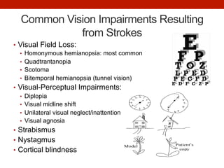
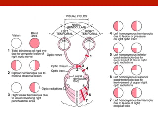
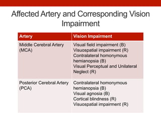
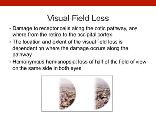
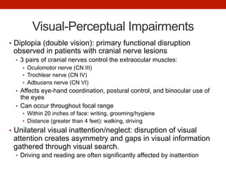
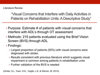
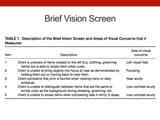
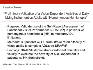
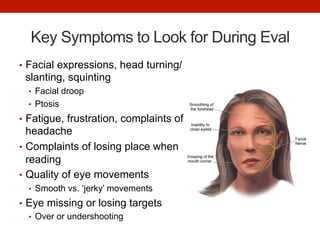
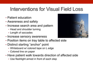
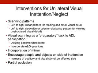
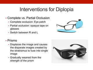
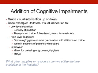
VISION DEFICITS
POST-STROKE
INCORPORATING VISION INTO INPATIENT
REHAB
Shannon Corcoran, OTS
Loma Linda University
Stanford Hospital
March 25, 2016
Guiding Research Question
Does the addition of visual scanning interventions
during OT treatment sessions improve functional
independence with ADL’s in patients recovering
from a stroke?
OT Practice Framework
Client Factors: Visual functions
“Quality of vision, visual acuity, visual stability, and visual field
functions to promote visual awareness of environment at various
distances for functioning”
Cerebrovascular Accidents (Strokes)
•  Acute neurologic dysfunction caused by a lesion in the
brain due to insufficient blood flow to the brain
•  Can occur via two mechanisms
•  Ischemic stroke: caused by blockage or clot of a blood vessel
•  ~87% of all strokes
•  Hemorrhagic stroke: bleeding in the brain
•  ~13%
•  Upper motor neuron dysfunction produces hemiplegia
(paralysis) of one side of the body contralateral to
hemisphere of brain with lesion
*Approximately what percent of stroke patients experience
some form of visual impairment?*
Common Vision Impairments Resulting
from Strokes
•  Visual Field Loss:
•  Homonymous hemianopsia: most common
•  Quadtrantanopia
•  Scotoma
•  Bitemporal hemianopsia (tunnel vision)
•  Visual-Perceptual Impairments:
•  Diplopia
•  Visual midline shift
•  Unilateral visual neglect/inattention
•  Visual agnosia
•  Strabismus
•  Nystagmus
•  Cortical blindness
Affected Artery and Corresponding Vision
Impairment
Artery Vision Impairment
Middle Cerebral Artery
(MCA)
Visual field impairment (B)
Visuospatial impairment (R)
Contralateral homonymous
hemianopsia (B)
Visual Perceptual and Unilateral
Neglect (R)
Posterior Cerebral Artery
(PCA)
Contralateral homonymous
hemianopsia (B)
Visual agnosia (B)
Cortical blindness (R)
Visuospatial impairment (R)
Visual Field Loss
•  Damage to receptor cells along the optic pathway, any
where from the retina to the occipital cortex
•  The location and extent of the visual field loss is
dependent on where the damage occurs along the
pathway
•  Homonymous hemianopsia: loss of half of the field of view
on the same side in both eyes
Visual-Perceptual Impairments
•  Diplopia (double vision): primary functional disruption
observed in patients with cranial nerve lesions
•  3 pairs of cranial nerves control the extraocular muscles:
•  Oculomotor nerve (CN III)
•  Trochlear nerve (CN IV)
•  Adbucens nerve (CN VI)
•  Affects eye-hand coordination, postural control, and binocular use of
the eyes
•  Can occur throughout focal range
•  Within 20 inches of face: writing, grooming/hygiene
•  Distance (greater than 4 feet): walking, driving
•  Unilateral visual inattention/neglect: disruption of visual
attention creates asymmetry and gaps in visual information
gathered through visual search.
•  Driving and reading are often significantly affected by inattention
“Visual Concerns that Interfere with DailyActivities in
Patients on Rehabilitation Units:ADescriptive Study”
•  Purpose: Estimate # of patients with visual concerns that
interfere with ADL’s through OT assessment
•  Methods: 215 patients evaluated using the Brief Vision
Screen (BVS) through ADL
•  Findings:
•  Largest proportion of patients (55%) with visual concerns were
diagnosed with stroke.
•  Results consistent with previous literature which suggests visual
impairment is common among patients in rehabilitation units
•  Further validation of the BVS is needed
Literature Review
(Grider, S.L, Yuen, H.K., Vogtle, L.K, & Warren, M, 2014)
Brief Vision Screen
“Preliminary Validation of a Vision-DependentActivities of Daily
Living Instrument onAdults with Homonymous Hemianopia”
•  Purpose: Validate use of the Self-Report Assessment of
Functional Visual Performance (SRAFVP) in patients w/
homonymous hemianopia (HH) to measure ADL
limitations
•  Methods: 30 patients w/ HH from stroke rated difficulty of
visual ability to complete ADL’s on SRAFVP
•  Findings: SRAFVP demonstrates sufficient reliability and
validity to evaluate the severity of ADL impairment in
patients w/ HH from stroke.
Literature Review
(Mennem, T. A., Warren, M., & Yuen, H. K., 2012)
“Compensatory Strategies Following Visual Search
Training in Patients with Homonymous Hemianopia: An
Eye Movement Study”
•  Purpose: Characterize changes in oculomotor scanning
with addition of practiced visual search in patients with
homonymous visual field defect.
•  Methods: 31 patients performed visual search training for
20 sessions over 4 weeks
•  Findings: Post training, patients improved search
efficiency and quickness with locating targets within their
hemispace
Literature Review
(Mannan, S., Pambakian, A., & Kennard, C., 2010)
Key Symptoms to Look for During Eval
•  Facial expressions, head turning/
slanting, squinting
•  Facial droop
•  Ptosis
•  Fatigue, frustration, complaints of
headache
•  Complaints of losing place when
reading
•  Quality of eye movements
•  Smooth vs. ‘jerky’ movements
•  Eye missing or losing targets
•  Over or undershooting
Interventions for Visual Field Loss
•  Patient education
•  Awareness and safety
•  Increase search area and pattern
•  Head and shoulder turning
•  Length of saccades
•  Increase sensory awareness
•  Position items on tray table to affected side
•  Distinct starting “anchor” point
•  Whiteboard w/ colored tape on L edge
•  Colored line on paper
•  Have patient walk towards direction of affected side
•  Use flashlight aimed in front of each step
Interventions for Unilateral Visual
Inattention/Neglect
•  Scanning patterns
•  Left to right linear pattern for reading and small visual detail
•  Left to right clockwise or counter-clockwise pattern for viewing
unstructured visual details
•  Visual scanning as a “preparatory” task to ADL
participation
•  Utilizing patients whiteboard
•  Incorporate A&O questions
•  Incorporation of mirror
•  Encourage people and objects on side of inattention
•  Increase of auditory and visual stimuli on affected side
•  Partial occlusion
Interventions for Diplopia
•  Complete vs. Partial Occlusion
•  Complete occlusion: Eye patch
•  Partial occlusion: opaque tape on
glasses
•  Switch between R and L
•  Prisms
•  Displaces the image and causes
the disparate images created by
the strabismus to fuse into single
image
•  Gradually weaned from the
strength of the prism
Addition of Cognitive Impairments
•  Grade visual intervention up or down
•  Case example: Unilateral visual inattention to L
•  Low level cognition:
•  Sensory stimulation
•  Therapist on L side: follow hand, reach for washcloth
•  High level cognition:
•  Grooming/hygiene or meal preparation with all items on L side
•  Write in sections of patient’s whiteboard
•  In between
•  Mirror for dressing or grooming/hygiene
•  MoCA
What other supplies or resources can we utilize that are
available in the hospital?
Looking ahead…
•  Utilize resources:
•  Outpatient neuro rehabilitation
•  Referral/recommendation to vision care specialists
•  Optometrist
•  Ophthalmologist
•  Low vision specialist
•  Literature Review: “Impact of visual impairment assessment
on functional recovery in stroke patients”
•  Quantitative findings: Visual assessment did not influence functional
recovery
•  Qualitative findings: Perceived benefits noted from the vision
assessment service
•  Continued research on BVP and SRAVFP assessments
Conclusion
•  Factors impacting conclusion/answer of research
question…
•  Patient’s short length of stay and limited interactions
•  Standardized Vision Assessments
•  Complex patient cases including impaired cognition and
communication
References
•  Grider, S.L, Yuen, H.K., Vogtle, L.K, & Warren, M. (2014). Visual concerns that interfere with
daily activities in patients on rehabilitation units: A descriptive study. Occupational Therapy
in Health Care, 28(4), 362-370. doi:10.3109/07380577.2014.933946
•  Hillier, R., & Tarbutton, N. M. (2014). Vision deficits following stroke: Implications for
occupational therapy practice. OT Practice 19(21), 13–16.
•  Jarvis, K., Grant, E., Rowe, F., Evans, J., & Cristino-Amenos, M. (2012). Impact of visual
impairment assessment on functional recovery in stroke patients: a pilot randomized
controlled trial. International Journal of Therapy and Rehabilitation. 19(1), 11-22.
•  Mannan, S., Pambakian, A., & Kennard, C. (2010). Compensatory strategies following visual
search training in patients with homonymous hemianopia: An eye movement study. Journal
of Neurology, 257, 1812–1821.
•  Mennem, T. A., Warren, M., & Yuen, H. K. (2012). Preliminary validation of a vision-
dependent activities of daily living instrument on adults with homonymous hemianopia.
American Journal of Occupational Therapy, 66, 478–482.
http://dx.doi.org/10.5014/ajot.2012.004762
•  Pendleton, H. M., & Schultz-Krohn, W. (2013). Pedretti’s occupational therapy: Practice
skills for physical dysfunction (7th ed.), St. Louis, MO: Elsevier.
•  Smith-Gabai, H. (2011). Occupational therapy in acute care. Bethesda, MD: The American
Occupational Therapy Association, Inc.
THANK YOU!

Vision Deficits Post-Stroke In-Service

  • 1.
    VISION DEFICITS POST-STROKE INCORPORATING VISIONINTO INPATIENT REHAB Shannon Corcoran, OTS Loma Linda University Stanford Hospital March 25, 2016
  • 2.
    Guiding Research Question Doesthe addition of visual scanning interventions during OT treatment sessions improve functional independence with ADL’s in patients recovering from a stroke? OT Practice Framework Client Factors: Visual functions “Quality of vision, visual acuity, visual stability, and visual field functions to promote visual awareness of environment at various distances for functioning”
  • 3.
    Cerebrovascular Accidents (Strokes) • Acute neurologic dysfunction caused by a lesion in the brain due to insufficient blood flow to the brain •  Can occur via two mechanisms •  Ischemic stroke: caused by blockage or clot of a blood vessel •  ~87% of all strokes •  Hemorrhagic stroke: bleeding in the brain •  ~13% •  Upper motor neuron dysfunction produces hemiplegia (paralysis) of one side of the body contralateral to hemisphere of brain with lesion *Approximately what percent of stroke patients experience some form of visual impairment?*
  • 4.
    Common Vision ImpairmentsResulting from Strokes •  Visual Field Loss: •  Homonymous hemianopsia: most common •  Quadtrantanopia •  Scotoma •  Bitemporal hemianopsia (tunnel vision) •  Visual-Perceptual Impairments: •  Diplopia •  Visual midline shift •  Unilateral visual neglect/inattention •  Visual agnosia •  Strabismus •  Nystagmus •  Cortical blindness
  • 6.
    Affected Artery andCorresponding Vision Impairment Artery Vision Impairment Middle Cerebral Artery (MCA) Visual field impairment (B) Visuospatial impairment (R) Contralateral homonymous hemianopsia (B) Visual Perceptual and Unilateral Neglect (R) Posterior Cerebral Artery (PCA) Contralateral homonymous hemianopsia (B) Visual agnosia (B) Cortical blindness (R) Visuospatial impairment (R)
  • 7.
    Visual Field Loss • Damage to receptor cells along the optic pathway, any where from the retina to the occipital cortex •  The location and extent of the visual field loss is dependent on where the damage occurs along the pathway •  Homonymous hemianopsia: loss of half of the field of view on the same side in both eyes
  • 8.
    Visual-Perceptual Impairments •  Diplopia(double vision): primary functional disruption observed in patients with cranial nerve lesions •  3 pairs of cranial nerves control the extraocular muscles: •  Oculomotor nerve (CN III) •  Trochlear nerve (CN IV) •  Adbucens nerve (CN VI) •  Affects eye-hand coordination, postural control, and binocular use of the eyes •  Can occur throughout focal range •  Within 20 inches of face: writing, grooming/hygiene •  Distance (greater than 4 feet): walking, driving •  Unilateral visual inattention/neglect: disruption of visual attention creates asymmetry and gaps in visual information gathered through visual search. •  Driving and reading are often significantly affected by inattention
  • 9.
    “Visual Concerns thatInterfere with DailyActivities in Patients on Rehabilitation Units:ADescriptive Study” •  Purpose: Estimate # of patients with visual concerns that interfere with ADL’s through OT assessment •  Methods: 215 patients evaluated using the Brief Vision Screen (BVS) through ADL •  Findings: •  Largest proportion of patients (55%) with visual concerns were diagnosed with stroke. •  Results consistent with previous literature which suggests visual impairment is common among patients in rehabilitation units •  Further validation of the BVS is needed Literature Review (Grider, S.L, Yuen, H.K., Vogtle, L.K, & Warren, M, 2014)
  • 10.
  • 11.
    “Preliminary Validation ofa Vision-DependentActivities of Daily Living Instrument onAdults with Homonymous Hemianopia” •  Purpose: Validate use of the Self-Report Assessment of Functional Visual Performance (SRAFVP) in patients w/ homonymous hemianopia (HH) to measure ADL limitations •  Methods: 30 patients w/ HH from stroke rated difficulty of visual ability to complete ADL’s on SRAFVP •  Findings: SRAFVP demonstrates sufficient reliability and validity to evaluate the severity of ADL impairment in patients w/ HH from stroke. Literature Review (Mennem, T. A., Warren, M., & Yuen, H. K., 2012)
  • 12.
    “Compensatory Strategies FollowingVisual Search Training in Patients with Homonymous Hemianopia: An Eye Movement Study” •  Purpose: Characterize changes in oculomotor scanning with addition of practiced visual search in patients with homonymous visual field defect. •  Methods: 31 patients performed visual search training for 20 sessions over 4 weeks •  Findings: Post training, patients improved search efficiency and quickness with locating targets within their hemispace Literature Review (Mannan, S., Pambakian, A., & Kennard, C., 2010)
  • 13.
    Key Symptoms toLook for During Eval •  Facial expressions, head turning/ slanting, squinting •  Facial droop •  Ptosis •  Fatigue, frustration, complaints of headache •  Complaints of losing place when reading •  Quality of eye movements •  Smooth vs. ‘jerky’ movements •  Eye missing or losing targets •  Over or undershooting
  • 14.
    Interventions for VisualField Loss •  Patient education •  Awareness and safety •  Increase search area and pattern •  Head and shoulder turning •  Length of saccades •  Increase sensory awareness •  Position items on tray table to affected side •  Distinct starting “anchor” point •  Whiteboard w/ colored tape on L edge •  Colored line on paper •  Have patient walk towards direction of affected side •  Use flashlight aimed in front of each step
  • 15.
    Interventions for UnilateralVisual Inattention/Neglect •  Scanning patterns •  Left to right linear pattern for reading and small visual detail •  Left to right clockwise or counter-clockwise pattern for viewing unstructured visual details •  Visual scanning as a “preparatory” task to ADL participation •  Utilizing patients whiteboard •  Incorporate A&O questions •  Incorporation of mirror •  Encourage people and objects on side of inattention •  Increase of auditory and visual stimuli on affected side •  Partial occlusion
  • 16.
    Interventions for Diplopia • Complete vs. Partial Occlusion •  Complete occlusion: Eye patch •  Partial occlusion: opaque tape on glasses •  Switch between R and L •  Prisms •  Displaces the image and causes the disparate images created by the strabismus to fuse into single image •  Gradually weaned from the strength of the prism
  • 17.
    Addition of CognitiveImpairments •  Grade visual intervention up or down •  Case example: Unilateral visual inattention to L •  Low level cognition: •  Sensory stimulation •  Therapist on L side: follow hand, reach for washcloth •  High level cognition: •  Grooming/hygiene or meal preparation with all items on L side •  Write in sections of patient’s whiteboard •  In between •  Mirror for dressing or grooming/hygiene •  MoCA What other supplies or resources can we utilize that are available in the hospital?
  • 18.
    Looking ahead… •  Utilizeresources: •  Outpatient neuro rehabilitation •  Referral/recommendation to vision care specialists •  Optometrist •  Ophthalmologist •  Low vision specialist •  Literature Review: “Impact of visual impairment assessment on functional recovery in stroke patients” •  Quantitative findings: Visual assessment did not influence functional recovery •  Qualitative findings: Perceived benefits noted from the vision assessment service •  Continued research on BVP and SRAVFP assessments
  • 19.
    Conclusion •  Factors impactingconclusion/answer of research question… •  Patient’s short length of stay and limited interactions •  Standardized Vision Assessments •  Complex patient cases including impaired cognition and communication
  • 20.
    References •  Grider, S.L,Yuen, H.K., Vogtle, L.K, & Warren, M. (2014). Visual concerns that interfere with daily activities in patients on rehabilitation units: A descriptive study. Occupational Therapy in Health Care, 28(4), 362-370. doi:10.3109/07380577.2014.933946 •  Hillier, R., & Tarbutton, N. M. (2014). Vision deficits following stroke: Implications for occupational therapy practice. OT Practice 19(21), 13–16. •  Jarvis, K., Grant, E., Rowe, F., Evans, J., & Cristino-Amenos, M. (2012). Impact of visual impairment assessment on functional recovery in stroke patients: a pilot randomized controlled trial. International Journal of Therapy and Rehabilitation. 19(1), 11-22. •  Mannan, S., Pambakian, A., & Kennard, C. (2010). Compensatory strategies following visual search training in patients with homonymous hemianopia: An eye movement study. Journal of Neurology, 257, 1812–1821. •  Mennem, T. A., Warren, M., & Yuen, H. K. (2012). Preliminary validation of a vision- dependent activities of daily living instrument on adults with homonymous hemianopia. American Journal of Occupational Therapy, 66, 478–482. http://dx.doi.org/10.5014/ajot.2012.004762 •  Pendleton, H. M., & Schultz-Krohn, W. (2013). Pedretti’s occupational therapy: Practice skills for physical dysfunction (7th ed.), St. Louis, MO: Elsevier. •  Smith-Gabai, H. (2011). Occupational therapy in acute care. Bethesda, MD: The American Occupational Therapy Association, Inc.
  • 21.