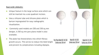
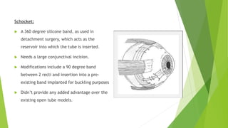
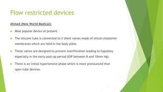
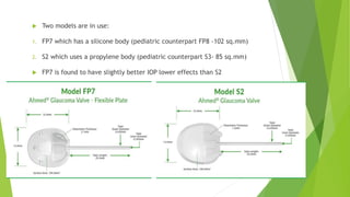
2. Introduction
Long history of various devices designed to facilitate aqueous flow out of the
anterior chamber to control IOP.
Setons: solid threads, wires or hairs placed to drain aqueous, which was
allowed to run alongside these structures.
In 1906, Rollet used a horse hair; and in 1912, Zorab used a silk thread.
In 1969, Molteno devised the first proper glaucoma drainage device (GDD).
3. Physiology
Consist of a silicone tube extending from the anterior chamber(AC), to a
plate, disc or ring beneath the conjunctiva and Tenons capsule
Mechanism:
After insertion,
collagenous capsule
forms around it
Surrounded by a
granulomatous
reaction
Reactio
n
resolves
in 4
months
Capsule
remains
stable
Matures
over
time,
becomes
thinner
after 6
months
Filtering
bleb
4. Ouflow α Surface area of the implant.
Outflow α 1/Bleb Diameter. This is because the surface tension increases as
diameter increases, which in turn will decrease the outflow. Same goes for
the bleb thickness.
The edge of the external plate usually has a ridge, through which the tube is
inserted. This decreases the chances of obstruction of the posterior opening
of the tube.
5. Indications
Generally reserved for cases wherein filtering surgery has failed or is likely to
fail.
Recent studies have revealed its use as a primary procedure in specific
conditions.
Classically considered in the following scenarios:
1. Failed trabeculectomies with antifibrotics
2. Active uveitis
3. Neovascular glaucoma
4. Inadequate conjunctiva
5. Aphakia
6. Implant designs
Subdivided into open tube (non-valved) and flow restricted (valved) designs
All types have a tube invariably made of silicone. The external material is
usually proplylene or silicone.
Propylene, being more rigid, has a higher tendency to cause inflammation
than silicone.
7. Implant designs
Open tube designs
1. Molteno
2. Baerveldt
3. Schocket
Flow restricted designs
1. Ahmed
2. Krupin
Other designs: Ex-PRESS device, iStent
8. Open tube devices
Molteno
Prototype implant.
Original design had single acrylic plate with
silicone tube. Further modifications lead to using
a propylene body.
Single plate (surface area 135 sq.mm) designs
were followed by double plates thereby
increasing the surface area (270 sq.mm). Second
plate is connected to the first by means of
another tube.
9. Single vs double plates: latter provides better IOP
control, but more complications, especially
hypotony.
Another modification involved the addition of a ‘V’
shaped pressure ridge on top of the plate enclosing
an area of 10.55 sq.mm.
This helps regulate aqueous flow and is supposed
to help with the problem of hypotony.
10. Baerveldt (Abbott):
Unique feature is the large surface area which can
still be inserted via a one quadrant incision.
Uses a silicone tube and silicone plate which is
barium impregnated for easy radiographic
visualization.
Commonly used models are 250 and 350 sq.mm
designs. A 350 sq.mm pars plana model is also
available.
The plate has fenestrations into which fibrous
growth occurs. This serves to reduce the bleb height
and prevent its complications including diplopia.
11. Schocket:
A 360 degree silicone band, as used in
detachment surgery, which acts as the
reservoir into which the tube is inserted.
Needs a large conjunctival incision.
Modifications include a 90 degree band
between 2 recti and insertion into a pre-
existing band implanted for buckling purposes
Didn’t provide any added advantage over the
existing open tube models.
12. Flow restricted devices
Ahmed (New World Medical):
Most popular device at present.
The silicone tube is connected to 2 sheet valves made of silicon elastomer
membranes which are held in the body plate.
These valves are designed to prevent overfiltration leading to hypotony
especially in the early post-op period (IOP between 8 and 10mm Hg).
There is an initial hypertensive phase which is more pronounced than
open tube devices.
13. Two models are in use:
1. FP7 which has a silicone body (pediatric counterpart FP8 -102 sq.mm)
2. S2 which uses a propylene body (pediatric counterpart S3- 85 sq.mm)
FP7 is found to have slightly better IOP lower effects than S2
14. Ahmed vs Molteno:
The IOP lowering effect of Molteno implants were higher.
Success rates of double plated Molteno devices were around 73% after 1 year
and 56% after 5 years, compared to around 60% at 1 year and 50% at 5 years
for Ahmed devices.
However, Ahmed devices were less likely to cause complications requiring
another surgery.
15. Krupin Implants:
The Krupin-Denver device first developed, was attached to a 180
degree Schocket type explant.
Subsequently the Krupin eye valve with disc was developed.
The valves at the distal end of the tube opens only at IOPs above
10 or 12 mm Hg .
But these devices exposed the valves directly to sub-conjunctival
tissue, leading higher rates of failure.
16. Other devices
Ex-PRESS shunt:
Made of a stainless steel tube with a barbed end,
to anchor it into the trabecular meshwork,
through which a stent is placed.
This is drained underneath a traditional
trabeculectomy flap
Theoretical advantages over a trabeculectomy of
no tissue excision with less chance of bleeding,
quieter eyes postoperatively and simpler
operative technique.
17. iStent:
It is a stainless steel stent that is
implanted from an ab interno
approach.
The idea is to bypass the trabecular
meshwork and drain aqueous from the
AC straight into the Schelmm canal.
Early studies are showing promising
results.
18. Basic surgical principles
Although different devices have variations in the surgical procedures, the
basic techniques apply in general.
All procedures start with adequate surgical exposure with the help of a
traction suture.
Superotemporal quadrant is preferred, as it offers better exposure and is less
likely to cause diploplia (protects the superior oblique muscle).
Fornix based flap is commonly fashioned, so that the foreign body plate does
not come in contact with the suture line as in a limbus based flap.
19. After exposing the scleral bed, a muscle hook is used to isolate 2 adjacent
recti muscles (usually superior and lateral recti).
Valved tubes need to be irrigated, before placing them, to open them up
(Priming).
The external plate is tucked posteriorly into the sub-Tenon space, underneath
the recti and sutured with non-absorbable 8-0 or 9-0 nylon to the sclera.
The anterior border must be atleast 8 to 10 mm away from the limbus, to
keep them away from the palpebral area and the insertion of the recti.
20. Tube is then cut, bevel up, so as to extend 2-3 mm into the anterior chamber
(AC).
Then a 23-gauge needle is used to create a tract, parallel to the iris plane, to
enter the AC, without touching the cornea.
Tube is entered via the needle track into the AC
To prevent erosion of the tube at the limbus, pericardium, donor sclera or
cornea is sutured over the tube at this site.
The conjunctiva is sutured using 6-0 Vicryl.
21. Special considerations
To prevent hypotony in open tube models:
1. Placement of tube 3 to 4 weeks after inserting the plate.
2. Ligature of tube with Vicryl to prevent drainage till the suture dissolves.
3. Usage of stents.
Pars plana insertion:
1. In eyes that have undergone vitrectomy such as aphakics.
2. Tube doesn’t touch cornea. Useful in pre-existing corneal grafts
3. In cases with extensive peripheral anterior synechiae.
22. Post-op management
As with trabeculectomy, patient is treated with topical steroid, antibiotics ad
cycloplegic agents.
Early IOP spikes are best managed medically, with the help of aqueous
suppressants and this usually resolves in 1 to 6 months.
Occluding sutures, if applied, may also be removed or left to dissolve on its
own.
23. Complications
Hypotony:
Most common in open tube devices. Treatment as discussed
Another cause maybe due to a leakage around the tube. Avoided by creating
water tight entries. Management is by forming the AC, repositioning the tube.
Elevated IOP:
As a result of inflammation. This is treated medically and usually resolves in 1-6
months
Early causes include occlusion by blood clot, iris, vitreous membrane.
Treated by irrigating the tube with a paracentesis or opening with Nd-Yag laser.
24. Migration, extrusion and erosion:
Posterior migration out of the AC may occur if the tube is not secured, or
anterior migration due to plate migration may occur.
Migration is common pediatric patients because of the growth of the eyeball.
Treatment involves securing the tube or plate, if necessary, or repositioning
the tube.
Erosion of the tube through the overlying conjunctiva is another possibility.
Cover with pericardium, patch sclera or cornea minimizes the occurrence.
25. Diplopia and ocular motility disturbance:
Risk factors: superonasal incision, large plates like Baerveldt, large bleb
height/diameter.
Corrective measures include removal of the device, placement of smaller
devices and/or shifting to the supero-temporal quadrant.
Corneal decompensation:
Tube-cornea touchis the major cause. May also be due to retrograde flow into
the AC especially in non-valved shunts.
Reforming the AC or repositioning the tube maybe necessary.
26. Endophthalmitis:
As with any invasive procedure, there is a risk of endophthalmitis.
Early diagnosis and treatment as per protocol is essential.
May require immediate removal of the contaminated device.
27. Outcomes
Long term outcome studies revealed success rates ranging from 65 to 85% in
achieving IOP control.
Variations between different study models and among different devices exist.
Molteno implants, in general, lower IOP more than Ahmed implants but have
higher risk of complications and need for resurgery.
Success is lower in pediatric glaucomas, with failure rates of more than 30%,
and most of them requiring other surgeries.
Drainage devices have been particularly successful in neovascular glaucoma
(success rates 62% at 1 yr but diminished with time).
28. Tube versus Trabeculectomy (TVT) study
Multicenter RCT comparing tube shunts with trabeculectomy with mitomycin-
C(MMC) in eyes with previous cataract and/failed glaucoma surgery.
A 350 sq.mm Baerveldt implant was used.
During the 1st year of follow-up, the tube patients were more likely to maintain
IOP and avoid hypotony or loss of light perception or resurgery as compared to
trabeculectomy patients. However tube patients needed more supplemental
therapy.
At the end of 5 yrs of follow-up, IOP control was found to be similar in both groups
but the trabeculectomy group needed additional surgery more than the tube
group.
29. Primary TVT study
Recently concluded RCT, comparing tube and trabeculectomy with MMC as a
primary procedure in patients with uncontrolled glaucoma.
Used the 350 sq.mm Baerveldt implant.
At the end of 1 yr of follow-up, it was found that greater IOP reduction was
found in the trabeculectomy group.
Failure rates were also higher in the tube group (20% in tube vs 8% in
trabeculectomy).
No significant difference in the rates of complications and interventions.