Downloaded 204 times


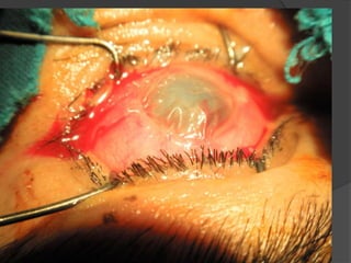
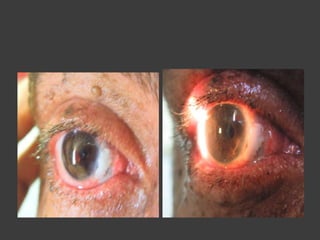


The amniotic membrane (AM) is a resilient tissue with antiadhesive, bacteriostatic, and epithelialization properties, used in various ophthalmic procedures such as conjunctival reconstruction and treatment of corneal defects. Its application techniques include graft, patch, and layered methods, though its limitations are noted in specific surgeries like pterygium and acute alkali burns. Case studies highlighted the necessity for case selection due to variable effectiveness and potential complications associated with AM treatment.


