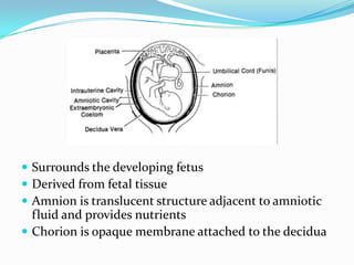
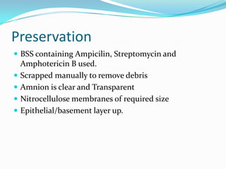
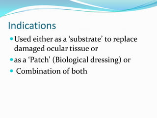
Dr Abhijit Wadekar
M.S., D.N.B, (Ophthal)
Cornea Fellow L.V.P.E.I, Hyderabad
Leela Eye Institute
Kalyan
 Surrounds the developing fetus
 Derived from fetal tissue
 Amnion is translucent structure adjacent to amniotic
fluid and provides nutrients
 Chorion is opaque membrane attached to the decidua
Histology
 Single layer of cuboidal epithelial cells
 A thick Basement Membrane
 Avascular stromal matrix , loosely attached to the
chorion
Introduction
 Innermost layer forming the fetal membrane.
 First used by Davis in 1910 in skin transplant.
 First documented ophthalmic application for
conjunctival surface reconstruction in 1940 by De
Roth.
 Sorsby et al in 1946 reported its use in acute ocular
burns.
 Kim and Tseng successfully reintroduced usage in 1995
Properties of Amniotic membrane
Facilitates migration of epithelial cells
Reinforcement of basal cell adhesion
Induction of epithelial cell differentiation and promotes goblet cell
differentiation
Anti-inflammatory action reduces scarring and vascularization
bacteriostatic properties
Lack of Immunogenicity
 Cytokines, growth factors and protease inhibitors
(IL4,6) EPG, FGF, TGF, HGF and 2- microglobulin.
 Exclusion of inflammatory cells with anti-protease
activities
 Suppression of TGF- signalling system and
myofibroblast differentiation of normal fibroblasts.
 Prolongation of life span and clonogenicity of
epithelial progenitor cells.
 Promotion of non-goblet cells epithelial
differentiation
 Promotion of goblet cells differentiation when
combined with epithelial fibroblast
Collection
 Consenting Sero negative (Hepatitis B and C virus,
Syphilis and HIV) maternal donors determined pre
surgery
 elective caesarian section
 Under sterile conditions
 An antibiotic cocktail covering most of Gram –positive
and Negative bacteria and fungi in Balanced Salt
Solution for 24 hours
 Second wash of BSS
 Amnion separated from chorion
Harvesting Amniotic membrane
Preservation
 BSS containing Ampicilin, Streptomycin and
Amphotericin B used.
 Scrapped manually to remove debris
 Amnion is clear and Transparent
 Nitrocellulose membranes of required size
 Epithelial/basement layer up.
 Preserved in solution containing Modified
Eagle’s Medium (MEM) and glycerol in 1:1 ratio
with 3.3% l-glutamine, 25 ug/ml Gentamicin, 50
units/ml penicillin, 100ug/ml ciprofloxacin and
0.5 mg/ml Amphotericin B
 Stored in 50 ML wide mouthed screw capped
irradiated transparent bottles at -80 Degrees
Indications
Used either as a ‘substrate’ to replace
damaged ocular tissue or
as a ‘Patch’ (Biological dressing) or
 Combination of both
Corneal diseases
Symptomatic Bullous keratopathy
Chemicalinjury
Limbal stem cell
deficiency (Partial
or total): combined
with stem cell graft
Persistent epithelial defects
Corneal ulceration
Conjunctival diseases
Stevens-Johnson Syndrome
Symblepharon lysis
Conjunctival surface reconstruction
Pterygium surgery
Post Trabeculectomy:bleb leakage or
revision
Conjunctival cicatrisation/scar
Limbal stem cell transplant on
Amniotic membrane
Surgical techniques
Thawed at room temperature
Rinsed three times in BSS
Gently separated from the
nitrocellulose paper
Epithelium-basement membrane side
up
Stromal side up, away from the eye.
The stromal side of the membrane is
sticky
Trimmed to the appropriate shape
10-0 nylon interrupted sutures
Intercepted 10-0 nylon sutures
A large therapeutic contact lens
Occasionally, a tarsorrhaphy
Sutures and contact lens are often
removed after 2 to 4 weeks
Post-operative topical treatment
Tissue-cultured limbal stem cells and
amniotic membrane
Disintegrates within 2 to 4 weeks
Storage medium should be light pink
Complications
Suture granuloma
Persist ant inflammation
Hematoma dehiscence
Shrinkage of the graft
Failure to achieve the intended effect
Infection
Dislodged or loose AM
Hemorrhage
Early disintegration
Thank You

Amniotic Membrane Transplant

  • 1.
    Dr Abhijit Wadekar M.S.,D.N.B, (Ophthal) Cornea Fellow L.V.P.E.I, Hyderabad Leela Eye Institute Kalyan
  • 2.
     Surrounds thedeveloping fetus  Derived from fetal tissue  Amnion is translucent structure adjacent to amniotic fluid and provides nutrients  Chorion is opaque membrane attached to the decidua
  • 3.
    Histology  Single layerof cuboidal epithelial cells  A thick Basement Membrane  Avascular stromal matrix , loosely attached to the chorion
  • 4.
    Introduction  Innermost layerforming the fetal membrane.  First used by Davis in 1910 in skin transplant.  First documented ophthalmic application for conjunctival surface reconstruction in 1940 by De Roth.  Sorsby et al in 1946 reported its use in acute ocular burns.  Kim and Tseng successfully reintroduced usage in 1995
  • 5.
    Properties of Amnioticmembrane Facilitates migration of epithelial cells Reinforcement of basal cell adhesion Induction of epithelial cell differentiation and promotes goblet cell differentiation Anti-inflammatory action reduces scarring and vascularization bacteriostatic properties Lack of Immunogenicity
  • 6.
     Cytokines, growthfactors and protease inhibitors (IL4,6) EPG, FGF, TGF, HGF and 2- microglobulin.  Exclusion of inflammatory cells with anti-protease activities  Suppression of TGF- signalling system and myofibroblast differentiation of normal fibroblasts.  Prolongation of life span and clonogenicity of epithelial progenitor cells.  Promotion of non-goblet cells epithelial differentiation  Promotion of goblet cells differentiation when combined with epithelial fibroblast
  • 7.
    Collection  Consenting Seronegative (Hepatitis B and C virus, Syphilis and HIV) maternal donors determined pre surgery  elective caesarian section  Under sterile conditions  An antibiotic cocktail covering most of Gram –positive and Negative bacteria and fungi in Balanced Salt Solution for 24 hours  Second wash of BSS  Amnion separated from chorion
  • 8.
  • 9.
    Preservation  BSS containingAmpicilin, Streptomycin and Amphotericin B used.  Scrapped manually to remove debris  Amnion is clear and Transparent  Nitrocellulose membranes of required size  Epithelial/basement layer up.
  • 10.
     Preserved insolution containing Modified Eagle’s Medium (MEM) and glycerol in 1:1 ratio with 3.3% l-glutamine, 25 ug/ml Gentamicin, 50 units/ml penicillin, 100ug/ml ciprofloxacin and 0.5 mg/ml Amphotericin B  Stored in 50 ML wide mouthed screw capped irradiated transparent bottles at -80 Degrees
  • 11.
    Indications Used either asa ‘substrate’ to replace damaged ocular tissue or as a ‘Patch’ (Biological dressing) or  Combination of both
  • 12.
  • 13.
    Chemicalinjury Limbal stem cell deficiency(Partial or total): combined with stem cell graft
  • 14.
  • 15.
    Conjunctival diseases Stevens-Johnson Syndrome Symblepharonlysis Conjunctival surface reconstruction Pterygium surgery Post Trabeculectomy:bleb leakage or revision
  • 16.
  • 17.
    Limbal stem celltransplant on Amniotic membrane
  • 18.
    Surgical techniques Thawed atroom temperature Rinsed three times in BSS Gently separated from the nitrocellulose paper Epithelium-basement membrane side up
  • 19.
    Stromal side up,away from the eye. The stromal side of the membrane is sticky Trimmed to the appropriate shape 10-0 nylon interrupted sutures Intercepted 10-0 nylon sutures
  • 20.
    A large therapeuticcontact lens Occasionally, a tarsorrhaphy Sutures and contact lens are often removed after 2 to 4 weeks Post-operative topical treatment
  • 22.
    Tissue-cultured limbal stemcells and amniotic membrane Disintegrates within 2 to 4 weeks Storage medium should be light pink
  • 23.
    Complications Suture granuloma Persist antinflammation Hematoma dehiscence Shrinkage of the graft Failure to achieve the intended effect Infection Dislodged or loose AM Hemorrhage Early disintegration
  • 24.