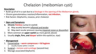
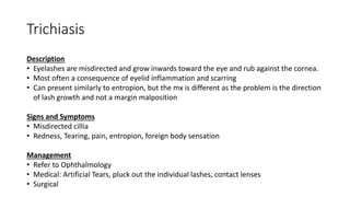
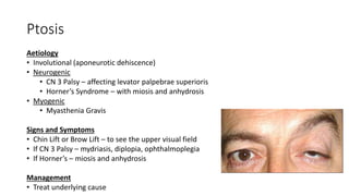
Ophthalmology SMSV lecture
2020
Kevin Liang
What are we doing today
• Quick anatomy revision
• General ophthal History + Exam
• Acute vision loss + red eye + pain
• Chronic vision loss
• Flashes and floaters
• Eyelid conditions
• Questions
Anatomy
Blood supply of retina
Internal carotid artery
Ophthalmic artery
Central retinal artery
Posterior ciliary arteries
Choroidal circulation
Inner 2/3 retina
• Ciliary body, iris
• Fovea
• Outer retina
• Rods and cones
• Retinal pigment
epithelium)
Via capillary plexus perfusion
Via diffusion
Lacrimal artery
Muscular arteries
Supra-orbital artery
Posterior ethmoidal
Anterior ethmoidal
Medial palpebral
Dorsal nasal
Supratrochleat
Anatomy of the cornea
R eye L eye
General ophthal history
• 1 vs 2 eyes affected?
• 1 eye visual loss or 1 sided visual loss (homonymous
hemianopia)
• Glasses/contacts?
• WWQQAA
• Acute vs gradual
• Ophthal specific history
• Vision changes
• Blurring of vision - central vs peripheral? Transient vs persistent?
• Diplopia
• Pain
• Location of pain – surface? Retro-orbital?
• Worse on movement? – optic neuritis
• Worse on blinking? – surface e.g. cornea issues
• Red eye
• Flashes and floaters
• Trauma/injury? Protective eyewear?
• Discharge, itch, watering, morning crusting – eyelid/ conjunctival
disorders
• Swelling of soft tissue
• Photophobia
• Systemic/others - Fever, rheumatological, neurological
• Medications
• Steroids (cataract,
glaucoma, infections)
• Allergies
• Family history
• Social history
• Activities and hobbies
(visual demands)
• Driving
General ophthal exam
• General – glasses/contact lens
• Vitals – regular pulse? – ?stroke
• Anterior eye exam
• Inspection
• Anatomy
• Redness, swelling
• Discharge, styes
• Ptosis
• Jaundice, anaemia
• Palpation
• Bony margins – trauma/fracture
• Evert eyelid - ?foreign body
• Tests (AAFFIRRM ST)
• Acuity
• Snellens chart  count fingers  hand
wave  light perception
• Accommodation
• Fields + blind spot
• Fundoscopy (do this last)
• Ishihara Colour Plates
• Reflexes (pupil)
• Red reflex
• Movements
• +/- Slit lamp +/- Fluorescein staining
• +/- Tonometry
• +/- cranial nerves
Slit lamp and fluorescein staining
Immediate referrals to ophthalmology
Orbital cellulitis
Traumatic subconjunctival hemorrhage - bullous elevation
Scleritis
Iritis
Endophthalmitis
Zoster opthalmicus
Acute close angle glaucoma
Corneal ulcer
Foreign body - if central cornea affected/ penetrating
injury/ rust ring present
Chemical injury
Retinal artery occlusion
Anterior ischaemic optic neuropathy - w/ GCA and PMR
Optic neuritis - w/ MS
Retinal detachment
Vitreous hemorrhage
Hyphaemia or Hypopyon
Severe eye pain
Significant vision loss
Pupil irregular, dilated or fixed
Differentials for red eye/BOV/pain
Acute Chronic
No blurring of vision Blurring of vision
Painful
• Preseptal cellulitis
• Conjunctivitis
(discomfort)
• Dry eye syndrome
(discomfort)
• Episcleritis
• Scleritis (unless
severe/posterior scleritis)
Painless
• Subconjunctival
haemorrhage (unless
a/w trauma)
Painful
• Foreign body/trauma
• Chemical injury
• Orbital cellulitis
• Corneal
ulcer/abrasions
• HSV/viral keratitis
• Photokeratitis
• Uveitis
• Acute closed angle
glaucoma
• Endophthalmitis
• Posterior scleritis
• Optic neuritis
• GCA
Painless
• Dry eye syndrome
• Lens subluxation/
dislocation
• Vitreous
haemorrhage
• Retinal artery
occlusion/Retinal
vein occlusion
• Retinal
tear/detachment
• TIA/stroke
Painless
• Dry eye syndrome
• Corneal opacity
• Refractive error
• Cataracts
• AMD
• Diabetic retinopathy
• Hypertensive
retinopathy
• Creeping retinal
detachment
• Open angle glaucoma
• Compressive optic
neuropathy
Think along:
Trauma
Soft tissue
Tear film
Cornea
Anterior chamber
Lens
Pupil
Posterior chamber
Lens
Vitreous
Retina
Optic nerve
Optic chiasm
Optic tract
Brain
Red eye conditions
(usually more anterior part of
eye)
Acute conditions
Acute conditions
Acute Chronic
No blurring of vision Blurring of vision
Painful
• Preseptal cellulitis
• Conjunctivitis
(discomfort)
• Dry eye syndrome
(discomfort)
• Episcleritis
• Scleritis (unless
severe/posterior scleritis)
Painless
• Subconjunctival
haemorrhage (unless
a/w trauma)
Painful
• Foreign body/trauma
• Chemical injury
• Orbital cellulitis
• Corneal
ulcer/abrasions
• HSV/viral keratitis
• Photokeratitis
• Uveitis
• Acute closed angle
glaucoma
• Endophthalmitis
• Posterior scleritis
• Optic neuritis
• GCA
Painless
• Dry eye syndrome
• Lens subluxation/
dislocation
• Vitreous
haemorrhage
• Retinal artery
occlusion/Retinal
vein occlusion
• Retinal
tear/detachment
• TIA/stroke
Painless
• Dry eye syndrome
• Corneal opacity
• Refractive error
• Cataracts
• AMD
• Diabetic retinopathy
• Hypertensive
retinopathy
• Creeping retinal
detachment
• Open angle glaucoma
• Compressive optic
neuropathy
Red eye conditions
(usually more anterior part of
eye)
Think along:
Trauma
Soft tissue
Tear film
Cornea
Anterior chamber
Lens
Pupil
Posterior chamber
Lens
Vitreous
Retina
Optic nerve
Optic chiasm
Optic tract
Brain
Foreign body
Signs and Symptoms
• “something in my eye”
• Metal Work (e.g. Grinding, Welding)
• No protective eye-wear
• Tear-drop pupil
• Indicates penetrating eye injury
• Tear drop almost always points towards foreign body
Investigations
• If penetrating  immediate referral to ophthalmology
• Slit Lamp + Fluorescein Stain + Blue Light
• Seidel’s Test – changes to fluorescence area colour/surface = aqueous leakage in the cornea
• Thin cut CT Orbit if suspected metal foreign body
Foreign body
Removal of corneal foreign body
• Topical anaesthetic (its painful!)
• Try irrigation first
• Try scrape off with a cotton wool swab if not embedded
• Try to use needle/foreign body spud to remove the object
• Under slit lamp magnification
• Any gauge will do, physician preference, but bigger needles better
usually
• If still cannot, REFER
Chemical injury
Signs and Symptoms
• Eye Pain + Redness + Tearing
• Photophobia
• Decreased Visual Acuity
• Cornea haze/cloudy
• Limbal ischemia
• Corneal Epithelial Defect
Management
• Immediate Irrigation – Morgan Lens
• Check litmus paper for initial pH
• Topical Anaesthetic + 2 Litre of Saline
• Repeat litmus paper to check if pH is back to 7.0-7.5, if not continue irrigation
• Immediate Referral to Ophthalmology after
Cellulitis
• Preseptal vs orbital cellulitis
Preseptal/periorbital cellulitis
Description
• Common infection of the eyelid and periorbital soft tissues anterior
to the orbital septum, often spread from the upper respiratory tract
• Not involving orbit or other intraocular structures
Organisms
• Majority Bacteria: S aureus, Strep pneumoniae, anaerobes
• Viruses
Pathophysiology
• Direct inoculation: after eyelid trauma and infected insect bites
• Spread from contiguous structure: paranasal sinuses
Preseptal/periorbital cellulitis
Signs and Symptoms
• Fever + Unilateral Red and Swollen eyelid – may be to the point of being shut
• +/- URTI Symptoms (e.g. rhinitis, cough, adenopathy)
• Normal conjunctiva
• NO visual loss or proptosis or ophthalmoplegia
• No pain on eye movements
Investigations
• FBE - leukocytosis
• CT if eyelid oedema so severe that precludes eye examination
• Distinguish orbital vs preorbital cellulitis
• Cultures of eyelid wound, conjunctiva, blood, abscess contents or paranasal sinus secretions
Management (see eTg)
• PO flucloxacillin/dicloxacillin as empirical therapy
• IV flucloxacillin (if severe)
Orbital cellulitis
Description
• Infection of the internal orbital structures such as the rectus muscles, usually from
extension of infection from paranasal sinusitis.
• Children < 4 y/o have an incomplete septum, so they are at increased risk of orbital
cellulitis from retrograde spread of infection from the preseptal to orbital space
Organisms
• Staph aureus, strep, HiB (if not vaccinated), anaerobic bacteria
Signs and Symptoms
• Fever + Unilateral Red and Swollen Shut
• Reduced Visual Acuity
• Proptosis + Ophthalmoplegia (vs. Preseptal Cellulitis – which has none of this)
• Use a cotton bud to open the eye to check eye movements
• Chemotic conjunctiva
• Pain on Eye Movements (vs preseptal)
Orbital cellulitis
Investigations
• Bloods: Blood culture
• Imaging: CT orbits and sinuses
(to assess posterior spread of infection)
Management
• IV Antibiotics
• IV Cefotaxime or
• IV Ceftriaxone + IV flucloxacillin (or vanc)
• PO antibiotics if clinically improving
• Amoxicillin + clavulanate
• Surgical Drainage of sinuses/abscess – if continue to progress even with antibiotics, or
vision loss, etc.
Preseptal cellulitis Orbital cellulitis
Visual acuity Normal May be decreased
Colour vision Normal May be decreased
Pupils Normal May have RAPD
Conjunctiva Normal Injected, chemosed
Eye movements Full Restricted
May have diplopia
Proptosis None Present
Conjunctivitis
Definition
Inflammation of conjunctiva (tarsal and bulbar)
Aetiology
• Infectious
• Viral (more common in adults)
• Bacterial (more common in paediatrics)
• Non infectious
• Allergic
• Non-allergic
Conjunctivitis
Allergic
Pathophysiology
• Type 1 immune response to an allergen, usually airborne that contacts the eye --> IgE --> mast cell
degranulation
Signs and symptoms
• Bilateral red eye + itching + watering
• Marked chemosis
• History of atopy, allergy
Treatment
• Hand hygiene
• Pharm
• Artificial tears, cold compresses
• PO antihistamine/decongestant drops – symptomatic relief
• Intranasal steroids
• Mast cell stabiliser/antihistamine eyedrops
Conjunctivitis
Bacterial
• Microbiology:
• S aureas, S pneumoniae, HIB, M catarrhalis
• Gonococcal, chlamydia (for sexually active)
• Signs and symptoms
• Red eye + purulent discharge (eye often stuck shut
in morning)
• Gritty, burning sensation
• Treatment
• Lifestyle: avoid spreading, discontinue contact lens
wear for >2 weeks, hand hygiene
• Pharm
• Artificial tears, cold compresses
• Chloramphenicol drops (chlorsig)
• PO azithromycin if chlamydia, + IM ceftriaxone
if gonococcal
Viral
• Microbiology:
• Adenovirus (65-90%)
• Signs and symptoms
• Prodrome: adenopathy, URTI, pharyngitis
• Red eye + watery/mucoserous discharge
• Gritty, burning sensation
• Exam:
• Follicular appearance to tarsal conjunctiva
• Treatment
• Lifestyle: avoid spreading, discontinue contact
lens wear for >2 weeks, hand hygiene
• Pharm (symptomatic relief)
• Artificial tears, cold compresses
• PO antihistamine/decongestant drops
Corneal abrasion
Description
• Often used to refer to any defect in the corneal surface epithelium.
• Corneal epithelium is richly innervated with sensory pain fibres from CNV1
Aetiology
• Traumatic – e.g. scratched by a branch, fingernail, paper
• Spontaneous (Recurrent Corneal Erosion Syndrome)
• No immediate antecedent injury/foreign body
• Entropion (Particularly in ATSI community)
Signs and Symptoms
• Eye Pain +/- Foreign Body Sensation +/- Blepharospasm
• Pain with blinking
• Redness
• Photophobia +/- Reduced Visual Acuity
Corneal abrasion
Evaluation
• Exclude an open globe and hyphema
• Evert lid
• Measure visual acuity
• Penlight and fluorescein examination
• If linear abrasions – Suspect subtarsal foreign body
Management
• Observation if small
• Debride any loose epithelium
• Chloramphenicol Drops (prophylaxis)
• Oral Analgesia
• Bandage contact lenses
• Review daily and don’t touch or rub the eye
• Should resolve by 1 week
Corneal ulcer (AKA microbial/bacterial) keratitis
Description
• Defect in the corneal surface epithelium and extending into the stroma.
Aetiology
• Contact Lens – (bacterial, viral, fungal, protozoa)
• Especially sleeping with contact lens, not washing lenses
• Monthlies > Weeklies > Dailies
• Foreign Body
• Immunosuppression
Signs and Symptoms
• Severe Eye Pain +/- Foreign Body Sensation +/- Blepharospasm
• Red
• Photophobia +/- Reduced Visual Acuity
• White spot on cornea on inspection
• +/- hypopyon
Corneal ulcer
Management
1. Discontinue contact lenses
2. Referral to Ophthalmology
a) Corneal Scrape MCS
b) Topical Ofloxacin/Ciprofloxacin
c) Surgical: corneal transplant or patch graft
Complications
1. Corneal Scarring
2. Perforation and Endophthalmitis
Photokeratitis/UV keratitis
Description
• Acute syndrome occurring after UV irradiation of the
eye. Akin to a sunburn of the cornea and conjunctiva
Pathophysiology
• UV light absorption from cornea results in surface
epithelial cell death and desquamation
Aetiology
• Welder’s arc (arc eye)
• Snowblindness - UV rays reflected off ice and snow
• Sunlamps
Signs and Symptoms
• Occurs 6-12h after exposure
• Bilateral eye pain - Severe
• Inability to open eyes
• Conjunctival injection
• Photophobia
Management
• Remove contact lenses, get out of sun into dark room
• Supportive – Analgesics (usually mild opioids
needed)
• Should recover in a few days (regeneration of
epithelial surface)
Viral keratitis
Description
• Dendritic ulcer of the cornea caused by HSV or HZV
• Commonly recurrent
Aetiology
• Herpes Simplex Virus
• Herpes Zoster Virus – Zoster Ophthalmicus
Signs and Symptoms
• Herpes symptoms
• Concomitant viral-like illness
• History of oral infections with the virus
• Eye symptoms
• Red eye +pain/FB sensation + Photophobia +
Watery Discharge
Examination
• Faint branching grey opacity on penlight
• Fluorescein - Branching dendrites with terminal
bulbs (Dendritic lesion)
Management
• NO steroid drops -> Uncontrolled infection
• Referral to ophthalmologist
• Topical aciclovir 3%
• PO valaciclovir or aciclovir (if eye ointment not
available)
HZO/zoster keratitis
Description
• Herpes zoster involvement of the ophthalmic division of the trigeminal nerve
Signs and Symptoms
• Prodrome – fever, headache, malaise
• Unilateral painful vesicular eruption – along V1 dermatome (eye, forehead)
• Red Eye: rash + hyperemic conjunctivitis, uveitis, episcleritis, keratitis
• Hutchinson’s Sign – if vesicular lesions on the nose, indicating nasociliary nerve involvement 
increased likelihood of ocular complications
• +/- Dendritic Ulcers (also seen with HSV Keratitis)
Management
• Refer Ophthalmology
• Oral Aciclovir (within 72hrs)
• IV if immunocompromised/sight threatening disease
• Oral Analgesia
Episcleritis
Description
• Abrupt onset of inflammation in the episclera of one or both eyes. It occurs most frequently in
young and middle-aged adults but may affect all age groups, and mostly females (70%).
• Most commonly due to dry eye syndrome. Sometimes associated with systemic disease e.g. RA
(11%), IBD, vasculitis, SLE
Signs and Symptoms
• Sudden Onset Segmental Red Eye
• Radial pattern, localised to one sector
• Redness, Irritation, Watering
• No pain/mild Ache
• NO change in vision
Management (topical treatment vs scleritis)
• Symptomatic Relief (self-resolving)
• Artificial Tears
• Topical NSAIDs (e.g. Diclofenac)
• Topical glucocorticoids
Scleritis
Description
• Painful, destructive, potentially blinding inflammatory disorder
• Highly symptomatic clinical presentation (vs episcleritis)
• Unknown why the eye is targeted
Subtypes
• Anterior scleritis (90% involves anterior portion)
• Posterior scleritis (inflammation posterior to the insertion of medial and lateral rectus muscles)
Aetiology
• Idiopathic
• Inflammatory and autoimmune process, esp vasculitis
• RA, Wegener's
• Infectious (HZO, HSV, HIV, lyme disease)
Scleritis
Signs and Symptoms
• Severe Pain and Tender globe – usually deep and ‘boring’ over several days
• Worse at night/early morning
• Might be worsened by eye movements (due to extraocular muscle insertions into sclera)
• Radiates to face and periorbital region
• Red Eye – usually diffuse (but can be localised) and radiates out from centre
• +/- Headache
• +/- Photophobia
• +/- Decreased Visual Acuity (esp in posterior scleritis)
Management - systemic treatment (vs episcleritis)
• Referral to Ophthalmology
• PO NSAIDs or corticosteroids or Biologics
Episcleritis Scleritis
Pain Absent or Mild Severe
Redness May be localised or diffuse
Vessels are disorganised
Violet-bluish hue
Usually diffuse
Vessels usually radiate outwards
Aetiology 1. Idiopathic – more likely in
episcleritis
2. Rheumatology (RA, SLE, GPA, etc.) –
more likely in scleritis
3. Syphilis, TB
1. Herpes Zoster Virus
2. Gout
1. Rheumatology (RA, SLE, GPA, etc.) –
more likely in scleritis
2. Infectious
Cotton Bud Sweep Vessels likely to move Vessels not likely to move
Topical
Phenylephrine
Vessels blanch (superficial episcleral
vessels)
Vessels do not blanch (deeper episcleral
vessels, more fixed to sclera)
Complications Rare • Necrosis
• Thinning +/- Perforation
Management • Self-limiting
• Lubricants
• Topical NSAIDs
• Refer to Ophthalmology
• PO NSAIDs +/- Steroids (cos systemic
disease)
Iritis/anterior uveitis
Description
• Basically inflammation limited to the uvea
• Anterior Uveitis: Anterior chamber, iris and ciliary body
Aetiology
• Idiopathic
• 50% HLA-B27 positive (most commonly ank spond)
• Viral (e.g. HSV, VZV, CMV)
• Behcet’s disease
Iritis/anterior uveitis
Signs and symptoms
• Painful unilateral red eye +/- photophobia
• Ciliary flush/Limbal flush/limbic flare (Peri-limbal
hyperaemia/circumciliary injection)
• Reduced visual acuity
• +/- Irregularly shaped pupil - Due to posterior synechiae
• "Tear-drop sign“. Adhesion of iris to lens.
• +/- Hypopyon
• Keratic precipitates (KP) on the cornea endothelium
Management
• Refer to ophthalmology
• Topical steroids (reduce inflammation)
• Topical cyclopentolate (mydriatic drops)
• Investigate for rheum causes if recurrent
Acute closed angle glaucoma
Definition
• An optic neuropathy, whereby optic nerve damage results in progressive
loss of retinal ganglion cells
• Characterised by narrowing of the anterior chamber angle
Risk factors
• Age >50, female
• Family history
• Asian
• Hyperopia (longsightedness)
Acute closed angle glaucoma
Symptoms (acute!)
• Usually evening/dark (when lower
light levels cause mydraisis)
• Headache
• Blurring of vision
• Halos around lights
• Severe eye pain
• +/- Unilateral red eye
• Nausea/vomiting
Signs
• Loss of vision
• Mid-dilated pupil, non reactive
• Cloudy cornea
• Cupping of optic disc
• +/- Red eye
DO NOT DILATE PUPIL AS IT CAN
WORSEN CLOSURE
Acute closed angle glaucoma
Management
• Immediate referral to ophthalmology or emergency
• Medical
a) Pressure Lowering Eye-drops – 0.5% Timolol, 1% Apraclonidine, 2%
Pilocarpine
b) 2nd line: Systemic – PO/IV Acetazolamide (reduces aqueous secretion)
c) 3rd line: IV mannitol
• Surgical
• Laser peripheral iridotomy
• Symptomatic: antiemetics, analgesia
Endopthalmitis
Description
• Endophthalmitis refers to bacterial or fungal infection including involvement of the vitreous
and/or aqueous humours.
Aetiology
• Acute postoperative (e.g. Cataract)
• Most within 1-2 weeks
• Post-intravitreal Injection
• Bleb related
• Post-Traumatic
Signs and Symptoms
• Red, Painful Eye, discharge
• Severely Reduced Visual Acuity
• Hypopyon, Hyphema
Management
• Immediate Referral to Ophthalmology - sight threatening
Retinal artery occlusion (RAO)
Description
• Rare event (1 in 100000) causing ischemia of the retina
• “stroke of the eye”
Risk factors/aetiology (mainly cardiovascular)
• Carotid Artery Atherosclerosis (especially age > 40)
• Cardiogenic Embolism (especially age < 40)
• Microvascular Disease (e.g. diabetes, hypertension)
• Haematologic disease (e.g. sickle cell, hypercoagulable states)
• Inflammatory disease (e.g. GCA)
Types
• Central retinal artery occlusion (CRAO)
• Branch retinal artery occlusion (BRAO)
Retinal artery occlusion
Symptoms
• Rapid onset, unilateral, painless, loss of vision
• Central – reduced visual acuity (VA) and usually almost whole field loss
• Branch – may have normal VA and partial field loss
Signs – pale area on retina (based on distribution of branch)
• Pizza pie (branch)
• Cherry red spot (only for central retinal artery occlusion)
• Cattletrucking
• May have carotid bruit, AF, cardiac murmurs
Investigations
• Urgent Carotid U/S, TTE
• MRI brain
• CRP/ESR (if >50 y/o + GCA sx) Cholesterol plaque in branch retinal artery.
Retinal artery occlusion
Management
• Immediate Referral to ED and Ophthalmology – attempt to restore blood flow
within 2 hours
• No difference between thrombolytic therapy vs conservative therapy
• Non-pharmacological
• Massage Globe – may dislodge the embolism
• Medical
• Decrease IOP – IV Mannitol or PO/IV Acetazolamide
• Surgical
• Anterior Chamber Paracentesis (drops intraocular pressure) – may dislodge embolism
• Prevention
• SNAPW, optimisation of atherosclerotic diseases
Prognosis for CRAO poor, but 80% BRAO recover normal vision
Retinal vein occlusion (RVO)
Description
• RVO is the second most common cause of vision loss from
retinal vascular disease, following diabetic retinopathy.
Types
• BRVO - usually compression of vein by atherosclerotic
arteries at AV crossings
• CRVO - usually thrombus
• Hemiretinal vein occlusion – may occur when there is
blockage of a vein draining the superior or inferior hemiretina
Risk factors
• Cardiovascular (e.g. HTN, T2DM, Lipids, BMI) – usually
embolic
• Hypercoagulable State
• Glaucoma – prevents retinal vein outflow and leads to stasis
Pathophysiology
Blockage ---> pressure ---> haemorrhage
May have neovascularisation after that,
may cause neovascular glaucoma
Retinal vein occlusion (RVO)
Symptoms
• BRVO
• May be asymptomatic, noted incidentally
• Relative scotoma, areas of blurred vision
• CRVO (almost always symptomatic)
• Sudden onset, painless, LOV unilaterally
Signs
• Dilated/tortuous veins, retinal haemorrhage (blood and thunder), oedema
• +/- Cotton Wool Spots
• +/- RAPD
BRVO
Optic neuritis
In MULTIPLE SCLEROSIS, it is the presenting feature in 15-20% of patients, occurs in
50% at some time during the course of their illness
Symptoms
• MONOCULAR (bilateral rare)
• Vision loss (develops over hours to days, peaking within 1-2 weeks)
• Eye pain, worsened by eye movement
• RAPD
• Loss of colour vision
• Central scotoma
• Photopsias (flashes), precipitated by eye movement
• Hyperemia and swelling of disk, blurring of disk margins, distended veins
Subconjunctival haemorrhage
Description
• Collection of blood between the sclera and the conjunctiva.
Aetiology
• Spontaneous – mostly
• Blunt Trauma – may cause globe rupture
• Contact Lens – may cause minor trauma
• Elevated Venous Pressure (e.g. coughing (e.g. pertussis), vomiting, Valsalva)
• Hypertension, Diabetes, Warfarin
Signs and Symptoms
• Focal, Flat, Red Lesion – changes to yellow then fades
• Painless; Normal Vision (unless the trauma caused other issues e.g. bleeding
behind the eye)
• Bullous Elevation – usually traumatic cause, severe!
Management
• Non-traumatic – self-resolves in 2-3 weeks
• Traumatic – immediate referral to ophthalmology
Pterygium
Description
• Triangular wedge of fibro-vascular conjunctival tissue that typically starts medially on the nasal
conjunctiva and extends laterally onto the cornea. Benign growth
• The prevalence of pterygium is associated with chronic sun exposure.
Signs and Symptoms
• Asymptomatic, just noticed a change in appearance
• Redness and Irritation – at most mild
Management
• Avoid UV Exposure
• Artificial Tears – if redness and irritation
• Surgical Excision – if visual acuity affected
Chronic/gradual BOV
(blurring of vision)
Differentials for chronic BOV
Acute Chronic
No blurring of vision Blurring of vision
Painful
• Preseptal cellulitis
• Conjunctivitis
(discomfort)
• Dry eye syndrome
(discomfort)
• Episcleritis
• Scleritis (unless
severe/posterior scleritis)
Painless
• Subconjunctival
haemorrhage (unless
a/w trauma)
Painful
• Foreign body/trauma
• Chemical injury
• Orbital cellulitis
• Corneal
ulcer/abrasions
• HSV/viral keratitis
• Photokeratitis
• Uveitis
• Acute closed angle
glaucoma
• Endophthalmitis
• Posterior scleritis
• Optic neuritis
• GCA
Painless
• Dry eye syndrome
• Lens subluxation/
dislocation
• Vitreous
haemorrhage
• Retinal artery
occlusion/Retinal
vein occlusion
• Retinal
tear/detachment
• TIA/stroke
Painless
• Dry eye syndrome
• Corneal opacity
• Refractive error
• Cataracts
• AMD
• Diabetic retinopathy
• Hypertensive
retinopathy
• Creeping retinal
detachment
• Open angle glaucoma
• Compressive optic
neuropathy
Red eye conditions
(usually more anterior part of
eye)
Think along:
Trauma
Soft tissue
Tear film
Cornea
Anterior chamber
Lens
Pupil
Posterior chamber
Lens
Vitreous
Retina
Optic nerve
Optic chiasm
Optic tract
Brain
Keratoconjunctivitis sicca (dry eye syndrome)
Aetiology
Usually due to loss of lipid production in Meibomian glands.
• Aqueous-deficient causes
• Sjogren syndrome (primary or secondary)
• Non-SS
• Lacrimal gland deficiency, obstruction, post-ocular procedures e.g. LASIK
• Lacrimal gland infiltration due to sarcoidosis, lymphoma, GVHD, episcleritis
• Evaporative causes
• Lid disorder
• Blepharitis, Meibomian gland dysfunction
• Low blink rate
• Ageing, low humidity/high wind velocity
Keratoconjunctivitis sicca (dry eye syndrome)
Investigations
• Bedside
• Fluorescein
• Schirmer Test – if decreased, suggests Sjogren’s Syndrome
• Bloods
• ANA, SS-A, SS-B
Management
• Lifestyle - Encourage frequent blinking, watch less TV
• Lubricants
• Artificial Tears – in the day; Ointment – in the night
• Surgical
• Punctal Occlusion (punctum plug)
Cataracts
Description
• Clouding of the crystalline lens
• Leading cause of curable blindness in the world
• Legal blindness = 6/60
Risk factors
• Age >60
• Congenital
• Modifiable
• Trauma
• UV light/radiation, smoking
• Diabetes mellitus, metabolic syndrome, cushings
• Corticosteroids (systemic, topical, inhaled?)
• Poor lifestyle habits, malnutrition, physical inactivity
Cataracts
Symptoms
• Typically bilateral
• Blurred vision/visual loss
• Glare, haloes around lights (esp driving at night)
• Increasing myopia (due to increase in refractive power of a lens)
• Lack of red reflex (+/-)
Management
• Lifestyle/non-surgical
• Driving
• Avoid UV light/wear sunglasses
• Glasses – to correct residual refractive error
• Surgery
• Phacoemulsification + intraocular lens implantation
Open-angle glaucoma
Description
• An optic neuropathy, loss of retinal ganglion cells
• Second leading cause of blindness (after cataracts)
Risk factors
• Age >50, family history
• Hypertension, diabetes
• Myopia
• Elevated Intra-ocular Pressure (IOP) > 20 mmHg – not specific
Signs and symptoms
1. Progressive Visual Loss (usually asymptomatic)
a) Progressive peripheral visual field loss, followed by central field loss
2. Increased Cup-to-Disc Ratio > 0.5
Glaucoma
Screening
• Ophthalmoscopy, tonometry, perimetry
Tonometry
Perimetry
Open-angle glaucoma
Management
1. Pharmacological (decreasing aqueous production + increase outflow)
a) 1st line Topical Prostaglandins (e.g. Latanoprost, Bimatoprost) – increases outflow
of aqueous
b) Topical Beta-blockers (e.g. Timolol) – decreases secretion of aqueous humour
c) Topical cholinergic (e.g. Pilocarpine)
d) Topical Alpha-agonists (e.g. Brimonidine)
e) Topical Acetazolamide – decrease production
2. Surgical
a) Trabeculoplasty (using laser)
a) Increases aqueous outflow
b) Trabeculectomy
a) Creating a surgical Filtration Bleb
Trabeculoplasty
Age related macular degeneration (AMD)
Description
• Acquired degeneration of retina that causes
central visual impairment
Pathophysiology
• Decreased choroidal or Bruch’s membrane
perfusion, RPE ischemia/atrophy
Types
• Dry 90% (atrophic or non-exudative)
• Can progress to wet AMD if advanced
• Wet 10% (neovascular or exudative)
• Causing macular oedema
Risk factors
• Age (>50)
• Smoking and Alcohol
• Family History
AMD
Optical coherence
tomography
(OCT) ↓
Drusen under RPE
Sub-retinal fluid Sub-RPE fluid
AMD
Symptoms
• Asymptomatic if early
• Gradual Progressive Visual Loss
• Dry – over decades | Wet – over months
• Central scotoma (black patch in central vision)
• Difficulty with central vision (e.g. reading, driving, making out faces)
Signs on fundoscopy
• Dry AMD: Drusen + RPE pigmentation, atrophy
• Drusen are yellow deposits (lipids under retina)
• Wet AMD: Subretinal haemorrhage
Dry AMD
Wet AMD – subretinal
haemorrhage
AMD
Management
• Referral to Ophthalmology
• Dry AMD = Lifestyle
• Stop smoking, as it is a risk factor for progression to wet macular
degeneration
• Anti-oxidants (e.g. Vitamin A, C, Zinc)
• Self-monitoring with Amsler Grid – referral if any new symptoms
• Metamorphopsia – distorted vision (see pic)
• Wet AMD = Medical
• Intra-vitreal VEGF Inhibitors
• Bevacizumab, ranibizumab, aflibercept
Diabetic retinopathy
• Non-proliferative
• Cotton wool spots (nerve fibre infarct)
• Dot & blot haemorrhages: dot = microaneurysms,
blot = ruptured microaneurysms
• Hard exudates (lipid deposits)
• Proliferative (Neovascularisation)
• Vitreous haemorrhage
• Tractional retinal detachment
• Neovascular glaucoma
• Macular oedema (retinal thickening and
oedema involving the macula, due to leaky
new vessels usually)
• Can occur at any stage of DR
• Presents as central vision loss
• Leading cause of blindness in DR
Diabetic retinopathy
Signs and Symptoms
1. Asymptomatic in early stages (hence important to screen!)
2. Floaters
3. Blurred Vision
a) Gradually Worsening
b) May be sudden if vitreous haemorrhage
Ophthal screening and follow up in diabetic patients (RACGP diabetes Australia book!):
• At diagnosis of diabetes, then every 2 years (or yearly if developed
retinopathy/complications)
Diabetic retinopathy
Management
1. Optimise Cardiovascular Risk and Glucose Control
a) Manage DM, HTN, Dyslipidaemia
b) Regular Screening and Follow-up (based on severity/staging)
2. Macular Oedema
a) Intravitreal Anti-VEGF Injections (Bevacizumab, Ranibizumab)
b) Laser Photocoagulation
3. Severe Non-proliferative to Proliferative
a) Laser photocoagulation (pan-retinal photocoagulation)
a) Laser burning to shrink and prevent vessel growth, and stop leaking vessels
b) Effective at stabilising vision, but does not reverse vision loss, and can reduce visual fields
4. Vitreous Haemorrhage or Retinal Detachment
a) Vitrectomy
Diabetic retinopathy
• Laser photocoagulation
Hypertensive retinopathy
Clinical features
• Usually asymptomatic
• Related to RAO and RVO
Classification
• Grade 1
• Retinal arteriolar narrowing
• Grade 2
• Copper wiring
• AV nicking
• Grade 3
• Grade 2 plus:
• Silver wiring
• Flame haemorrhages, exudates, cotton
wool spots
• Grade 4
• Grade 3 plus:
• Disc oedema
Management
• Treat hypertension
Copper wiring
Silver wiring
Vitreous haemorrhage
Aetiology
• Posterior Vitreous Detachment +/- Retinal Tear
• Proliferative Diabetic Retinopathy
• Trauma – suspect non-accidental injury in children
Signs and Symptoms
• Sudden, Persistent Visual Loss
• +/- Reduced Red Reflex
• Unable to visualise retina on ophthalmoscope
Management
• Referral to Ophthalmology
• Observation
• U/S to rule out retinal detachment
• CT if suspected head trauma
• If persistent – consider vitrectomy
Refractive errors
Refractive errors fun facts/definitions
Facts
• Cornea ---> 2/3 of eye's refractive power
• Lens ---> 1/3 of refractive power
Definitions
• Emmetropia (normal refraction)
• Ametropia (abnormal refraction)
• Myopia (short sighted), hyperopia (long sighted)
• Astigmatism
• Presbyopia
Myopia
Description
• Refractive disorder in which the axial length of the eye is either too long or the refractive power of the eye's optical system is too
great.
Risk Factors
• Genetic
• Prolonged reading/close range
• DM
• Trauma
• Excessive accomodation
• Increased IOP
• Cataract
• Maternal factors (greater maternal age, smoking)
• Light exposure (unclear role)
Management
• Refer to optometrist
• Spectacles or Contact Lens
• Diverging (minus) Lens
• Spherical lens
• Refractive Surgery
• LASIK (laser-assisted in situ keratomileusis)
• PRK (photorefractive keratectomy)
Hyperopia
Description
• Refractive disorder in which the axial length of the eye is either too short or the refractive power of the
eye's optical system is too weak.
Risk factors
• Trauma
• Mass effect of orbital tumour posterior to retina
• Scleral inflammation with subretinal thicekning
• Removal of crystalline les without replacing it with synthetic intraocular lens
Management
• Refer to optometrist
• Spectacles or Contact Lens
• Converging (plus) Lens
• Spherical lens
• Refractive Surgery
• LASIK or PRK
Astigmatism
Description
• Refractive condition in which a warped corneal surface (think rugby ball vs soccer ball) causes
light rays entering the eye along different planes to be focused unevenly.
Risk factors
• Unknown
• Genetic and/or developmental
Management
• Refer to optometrist
• Spectacles or Contact Lens
• Toric Lenses
• Cylindrical lens, different power in different meridians/planes
• Refractive Surgery
Presbyopia
Description
• Non-refractive error that occurs when the lens loses its elasticity and
therefore normal accommodating power and can no longer focus on
objects viewed at arm's length or closer. Presbyopia usually begins after
age 40 when the crystalline lens loses its elasticity.
Signs and Symptoms
• Progressive Hyperopia
• Unable to read at close distances
Management
• Bifocal Lenses or Reading Glasses
Flashes and floaters
Flashes (photopsias): perception of light in absence of external light
stimuli
• Comes from rods and cones being activated and irritated
Floaters: Sensation of dark spots in vision
• Caused by debris in vitreous from collagen clumping together
• Typically due to age related degeneration of vitreous
• At birth, vitreous is like jello. With age, vitreous undergoes "syneresis",
becoming more fluid or liquid like.
• Pathological if sudden onset, a/w visual loss
Flashes and floaters ddx
Flashes Floaters
Ophthalmic
• Posterior vitreous detachment
• Retinal tear
• Retinal detachment
• Optic neuritis
Non-ophthalmic
• Migraine
• Postural hypotension
• Occipital tumours
• Meningitis
• Psychogenic
• Seizures?
Ophthalmic
• Vitreous syneresis
• Vitreous haemorrhage
• Posterior vitreous detachment
• Retinal detachment
• Vitritis
• Tear film debris
Think along:
Trauma
Soft tissue
Tear film
Cornea
Anterior chamber
Lens
Pupil
Posterior chamber
Lens
Vitreous
Retina
Optic
nerve/chiasm/tract
Brain
Posterior vitreous detachment (PVD)
Common, age related, degeneration, usually nothing to worry about
Pathophysiology
• Liquefaction of vitreous occurs with age (syneresis) and shrinkage of
the vitreous. Liquefied vitreous fluid tracks between posterior hyaloid
face and retina (usually due to rupture in the posterior vitreous
cortex), separating the posterior hyaloid from the retina
Signs and Symptoms
• May be asymptomatic
• Flashes and Floaters
• Floaters often resolve over a period of 3-12 months; settle down
outside visual axis and/or become less noticeable
Examination
• Weiss Ring – ring of glial tissue avulsed from the optic disc – strong
indicator that PVD occured
Posterior vitreous detachment (PVD)
Risk Factors
• Age > 60
• Myopia (increased axial length of eye)
• FHx
• Intraocular Inflammation
• Blunt trauma, cataract surgery
Complications
• Retinal tears/holes in areas with abnormally strong vitreoretinal adhesion
• Retinal detachment
Management
• Floaters often resolve, might settle outside visual axis and/or become less noticeable
• Referral to Ophthalmology to rule out tear
• Non-sight-threatening, but 10-15% will have complications (e.g. retinal tear or detachment)
• If uncomplicated  Just follow up, no treatment
Retinal detachment
Description
• Retinal detachment occurs when the neurosensory retina separates from the
underlying retinal pigment epithelium and choroid → ischemia and rapid and
progressive photoreceptor degeneration
Main types
• Rhegmatogenous (break in retina) most common
• Risk factors: retinal tear, posterior vitreous detachment, traumatic
• Tractional
• Usually seen in proliferative diabetic retinopathy causing neovascularisation
• Exudative/serous
• Accumulation of fluid beneath retina, usually from inflammatory conditions e.g. sarcoidosis
or choroidal neoplasms
Retinal detachment
Signs and Symptoms
• Floaters (most common) and/or flashes
• Sudden dramatic increase i.e. “shower of floaters”
• Sudden Onset, Painless Visual Loss
• Shadow coming down
Examination (slit lamp + dilation)
• Rhegmatogenous = Horseshoe shape tears (retinal tear) + Corrugated appearance
(retinal detachment) and undulates with eye movements
• + signs of posterior vitreous detachment, weiss ring
• Tractional = smooth concave surfaces with minimal shifting with eye movements
• Exudative/serous = smooth retinal surface and shifting fluid depending on patient
positioning
Retinal detachment
Investigations
• OCT
• B-scan ultrasound
Management
• Immediate Referral to Emergency/Ophthalmology
• Laser/Pneumatic/Cryoretinopexy
• Pneumatic: Gas bubble into vitreous seals the tear and
reattaches macula
• Scleral buckle
• Vitrectomy
A progression..
Posterior vitreous detachment
• Liquefaction of vitreous occurs
with age (syneresis)  fluid
tracks between posterior
hyaloid face and retina
• Clinical features
• May be asymptomatic
• Flashes and floaters
• Weiss ring (ring of glial
tissue avulsed from the
optic disc)
• Mx
• Urgent referral for dilated
fundus examination
• 10-15% have retinal tears
Retinal Tear
• Must be ruled out in patients
with PVD
• 10-20% progress to
rhegmatogenous retinal
detachment
• Signs that indicate a high risk of a
tear
• ‘Tobacco dust’ in anterior
vitreous (Shafer’s sign)
• Vitreous haemorrhage
• Also can occur in absence of PVD:
high myopia, trauma, lattice
degeneration
• Mx (refer to ophthal)
• Laser retinopexy: reduces
risk of retinal detachment
Retinal Detachment
• Separation of the neurosensory retina
from the retinal pigment epithelium
• Types
• Rhegmatogenous (most common)
• Exudative/serous (eg choroidal
melanoma)
• Tractional (eg proliferative
diabetic retinopathy)
• Clinical features
• Flashes and floaters
• Visual field defect
• Decreased VA (macular-off)
• Elevated and corrugated
appearance of retina
• Mx (refer to ophthal)
• Vitrectomy and treatment of
retinal tears (laser retinopexy or
cryotherapy)
Credits: Dr Brandon Thia
Eyelid disease
Anatomy of eyelids
2 parts to remember:
1. Anterior portion
1. Eyelash, gland of Zeis, gland of Moll
2. Posterior portion
1. Meibomian glands
Anterior
Posterior (meibomian glands)
Blepharitis
Description
• Common chronic ophthalmologic condition characterized
by inflammation of the eyelid margin.
Types
• Anterior - eyelid skin, base of eyelashes, eyelash follicles,
includes traditional classifications of staph and seborrheic
blepharitis
• Posterior - Meibomian glands, gland orifices, rosacea
Blepharitis
Signs and Symptoms
• Generally chronic and recurrent, and bilateral
• Red, swollen, or itchy eyelid edges
• Crusting or scaling on the lashes or lid margins
• Sticky eyelids after waking up in the morning
• Recurrent styes
• Gritty, burning eye pain WITHOUT discharge
• Can be associated with rosacea and seborrheic dermatitis
Blepharitis
Management (eTg)
• Lid Hygiene
• Warm compresses to warm the eyelid and soften meibomian gland secretions
• 10-15 min, 3-5x/day
• Eyelid scrubs, crusting on the lashes and eyelid margin
• Mix small amounts of baby shampoo and water, then use a cotton bud to clean the
eyelids every morning
• Eyelid massage
• Dry eye: Topical lubricants
If not responding:
• Anterior: Topical antibiotics (chloramphenicol to eyelid margin, bacitracin or erythromycin)
• Posterior: Oral tetracycline for 2-3 months in severe cases
• Pregnant/child <8 years use erythromycin
• Topical glucocorticoids
Chalazion (meibomian cyst) and styes (hordeolum)
Chelazion (meibomian cyst)
Description
• Build-up of oil in a cyst due to a blockage in the opening of the Meibomian glands.
• Chronic granulomatous reaction/inflammation, not infection.
• Risk factors: blepharitis, rosacea, prior chalazion
• Signs and Symptoms
1. Chronic Painless Lump (in eyelid)
a) May acutely become inflamed and tender
b) May exert ocular pressure causing impaired vision or discomfort
2. More common on upper eyelid (as more glands above)
3. Usually single, firm, and deeper within the eyelid (vs. stye)
• Management
1. Conservative – warm compresses + lid hygiene
1. Usually resolve within 1 month
2. Surgical – incision and curettage (second-line)
3. Manage associated blepharitis
Stye (Hordeolum)
Description
• Acute focal infection (usually staphylococcal) of the eyelash root, and hence are
essentially abscesses.
• Similar to acne pimples
Aetiology = Staphylococcus aureus
Types
• External: Abscess of glands in eyelash follicle/lid-margin (Gland of Zeiss/Moll)
• Internal: Abscess of Meibomian gland
• Swelling just under conjunctival side of eyelid
Stye (Hordeolum)
Signs and Symptoms
• Painful, Erythematous, Swollen Lump (in eyelid)
• May cause impaired vision
• Usually found on the eyelid margin
Management
• Conservative – warm compresses + lid hygiene
• Usually will self-resolve in 1-2 weeks
• Surgical - Incision and drainage
• Antibiotic ointment poor evidence, unless progress to preseptal cellulitis
Trachoma
Description
• A form of chronic Chlamydia trachomatis conjunctivitis caused by repeated infections
with C. trachomatis serotypes A, B
• Most common infectious cause of blindness worldwide
• Super common in ATSI population
• Australia is only developed country with endemic trachoma (in ATSI communities)
Transmission/risk factors: POOR HYGIENE!!! Personal contact (via hands, clothes or
bedding) and by flies that have been in contact with discharge from the eyes or nose of an
infected person
Problem with trachoma  scarring of the cornea, eyelids, corneal ulceration, loss of vision
Trachoma
Signs and Symptoms
Active Trachoma (conjunctivitis)
• Mild, self-limited follicular conjunctivitis
• Majority relatively asymptomatic
• Characteristic follicles on superior tarsal conjunctiva
• Large white/pale yellow foci of inflammation 0.5-2mm diameter
Cicatricial Disease (conjunctival scarring)
Repeated episodes of infection causing marked conjunctival inflammation causes:
1. Eyelid scarring
2. Entropion (the scar contracts and distorts lid margin)
3. Trichiasis (Greatly increased risk of blindness)
1. This finding indicates surgical intervention
4. Corneal oedema, ulceration and scarring
1. Due to eyelid abrasion on cornea
Trachoma
Antibiotics
• Azithromycin
• Single PO dose 20mg/kg OR 1g STAT
• Preferred for community programme, but risk of developing abx resistance in other
bugs
• OR Topical Tetracycline 1% eye ointment bd for 6 weeks
Surgery (improves visual acuity, unsure if prevents blindness)
• Bimellar tarsal rotation  Directs lashes away from the globe
• Corneal transplant
Trachoma
• WHO grading system for trachoma
Entropion
Description
• Inversion of the eyelid margin, causing the eyelashes to point inwards, causing surface irritation.
Risk factors
• Age
Signs and Symptoms
• Foreign body sensation
• Redness, Tearing
• Involution of lower eyelid margin
Management
• Artificial Tears
• Taping lower eyelid and cutting eyelashes can provide temporary relief
• Refer to Ophthalmology for surgical management (also for ectropion)
Trichiasis
Description
• Eyelashes are misdirected and grow inwards toward the eye and rub against the cornea.
• Most often a consequence of eyelid inflammation and scarring
• Can present similarly to entropion, but the mx is different as the problem is the direction
of lash growth and not a margin malposition
Signs and Symptoms
• Misdirected cillia
• Redness, Tearing, pain, entropion, foreign body sensation
Management
• Refer to Ophthalmology
• Medical: Artificial Tears, pluck out the individual lashes, contact lenses
• Surgical
Ptosis
Aetiology
• Involutional (aponeurotic dehiscence)
• Neurogenic
• CN 3 Palsy – affecting levator palpebrae superioris
• Horner’s Syndrome – with miosis and anhydrosis
• Myogenic
• Myasthenia Gravis
Signs and Symptoms
• Chin Lift or Brow Lift – to see the upper visual field
• If CN 3 Palsy – mydriasis, diplopia, ophthalmoplegia
• If Horner’s – miosis and anhydrosis
Management
• Treat underlying cause
Epiphora (excessive tearing)
Aetiology
• Impaired Drainage
• Punctal Stenosis
• Nasolacrimal Duct Blockage
• Eyelid Malposition
• Increased Production
• Dry Eyes – leading to poor tear film quality and hence more production
• Corneal or Conjunctival or Lid Irritation
Investigations
• Syringing – confirm any blockage of flow
Management
• Treat underlying cause
• Surgery
Dacyroadenitis and dacyrocystitis
Description
• Dacryoadenitis – inflammation of the lacrimal gland
• Dacryocystitis – inflammation of the lacrimal sac
Case 1
An old man was in a dark room
when her left eye suddenly
became painful. She also
complains of a headache and loss
of vision in her left eye
Exam shows a mid-dilated fixed
pupil and cloudy cornea
70 year old obese female with history of
long term steroid use, comes in with
progressively blurred vision and glare
especially when driving at night
On examination, you note no red reflex on
ophthalmoscope and you note this (see pic)
80 year old male with long term history of
cardiovascular disease, 2 AMIs and
hypertension and hyperlipidaemia, comes
in with sudden onset loss of vision in one
eye. There is no pain and no redness of
eye.
On fundoscopy you see this:
30 year old female comes in to the ED with R) painful eye and sudden
vision loss. On further questioning, the pain is worsened with
movement and she also reports loss of colour vision.
Examination shows a relative afferent pupillary defect
85 year old man with history of
smoking comes in to GP complaining
of progressive vision loss. On further
questioning he states that he is issues
with his central vision. There is no
redness in eye or any pain.
On fundoscopy, you note yellow
deposits around the macular region.
You think these are drusen.
72 year old female with sudden painless
loss of vision in L eye. She has a history of
loss of sensation in lower limbs which has
been ongoing for a long time.
On examination, R eye you note cotton
wool spots and hard exudates with some
neovascularisation. In L eye, you note this:
84 year old man comes in 6 months
later, complaining of a shower of
floaters and flashes in eye. He has
previously had flashes and floaters but
not to this extent. This is what you see:
30 year old man comes into GP clinic
complaining of pain in both eyes. On
further questioning he was doing some
welding work just a while before and
was not wearing protective eyewear.
On examination his eyes are like this.
40 year old man comes in with red eye
and pain with photophobia. This is
what the fluorescein stain shows:
50 year old male with long history of lower
back pain comes in with a painful unilateral
red eye.
There is redness around the iris and
hypopyon. On ophthalmoscope, you see
white precipitates on the corneal epithelium
8 year old male comes in with bilateral itchy
and red eyes. He has a history of asthma and
eczema.
On examination the boy is scratching his eyes
and has a runny nose. His eyes look watery
and are puffy
• Spot diagnosis
35 year old female comes in with severe right eye
pain with foreign body sensation, red eye and
photophobia. On further questioning, she is a
contact lens user and does not change her
contact lenses
On examination, you notice a white round spot
on her cornea:
7 year old boy comes in with redness
and swelling in eyelids. He has had
some runny nose cold symptoms a few
days before.
On examination, eye movements are
normal and there is no loss of vision
65 year old female goes to GP complaining
of dry, itchy eye
On examination you see this:
Same 65 year old female, comes in a
few months later, with this painless
nodule in eye. What is this?

Ophthalmology lecture.pptx

  • 1.
  • 2.
    What are wedoing today • Quick anatomy revision • General ophthal History + Exam • Acute vision loss + red eye + pain • Chronic vision loss • Flashes and floaters • Eyelid conditions • Questions
  • 3.
  • 4.
    Blood supply ofretina Internal carotid artery Ophthalmic artery Central retinal artery Posterior ciliary arteries Choroidal circulation Inner 2/3 retina • Ciliary body, iris • Fovea • Outer retina • Rods and cones • Retinal pigment epithelium) Via capillary plexus perfusion Via diffusion Lacrimal artery Muscular arteries Supra-orbital artery Posterior ethmoidal Anterior ethmoidal Medial palpebral Dorsal nasal Supratrochleat
  • 5.
  • 6.
  • 7.
    General ophthal history •1 vs 2 eyes affected? • 1 eye visual loss or 1 sided visual loss (homonymous hemianopia) • Glasses/contacts? • WWQQAA • Acute vs gradual • Ophthal specific history • Vision changes • Blurring of vision - central vs peripheral? Transient vs persistent? • Diplopia • Pain • Location of pain – surface? Retro-orbital? • Worse on movement? – optic neuritis • Worse on blinking? – surface e.g. cornea issues • Red eye • Flashes and floaters • Trauma/injury? Protective eyewear? • Discharge, itch, watering, morning crusting – eyelid/ conjunctival disorders • Swelling of soft tissue • Photophobia • Systemic/others - Fever, rheumatological, neurological • Medications • Steroids (cataract, glaucoma, infections) • Allergies • Family history • Social history • Activities and hobbies (visual demands) • Driving
  • 9.
    General ophthal exam •General – glasses/contact lens • Vitals – regular pulse? – ?stroke • Anterior eye exam • Inspection • Anatomy • Redness, swelling • Discharge, styes • Ptosis • Jaundice, anaemia • Palpation • Bony margins – trauma/fracture • Evert eyelid - ?foreign body • Tests (AAFFIRRM ST) • Acuity • Snellens chart  count fingers  hand wave  light perception • Accommodation • Fields + blind spot • Fundoscopy (do this last) • Ishihara Colour Plates • Reflexes (pupil) • Red reflex • Movements • +/- Slit lamp +/- Fluorescein staining • +/- Tonometry • +/- cranial nerves
  • 10.
    Slit lamp andfluorescein staining
  • 11.
    Immediate referrals toophthalmology Orbital cellulitis Traumatic subconjunctival hemorrhage - bullous elevation Scleritis Iritis Endophthalmitis Zoster opthalmicus Acute close angle glaucoma Corneal ulcer Foreign body - if central cornea affected/ penetrating injury/ rust ring present Chemical injury Retinal artery occlusion Anterior ischaemic optic neuropathy - w/ GCA and PMR Optic neuritis - w/ MS Retinal detachment Vitreous hemorrhage Hyphaemia or Hypopyon Severe eye pain Significant vision loss Pupil irregular, dilated or fixed
  • 12.
    Differentials for redeye/BOV/pain Acute Chronic No blurring of vision Blurring of vision Painful • Preseptal cellulitis • Conjunctivitis (discomfort) • Dry eye syndrome (discomfort) • Episcleritis • Scleritis (unless severe/posterior scleritis) Painless • Subconjunctival haemorrhage (unless a/w trauma) Painful • Foreign body/trauma • Chemical injury • Orbital cellulitis • Corneal ulcer/abrasions • HSV/viral keratitis • Photokeratitis • Uveitis • Acute closed angle glaucoma • Endophthalmitis • Posterior scleritis • Optic neuritis • GCA Painless • Dry eye syndrome • Lens subluxation/ dislocation • Vitreous haemorrhage • Retinal artery occlusion/Retinal vein occlusion • Retinal tear/detachment • TIA/stroke Painless • Dry eye syndrome • Corneal opacity • Refractive error • Cataracts • AMD • Diabetic retinopathy • Hypertensive retinopathy • Creeping retinal detachment • Open angle glaucoma • Compressive optic neuropathy Think along: Trauma Soft tissue Tear film Cornea Anterior chamber Lens Pupil Posterior chamber Lens Vitreous Retina Optic nerve Optic chiasm Optic tract Brain Red eye conditions (usually more anterior part of eye)
  • 13.
  • 14.
    Acute conditions Acute Chronic Noblurring of vision Blurring of vision Painful • Preseptal cellulitis • Conjunctivitis (discomfort) • Dry eye syndrome (discomfort) • Episcleritis • Scleritis (unless severe/posterior scleritis) Painless • Subconjunctival haemorrhage (unless a/w trauma) Painful • Foreign body/trauma • Chemical injury • Orbital cellulitis • Corneal ulcer/abrasions • HSV/viral keratitis • Photokeratitis • Uveitis • Acute closed angle glaucoma • Endophthalmitis • Posterior scleritis • Optic neuritis • GCA Painless • Dry eye syndrome • Lens subluxation/ dislocation • Vitreous haemorrhage • Retinal artery occlusion/Retinal vein occlusion • Retinal tear/detachment • TIA/stroke Painless • Dry eye syndrome • Corneal opacity • Refractive error • Cataracts • AMD • Diabetic retinopathy • Hypertensive retinopathy • Creeping retinal detachment • Open angle glaucoma • Compressive optic neuropathy Red eye conditions (usually more anterior part of eye) Think along: Trauma Soft tissue Tear film Cornea Anterior chamber Lens Pupil Posterior chamber Lens Vitreous Retina Optic nerve Optic chiasm Optic tract Brain
  • 15.
    Foreign body Signs andSymptoms • “something in my eye” • Metal Work (e.g. Grinding, Welding) • No protective eye-wear • Tear-drop pupil • Indicates penetrating eye injury • Tear drop almost always points towards foreign body Investigations • If penetrating  immediate referral to ophthalmology • Slit Lamp + Fluorescein Stain + Blue Light • Seidel’s Test – changes to fluorescence area colour/surface = aqueous leakage in the cornea • Thin cut CT Orbit if suspected metal foreign body
  • 16.
    Foreign body Removal ofcorneal foreign body • Topical anaesthetic (its painful!) • Try irrigation first • Try scrape off with a cotton wool swab if not embedded • Try to use needle/foreign body spud to remove the object • Under slit lamp magnification • Any gauge will do, physician preference, but bigger needles better usually • If still cannot, REFER
  • 17.
    Chemical injury Signs andSymptoms • Eye Pain + Redness + Tearing • Photophobia • Decreased Visual Acuity • Cornea haze/cloudy • Limbal ischemia • Corneal Epithelial Defect Management • Immediate Irrigation – Morgan Lens • Check litmus paper for initial pH • Topical Anaesthetic + 2 Litre of Saline • Repeat litmus paper to check if pH is back to 7.0-7.5, if not continue irrigation • Immediate Referral to Ophthalmology after
  • 18.
    Cellulitis • Preseptal vsorbital cellulitis
  • 19.
    Preseptal/periorbital cellulitis Description • Commoninfection of the eyelid and periorbital soft tissues anterior to the orbital septum, often spread from the upper respiratory tract • Not involving orbit or other intraocular structures Organisms • Majority Bacteria: S aureus, Strep pneumoniae, anaerobes • Viruses Pathophysiology • Direct inoculation: after eyelid trauma and infected insect bites • Spread from contiguous structure: paranasal sinuses
  • 20.
    Preseptal/periorbital cellulitis Signs andSymptoms • Fever + Unilateral Red and Swollen eyelid – may be to the point of being shut • +/- URTI Symptoms (e.g. rhinitis, cough, adenopathy) • Normal conjunctiva • NO visual loss or proptosis or ophthalmoplegia • No pain on eye movements Investigations • FBE - leukocytosis • CT if eyelid oedema so severe that precludes eye examination • Distinguish orbital vs preorbital cellulitis • Cultures of eyelid wound, conjunctiva, blood, abscess contents or paranasal sinus secretions Management (see eTg) • PO flucloxacillin/dicloxacillin as empirical therapy • IV flucloxacillin (if severe)
  • 21.
    Orbital cellulitis Description • Infectionof the internal orbital structures such as the rectus muscles, usually from extension of infection from paranasal sinusitis. • Children < 4 y/o have an incomplete septum, so they are at increased risk of orbital cellulitis from retrograde spread of infection from the preseptal to orbital space Organisms • Staph aureus, strep, HiB (if not vaccinated), anaerobic bacteria Signs and Symptoms • Fever + Unilateral Red and Swollen Shut • Reduced Visual Acuity • Proptosis + Ophthalmoplegia (vs. Preseptal Cellulitis – which has none of this) • Use a cotton bud to open the eye to check eye movements • Chemotic conjunctiva • Pain on Eye Movements (vs preseptal)
  • 22.
    Orbital cellulitis Investigations • Bloods:Blood culture • Imaging: CT orbits and sinuses (to assess posterior spread of infection) Management • IV Antibiotics • IV Cefotaxime or • IV Ceftriaxone + IV flucloxacillin (or vanc) • PO antibiotics if clinically improving • Amoxicillin + clavulanate • Surgical Drainage of sinuses/abscess – if continue to progress even with antibiotics, or vision loss, etc.
  • 23.
    Preseptal cellulitis Orbitalcellulitis Visual acuity Normal May be decreased Colour vision Normal May be decreased Pupils Normal May have RAPD Conjunctiva Normal Injected, chemosed Eye movements Full Restricted May have diplopia Proptosis None Present
  • 24.
    Conjunctivitis Definition Inflammation of conjunctiva(tarsal and bulbar) Aetiology • Infectious • Viral (more common in adults) • Bacterial (more common in paediatrics) • Non infectious • Allergic • Non-allergic
  • 25.
    Conjunctivitis Allergic Pathophysiology • Type 1immune response to an allergen, usually airborne that contacts the eye --> IgE --> mast cell degranulation Signs and symptoms • Bilateral red eye + itching + watering • Marked chemosis • History of atopy, allergy Treatment • Hand hygiene • Pharm • Artificial tears, cold compresses • PO antihistamine/decongestant drops – symptomatic relief • Intranasal steroids • Mast cell stabiliser/antihistamine eyedrops
  • 26.
    Conjunctivitis Bacterial • Microbiology: • Saureas, S pneumoniae, HIB, M catarrhalis • Gonococcal, chlamydia (for sexually active) • Signs and symptoms • Red eye + purulent discharge (eye often stuck shut in morning) • Gritty, burning sensation • Treatment • Lifestyle: avoid spreading, discontinue contact lens wear for >2 weeks, hand hygiene • Pharm • Artificial tears, cold compresses • Chloramphenicol drops (chlorsig) • PO azithromycin if chlamydia, + IM ceftriaxone if gonococcal Viral • Microbiology: • Adenovirus (65-90%) • Signs and symptoms • Prodrome: adenopathy, URTI, pharyngitis • Red eye + watery/mucoserous discharge • Gritty, burning sensation • Exam: • Follicular appearance to tarsal conjunctiva • Treatment • Lifestyle: avoid spreading, discontinue contact lens wear for >2 weeks, hand hygiene • Pharm (symptomatic relief) • Artificial tears, cold compresses • PO antihistamine/decongestant drops
  • 27.
    Corneal abrasion Description • Oftenused to refer to any defect in the corneal surface epithelium. • Corneal epithelium is richly innervated with sensory pain fibres from CNV1 Aetiology • Traumatic – e.g. scratched by a branch, fingernail, paper • Spontaneous (Recurrent Corneal Erosion Syndrome) • No immediate antecedent injury/foreign body • Entropion (Particularly in ATSI community) Signs and Symptoms • Eye Pain +/- Foreign Body Sensation +/- Blepharospasm • Pain with blinking • Redness • Photophobia +/- Reduced Visual Acuity
  • 28.
    Corneal abrasion Evaluation • Excludean open globe and hyphema • Evert lid • Measure visual acuity • Penlight and fluorescein examination • If linear abrasions – Suspect subtarsal foreign body Management • Observation if small • Debride any loose epithelium • Chloramphenicol Drops (prophylaxis) • Oral Analgesia • Bandage contact lenses • Review daily and don’t touch or rub the eye • Should resolve by 1 week
  • 29.
    Corneal ulcer (AKAmicrobial/bacterial) keratitis Description • Defect in the corneal surface epithelium and extending into the stroma. Aetiology • Contact Lens – (bacterial, viral, fungal, protozoa) • Especially sleeping with contact lens, not washing lenses • Monthlies > Weeklies > Dailies • Foreign Body • Immunosuppression Signs and Symptoms • Severe Eye Pain +/- Foreign Body Sensation +/- Blepharospasm • Red • Photophobia +/- Reduced Visual Acuity • White spot on cornea on inspection • +/- hypopyon
  • 30.
    Corneal ulcer Management 1. Discontinuecontact lenses 2. Referral to Ophthalmology a) Corneal Scrape MCS b) Topical Ofloxacin/Ciprofloxacin c) Surgical: corneal transplant or patch graft Complications 1. Corneal Scarring 2. Perforation and Endophthalmitis
  • 31.
    Photokeratitis/UV keratitis Description • Acutesyndrome occurring after UV irradiation of the eye. Akin to a sunburn of the cornea and conjunctiva Pathophysiology • UV light absorption from cornea results in surface epithelial cell death and desquamation Aetiology • Welder’s arc (arc eye) • Snowblindness - UV rays reflected off ice and snow • Sunlamps Signs and Symptoms • Occurs 6-12h after exposure • Bilateral eye pain - Severe • Inability to open eyes • Conjunctival injection • Photophobia Management • Remove contact lenses, get out of sun into dark room • Supportive – Analgesics (usually mild opioids needed) • Should recover in a few days (regeneration of epithelial surface)
  • 32.
    Viral keratitis Description • Dendriticulcer of the cornea caused by HSV or HZV • Commonly recurrent Aetiology • Herpes Simplex Virus • Herpes Zoster Virus – Zoster Ophthalmicus Signs and Symptoms • Herpes symptoms • Concomitant viral-like illness • History of oral infections with the virus • Eye symptoms • Red eye +pain/FB sensation + Photophobia + Watery Discharge Examination • Faint branching grey opacity on penlight • Fluorescein - Branching dendrites with terminal bulbs (Dendritic lesion) Management • NO steroid drops -> Uncontrolled infection • Referral to ophthalmologist • Topical aciclovir 3% • PO valaciclovir or aciclovir (if eye ointment not available)
  • 33.
    HZO/zoster keratitis Description • Herpeszoster involvement of the ophthalmic division of the trigeminal nerve Signs and Symptoms • Prodrome – fever, headache, malaise • Unilateral painful vesicular eruption – along V1 dermatome (eye, forehead) • Red Eye: rash + hyperemic conjunctivitis, uveitis, episcleritis, keratitis • Hutchinson’s Sign – if vesicular lesions on the nose, indicating nasociliary nerve involvement  increased likelihood of ocular complications • +/- Dendritic Ulcers (also seen with HSV Keratitis) Management • Refer Ophthalmology • Oral Aciclovir (within 72hrs) • IV if immunocompromised/sight threatening disease • Oral Analgesia
  • 34.
    Episcleritis Description • Abrupt onsetof inflammation in the episclera of one or both eyes. It occurs most frequently in young and middle-aged adults but may affect all age groups, and mostly females (70%). • Most commonly due to dry eye syndrome. Sometimes associated with systemic disease e.g. RA (11%), IBD, vasculitis, SLE Signs and Symptoms • Sudden Onset Segmental Red Eye • Radial pattern, localised to one sector • Redness, Irritation, Watering • No pain/mild Ache • NO change in vision Management (topical treatment vs scleritis) • Symptomatic Relief (self-resolving) • Artificial Tears • Topical NSAIDs (e.g. Diclofenac) • Topical glucocorticoids
  • 35.
    Scleritis Description • Painful, destructive,potentially blinding inflammatory disorder • Highly symptomatic clinical presentation (vs episcleritis) • Unknown why the eye is targeted Subtypes • Anterior scleritis (90% involves anterior portion) • Posterior scleritis (inflammation posterior to the insertion of medial and lateral rectus muscles) Aetiology • Idiopathic • Inflammatory and autoimmune process, esp vasculitis • RA, Wegener's • Infectious (HZO, HSV, HIV, lyme disease)
  • 36.
    Scleritis Signs and Symptoms •Severe Pain and Tender globe – usually deep and ‘boring’ over several days • Worse at night/early morning • Might be worsened by eye movements (due to extraocular muscle insertions into sclera) • Radiates to face and periorbital region • Red Eye – usually diffuse (but can be localised) and radiates out from centre • +/- Headache • +/- Photophobia • +/- Decreased Visual Acuity (esp in posterior scleritis) Management - systemic treatment (vs episcleritis) • Referral to Ophthalmology • PO NSAIDs or corticosteroids or Biologics
  • 37.
    Episcleritis Scleritis Pain Absentor Mild Severe Redness May be localised or diffuse Vessels are disorganised Violet-bluish hue Usually diffuse Vessels usually radiate outwards Aetiology 1. Idiopathic – more likely in episcleritis 2. Rheumatology (RA, SLE, GPA, etc.) – more likely in scleritis 3. Syphilis, TB 1. Herpes Zoster Virus 2. Gout 1. Rheumatology (RA, SLE, GPA, etc.) – more likely in scleritis 2. Infectious Cotton Bud Sweep Vessels likely to move Vessels not likely to move Topical Phenylephrine Vessels blanch (superficial episcleral vessels) Vessels do not blanch (deeper episcleral vessels, more fixed to sclera) Complications Rare • Necrosis • Thinning +/- Perforation Management • Self-limiting • Lubricants • Topical NSAIDs • Refer to Ophthalmology • PO NSAIDs +/- Steroids (cos systemic disease)
  • 38.
    Iritis/anterior uveitis Description • Basicallyinflammation limited to the uvea • Anterior Uveitis: Anterior chamber, iris and ciliary body Aetiology • Idiopathic • 50% HLA-B27 positive (most commonly ank spond) • Viral (e.g. HSV, VZV, CMV) • Behcet’s disease
  • 39.
    Iritis/anterior uveitis Signs andsymptoms • Painful unilateral red eye +/- photophobia • Ciliary flush/Limbal flush/limbic flare (Peri-limbal hyperaemia/circumciliary injection) • Reduced visual acuity • +/- Irregularly shaped pupil - Due to posterior synechiae • "Tear-drop sign“. Adhesion of iris to lens. • +/- Hypopyon • Keratic precipitates (KP) on the cornea endothelium Management • Refer to ophthalmology • Topical steroids (reduce inflammation) • Topical cyclopentolate (mydriatic drops) • Investigate for rheum causes if recurrent
  • 40.
    Acute closed angleglaucoma Definition • An optic neuropathy, whereby optic nerve damage results in progressive loss of retinal ganglion cells • Characterised by narrowing of the anterior chamber angle Risk factors • Age >50, female • Family history • Asian • Hyperopia (longsightedness)
  • 41.
    Acute closed angleglaucoma Symptoms (acute!) • Usually evening/dark (when lower light levels cause mydraisis) • Headache • Blurring of vision • Halos around lights • Severe eye pain • +/- Unilateral red eye • Nausea/vomiting Signs • Loss of vision • Mid-dilated pupil, non reactive • Cloudy cornea • Cupping of optic disc • +/- Red eye DO NOT DILATE PUPIL AS IT CAN WORSEN CLOSURE
  • 43.
    Acute closed angleglaucoma Management • Immediate referral to ophthalmology or emergency • Medical a) Pressure Lowering Eye-drops – 0.5% Timolol, 1% Apraclonidine, 2% Pilocarpine b) 2nd line: Systemic – PO/IV Acetazolamide (reduces aqueous secretion) c) 3rd line: IV mannitol • Surgical • Laser peripheral iridotomy • Symptomatic: antiemetics, analgesia
  • 44.
    Endopthalmitis Description • Endophthalmitis refersto bacterial or fungal infection including involvement of the vitreous and/or aqueous humours. Aetiology • Acute postoperative (e.g. Cataract) • Most within 1-2 weeks • Post-intravitreal Injection • Bleb related • Post-Traumatic Signs and Symptoms • Red, Painful Eye, discharge • Severely Reduced Visual Acuity • Hypopyon, Hyphema Management • Immediate Referral to Ophthalmology - sight threatening
  • 45.
    Retinal artery occlusion(RAO) Description • Rare event (1 in 100000) causing ischemia of the retina • “stroke of the eye” Risk factors/aetiology (mainly cardiovascular) • Carotid Artery Atherosclerosis (especially age > 40) • Cardiogenic Embolism (especially age < 40) • Microvascular Disease (e.g. diabetes, hypertension) • Haematologic disease (e.g. sickle cell, hypercoagulable states) • Inflammatory disease (e.g. GCA) Types • Central retinal artery occlusion (CRAO) • Branch retinal artery occlusion (BRAO)
  • 46.
    Retinal artery occlusion Symptoms •Rapid onset, unilateral, painless, loss of vision • Central – reduced visual acuity (VA) and usually almost whole field loss • Branch – may have normal VA and partial field loss Signs – pale area on retina (based on distribution of branch) • Pizza pie (branch) • Cherry red spot (only for central retinal artery occlusion) • Cattletrucking • May have carotid bruit, AF, cardiac murmurs Investigations • Urgent Carotid U/S, TTE • MRI brain • CRP/ESR (if >50 y/o + GCA sx) Cholesterol plaque in branch retinal artery.
  • 47.
    Retinal artery occlusion Management •Immediate Referral to ED and Ophthalmology – attempt to restore blood flow within 2 hours • No difference between thrombolytic therapy vs conservative therapy • Non-pharmacological • Massage Globe – may dislodge the embolism • Medical • Decrease IOP – IV Mannitol or PO/IV Acetazolamide • Surgical • Anterior Chamber Paracentesis (drops intraocular pressure) – may dislodge embolism • Prevention • SNAPW, optimisation of atherosclerotic diseases Prognosis for CRAO poor, but 80% BRAO recover normal vision
  • 48.
    Retinal vein occlusion(RVO) Description • RVO is the second most common cause of vision loss from retinal vascular disease, following diabetic retinopathy. Types • BRVO - usually compression of vein by atherosclerotic arteries at AV crossings • CRVO - usually thrombus • Hemiretinal vein occlusion – may occur when there is blockage of a vein draining the superior or inferior hemiretina Risk factors • Cardiovascular (e.g. HTN, T2DM, Lipids, BMI) – usually embolic • Hypercoagulable State • Glaucoma – prevents retinal vein outflow and leads to stasis Pathophysiology Blockage ---> pressure ---> haemorrhage May have neovascularisation after that, may cause neovascular glaucoma
  • 49.
    Retinal vein occlusion(RVO) Symptoms • BRVO • May be asymptomatic, noted incidentally • Relative scotoma, areas of blurred vision • CRVO (almost always symptomatic) • Sudden onset, painless, LOV unilaterally Signs • Dilated/tortuous veins, retinal haemorrhage (blood and thunder), oedema • +/- Cotton Wool Spots • +/- RAPD BRVO
  • 50.
    Optic neuritis In MULTIPLESCLEROSIS, it is the presenting feature in 15-20% of patients, occurs in 50% at some time during the course of their illness Symptoms • MONOCULAR (bilateral rare) • Vision loss (develops over hours to days, peaking within 1-2 weeks) • Eye pain, worsened by eye movement • RAPD • Loss of colour vision • Central scotoma • Photopsias (flashes), precipitated by eye movement • Hyperemia and swelling of disk, blurring of disk margins, distended veins
  • 51.
    Subconjunctival haemorrhage Description • Collectionof blood between the sclera and the conjunctiva. Aetiology • Spontaneous – mostly • Blunt Trauma – may cause globe rupture • Contact Lens – may cause minor trauma • Elevated Venous Pressure (e.g. coughing (e.g. pertussis), vomiting, Valsalva) • Hypertension, Diabetes, Warfarin Signs and Symptoms • Focal, Flat, Red Lesion – changes to yellow then fades • Painless; Normal Vision (unless the trauma caused other issues e.g. bleeding behind the eye) • Bullous Elevation – usually traumatic cause, severe! Management • Non-traumatic – self-resolves in 2-3 weeks • Traumatic – immediate referral to ophthalmology
  • 52.
    Pterygium Description • Triangular wedgeof fibro-vascular conjunctival tissue that typically starts medially on the nasal conjunctiva and extends laterally onto the cornea. Benign growth • The prevalence of pterygium is associated with chronic sun exposure. Signs and Symptoms • Asymptomatic, just noticed a change in appearance • Redness and Irritation – at most mild Management • Avoid UV Exposure • Artificial Tears – if redness and irritation • Surgical Excision – if visual acuity affected
  • 53.
  • 54.
    Differentials for chronicBOV Acute Chronic No blurring of vision Blurring of vision Painful • Preseptal cellulitis • Conjunctivitis (discomfort) • Dry eye syndrome (discomfort) • Episcleritis • Scleritis (unless severe/posterior scleritis) Painless • Subconjunctival haemorrhage (unless a/w trauma) Painful • Foreign body/trauma • Chemical injury • Orbital cellulitis • Corneal ulcer/abrasions • HSV/viral keratitis • Photokeratitis • Uveitis • Acute closed angle glaucoma • Endophthalmitis • Posterior scleritis • Optic neuritis • GCA Painless • Dry eye syndrome • Lens subluxation/ dislocation • Vitreous haemorrhage • Retinal artery occlusion/Retinal vein occlusion • Retinal tear/detachment • TIA/stroke Painless • Dry eye syndrome • Corneal opacity • Refractive error • Cataracts • AMD • Diabetic retinopathy • Hypertensive retinopathy • Creeping retinal detachment • Open angle glaucoma • Compressive optic neuropathy Red eye conditions (usually more anterior part of eye) Think along: Trauma Soft tissue Tear film Cornea Anterior chamber Lens Pupil Posterior chamber Lens Vitreous Retina Optic nerve Optic chiasm Optic tract Brain
  • 55.
    Keratoconjunctivitis sicca (dryeye syndrome) Aetiology Usually due to loss of lipid production in Meibomian glands. • Aqueous-deficient causes • Sjogren syndrome (primary or secondary) • Non-SS • Lacrimal gland deficiency, obstruction, post-ocular procedures e.g. LASIK • Lacrimal gland infiltration due to sarcoidosis, lymphoma, GVHD, episcleritis • Evaporative causes • Lid disorder • Blepharitis, Meibomian gland dysfunction • Low blink rate • Ageing, low humidity/high wind velocity
  • 56.
    Keratoconjunctivitis sicca (dryeye syndrome) Investigations • Bedside • Fluorescein • Schirmer Test – if decreased, suggests Sjogren’s Syndrome • Bloods • ANA, SS-A, SS-B Management • Lifestyle - Encourage frequent blinking, watch less TV • Lubricants • Artificial Tears – in the day; Ointment – in the night • Surgical • Punctal Occlusion (punctum plug)
  • 57.
    Cataracts Description • Clouding ofthe crystalline lens • Leading cause of curable blindness in the world • Legal blindness = 6/60 Risk factors • Age >60 • Congenital • Modifiable • Trauma • UV light/radiation, smoking • Diabetes mellitus, metabolic syndrome, cushings • Corticosteroids (systemic, topical, inhaled?) • Poor lifestyle habits, malnutrition, physical inactivity
  • 58.
    Cataracts Symptoms • Typically bilateral •Blurred vision/visual loss • Glare, haloes around lights (esp driving at night) • Increasing myopia (due to increase in refractive power of a lens) • Lack of red reflex (+/-) Management • Lifestyle/non-surgical • Driving • Avoid UV light/wear sunglasses • Glasses – to correct residual refractive error • Surgery • Phacoemulsification + intraocular lens implantation
  • 60.
    Open-angle glaucoma Description • Anoptic neuropathy, loss of retinal ganglion cells • Second leading cause of blindness (after cataracts) Risk factors • Age >50, family history • Hypertension, diabetes • Myopia • Elevated Intra-ocular Pressure (IOP) > 20 mmHg – not specific Signs and symptoms 1. Progressive Visual Loss (usually asymptomatic) a) Progressive peripheral visual field loss, followed by central field loss 2. Increased Cup-to-Disc Ratio > 0.5
  • 61.
  • 62.
    Open-angle glaucoma Management 1. Pharmacological(decreasing aqueous production + increase outflow) a) 1st line Topical Prostaglandins (e.g. Latanoprost, Bimatoprost) – increases outflow of aqueous b) Topical Beta-blockers (e.g. Timolol) – decreases secretion of aqueous humour c) Topical cholinergic (e.g. Pilocarpine) d) Topical Alpha-agonists (e.g. Brimonidine) e) Topical Acetazolamide – decrease production 2. Surgical a) Trabeculoplasty (using laser) a) Increases aqueous outflow b) Trabeculectomy a) Creating a surgical Filtration Bleb Trabeculoplasty
  • 63.
    Age related maculardegeneration (AMD) Description • Acquired degeneration of retina that causes central visual impairment Pathophysiology • Decreased choroidal or Bruch’s membrane perfusion, RPE ischemia/atrophy Types • Dry 90% (atrophic or non-exudative) • Can progress to wet AMD if advanced • Wet 10% (neovascular or exudative) • Causing macular oedema Risk factors • Age (>50) • Smoking and Alcohol • Family History
  • 64.
    AMD Optical coherence tomography (OCT) ↓ Drusenunder RPE Sub-retinal fluid Sub-RPE fluid
  • 65.
    AMD Symptoms • Asymptomatic ifearly • Gradual Progressive Visual Loss • Dry – over decades | Wet – over months • Central scotoma (black patch in central vision) • Difficulty with central vision (e.g. reading, driving, making out faces) Signs on fundoscopy • Dry AMD: Drusen + RPE pigmentation, atrophy • Drusen are yellow deposits (lipids under retina) • Wet AMD: Subretinal haemorrhage Dry AMD Wet AMD – subretinal haemorrhage
  • 66.
    AMD Management • Referral toOphthalmology • Dry AMD = Lifestyle • Stop smoking, as it is a risk factor for progression to wet macular degeneration • Anti-oxidants (e.g. Vitamin A, C, Zinc) • Self-monitoring with Amsler Grid – referral if any new symptoms • Metamorphopsia – distorted vision (see pic) • Wet AMD = Medical • Intra-vitreal VEGF Inhibitors • Bevacizumab, ranibizumab, aflibercept
  • 67.
    Diabetic retinopathy • Non-proliferative •Cotton wool spots (nerve fibre infarct) • Dot & blot haemorrhages: dot = microaneurysms, blot = ruptured microaneurysms • Hard exudates (lipid deposits) • Proliferative (Neovascularisation) • Vitreous haemorrhage • Tractional retinal detachment • Neovascular glaucoma • Macular oedema (retinal thickening and oedema involving the macula, due to leaky new vessels usually) • Can occur at any stage of DR • Presents as central vision loss • Leading cause of blindness in DR
  • 68.
    Diabetic retinopathy Signs andSymptoms 1. Asymptomatic in early stages (hence important to screen!) 2. Floaters 3. Blurred Vision a) Gradually Worsening b) May be sudden if vitreous haemorrhage Ophthal screening and follow up in diabetic patients (RACGP diabetes Australia book!): • At diagnosis of diabetes, then every 2 years (or yearly if developed retinopathy/complications)
  • 69.
    Diabetic retinopathy Management 1. OptimiseCardiovascular Risk and Glucose Control a) Manage DM, HTN, Dyslipidaemia b) Regular Screening and Follow-up (based on severity/staging) 2. Macular Oedema a) Intravitreal Anti-VEGF Injections (Bevacizumab, Ranibizumab) b) Laser Photocoagulation 3. Severe Non-proliferative to Proliferative a) Laser photocoagulation (pan-retinal photocoagulation) a) Laser burning to shrink and prevent vessel growth, and stop leaking vessels b) Effective at stabilising vision, but does not reverse vision loss, and can reduce visual fields 4. Vitreous Haemorrhage or Retinal Detachment a) Vitrectomy
  • 70.
  • 71.
    Hypertensive retinopathy Clinical features •Usually asymptomatic • Related to RAO and RVO Classification • Grade 1 • Retinal arteriolar narrowing • Grade 2 • Copper wiring • AV nicking • Grade 3 • Grade 2 plus: • Silver wiring • Flame haemorrhages, exudates, cotton wool spots • Grade 4 • Grade 3 plus: • Disc oedema Management • Treat hypertension Copper wiring Silver wiring
  • 72.
    Vitreous haemorrhage Aetiology • PosteriorVitreous Detachment +/- Retinal Tear • Proliferative Diabetic Retinopathy • Trauma – suspect non-accidental injury in children Signs and Symptoms • Sudden, Persistent Visual Loss • +/- Reduced Red Reflex • Unable to visualise retina on ophthalmoscope Management • Referral to Ophthalmology • Observation • U/S to rule out retinal detachment • CT if suspected head trauma • If persistent – consider vitrectomy
  • 73.
  • 74.
    Refractive errors funfacts/definitions Facts • Cornea ---> 2/3 of eye's refractive power • Lens ---> 1/3 of refractive power Definitions • Emmetropia (normal refraction) • Ametropia (abnormal refraction) • Myopia (short sighted), hyperopia (long sighted) • Astigmatism • Presbyopia
  • 75.
    Myopia Description • Refractive disorderin which the axial length of the eye is either too long or the refractive power of the eye's optical system is too great. Risk Factors • Genetic • Prolonged reading/close range • DM • Trauma • Excessive accomodation • Increased IOP • Cataract • Maternal factors (greater maternal age, smoking) • Light exposure (unclear role) Management • Refer to optometrist • Spectacles or Contact Lens • Diverging (minus) Lens • Spherical lens • Refractive Surgery • LASIK (laser-assisted in situ keratomileusis) • PRK (photorefractive keratectomy)
  • 76.
    Hyperopia Description • Refractive disorderin which the axial length of the eye is either too short or the refractive power of the eye's optical system is too weak. Risk factors • Trauma • Mass effect of orbital tumour posterior to retina • Scleral inflammation with subretinal thicekning • Removal of crystalline les without replacing it with synthetic intraocular lens Management • Refer to optometrist • Spectacles or Contact Lens • Converging (plus) Lens • Spherical lens • Refractive Surgery • LASIK or PRK
  • 77.
    Astigmatism Description • Refractive conditionin which a warped corneal surface (think rugby ball vs soccer ball) causes light rays entering the eye along different planes to be focused unevenly. Risk factors • Unknown • Genetic and/or developmental Management • Refer to optometrist • Spectacles or Contact Lens • Toric Lenses • Cylindrical lens, different power in different meridians/planes • Refractive Surgery
  • 78.
    Presbyopia Description • Non-refractive errorthat occurs when the lens loses its elasticity and therefore normal accommodating power and can no longer focus on objects viewed at arm's length or closer. Presbyopia usually begins after age 40 when the crystalline lens loses its elasticity. Signs and Symptoms • Progressive Hyperopia • Unable to read at close distances Management • Bifocal Lenses or Reading Glasses
  • 79.
  • 80.
    Flashes and floaters Flashes(photopsias): perception of light in absence of external light stimuli • Comes from rods and cones being activated and irritated Floaters: Sensation of dark spots in vision • Caused by debris in vitreous from collagen clumping together • Typically due to age related degeneration of vitreous • At birth, vitreous is like jello. With age, vitreous undergoes "syneresis", becoming more fluid or liquid like. • Pathological if sudden onset, a/w visual loss
  • 81.
    Flashes and floatersddx Flashes Floaters Ophthalmic • Posterior vitreous detachment • Retinal tear • Retinal detachment • Optic neuritis Non-ophthalmic • Migraine • Postural hypotension • Occipital tumours • Meningitis • Psychogenic • Seizures? Ophthalmic • Vitreous syneresis • Vitreous haemorrhage • Posterior vitreous detachment • Retinal detachment • Vitritis • Tear film debris Think along: Trauma Soft tissue Tear film Cornea Anterior chamber Lens Pupil Posterior chamber Lens Vitreous Retina Optic nerve/chiasm/tract Brain
  • 83.
    Posterior vitreous detachment(PVD) Common, age related, degeneration, usually nothing to worry about Pathophysiology • Liquefaction of vitreous occurs with age (syneresis) and shrinkage of the vitreous. Liquefied vitreous fluid tracks between posterior hyaloid face and retina (usually due to rupture in the posterior vitreous cortex), separating the posterior hyaloid from the retina Signs and Symptoms • May be asymptomatic • Flashes and Floaters • Floaters often resolve over a period of 3-12 months; settle down outside visual axis and/or become less noticeable Examination • Weiss Ring – ring of glial tissue avulsed from the optic disc – strong indicator that PVD occured
  • 84.
    Posterior vitreous detachment(PVD) Risk Factors • Age > 60 • Myopia (increased axial length of eye) • FHx • Intraocular Inflammation • Blunt trauma, cataract surgery Complications • Retinal tears/holes in areas with abnormally strong vitreoretinal adhesion • Retinal detachment Management • Floaters often resolve, might settle outside visual axis and/or become less noticeable • Referral to Ophthalmology to rule out tear • Non-sight-threatening, but 10-15% will have complications (e.g. retinal tear or detachment) • If uncomplicated  Just follow up, no treatment
  • 85.
    Retinal detachment Description • Retinaldetachment occurs when the neurosensory retina separates from the underlying retinal pigment epithelium and choroid → ischemia and rapid and progressive photoreceptor degeneration Main types • Rhegmatogenous (break in retina) most common • Risk factors: retinal tear, posterior vitreous detachment, traumatic • Tractional • Usually seen in proliferative diabetic retinopathy causing neovascularisation • Exudative/serous • Accumulation of fluid beneath retina, usually from inflammatory conditions e.g. sarcoidosis or choroidal neoplasms
  • 87.
    Retinal detachment Signs andSymptoms • Floaters (most common) and/or flashes • Sudden dramatic increase i.e. “shower of floaters” • Sudden Onset, Painless Visual Loss • Shadow coming down Examination (slit lamp + dilation) • Rhegmatogenous = Horseshoe shape tears (retinal tear) + Corrugated appearance (retinal detachment) and undulates with eye movements • + signs of posterior vitreous detachment, weiss ring • Tractional = smooth concave surfaces with minimal shifting with eye movements • Exudative/serous = smooth retinal surface and shifting fluid depending on patient positioning
  • 88.
    Retinal detachment Investigations • OCT •B-scan ultrasound Management • Immediate Referral to Emergency/Ophthalmology • Laser/Pneumatic/Cryoretinopexy • Pneumatic: Gas bubble into vitreous seals the tear and reattaches macula • Scleral buckle • Vitrectomy
  • 89.
    A progression.. Posterior vitreousdetachment • Liquefaction of vitreous occurs with age (syneresis)  fluid tracks between posterior hyaloid face and retina • Clinical features • May be asymptomatic • Flashes and floaters • Weiss ring (ring of glial tissue avulsed from the optic disc) • Mx • Urgent referral for dilated fundus examination • 10-15% have retinal tears Retinal Tear • Must be ruled out in patients with PVD • 10-20% progress to rhegmatogenous retinal detachment • Signs that indicate a high risk of a tear • ‘Tobacco dust’ in anterior vitreous (Shafer’s sign) • Vitreous haemorrhage • Also can occur in absence of PVD: high myopia, trauma, lattice degeneration • Mx (refer to ophthal) • Laser retinopexy: reduces risk of retinal detachment Retinal Detachment • Separation of the neurosensory retina from the retinal pigment epithelium • Types • Rhegmatogenous (most common) • Exudative/serous (eg choroidal melanoma) • Tractional (eg proliferative diabetic retinopathy) • Clinical features • Flashes and floaters • Visual field defect • Decreased VA (macular-off) • Elevated and corrugated appearance of retina • Mx (refer to ophthal) • Vitrectomy and treatment of retinal tears (laser retinopexy or cryotherapy) Credits: Dr Brandon Thia
  • 91.
  • 92.
    Anatomy of eyelids 2parts to remember: 1. Anterior portion 1. Eyelash, gland of Zeis, gland of Moll 2. Posterior portion 1. Meibomian glands Anterior Posterior (meibomian glands)
  • 93.
    Blepharitis Description • Common chronicophthalmologic condition characterized by inflammation of the eyelid margin. Types • Anterior - eyelid skin, base of eyelashes, eyelash follicles, includes traditional classifications of staph and seborrheic blepharitis • Posterior - Meibomian glands, gland orifices, rosacea
  • 94.
    Blepharitis Signs and Symptoms •Generally chronic and recurrent, and bilateral • Red, swollen, or itchy eyelid edges • Crusting or scaling on the lashes or lid margins • Sticky eyelids after waking up in the morning • Recurrent styes • Gritty, burning eye pain WITHOUT discharge • Can be associated with rosacea and seborrheic dermatitis
  • 95.
    Blepharitis Management (eTg) • LidHygiene • Warm compresses to warm the eyelid and soften meibomian gland secretions • 10-15 min, 3-5x/day • Eyelid scrubs, crusting on the lashes and eyelid margin • Mix small amounts of baby shampoo and water, then use a cotton bud to clean the eyelids every morning • Eyelid massage • Dry eye: Topical lubricants If not responding: • Anterior: Topical antibiotics (chloramphenicol to eyelid margin, bacitracin or erythromycin) • Posterior: Oral tetracycline for 2-3 months in severe cases • Pregnant/child <8 years use erythromycin • Topical glucocorticoids
  • 97.
    Chalazion (meibomian cyst)and styes (hordeolum)
  • 98.
    Chelazion (meibomian cyst) Description •Build-up of oil in a cyst due to a blockage in the opening of the Meibomian glands. • Chronic granulomatous reaction/inflammation, not infection. • Risk factors: blepharitis, rosacea, prior chalazion • Signs and Symptoms 1. Chronic Painless Lump (in eyelid) a) May acutely become inflamed and tender b) May exert ocular pressure causing impaired vision or discomfort 2. More common on upper eyelid (as more glands above) 3. Usually single, firm, and deeper within the eyelid (vs. stye) • Management 1. Conservative – warm compresses + lid hygiene 1. Usually resolve within 1 month 2. Surgical – incision and curettage (second-line) 3. Manage associated blepharitis
  • 99.
    Stye (Hordeolum) Description • Acutefocal infection (usually staphylococcal) of the eyelash root, and hence are essentially abscesses. • Similar to acne pimples Aetiology = Staphylococcus aureus Types • External: Abscess of glands in eyelash follicle/lid-margin (Gland of Zeiss/Moll) • Internal: Abscess of Meibomian gland • Swelling just under conjunctival side of eyelid
  • 100.
    Stye (Hordeolum) Signs andSymptoms • Painful, Erythematous, Swollen Lump (in eyelid) • May cause impaired vision • Usually found on the eyelid margin Management • Conservative – warm compresses + lid hygiene • Usually will self-resolve in 1-2 weeks • Surgical - Incision and drainage • Antibiotic ointment poor evidence, unless progress to preseptal cellulitis
  • 101.
    Trachoma Description • A formof chronic Chlamydia trachomatis conjunctivitis caused by repeated infections with C. trachomatis serotypes A, B • Most common infectious cause of blindness worldwide • Super common in ATSI population • Australia is only developed country with endemic trachoma (in ATSI communities) Transmission/risk factors: POOR HYGIENE!!! Personal contact (via hands, clothes or bedding) and by flies that have been in contact with discharge from the eyes or nose of an infected person Problem with trachoma  scarring of the cornea, eyelids, corneal ulceration, loss of vision
  • 102.
    Trachoma Signs and Symptoms ActiveTrachoma (conjunctivitis) • Mild, self-limited follicular conjunctivitis • Majority relatively asymptomatic • Characteristic follicles on superior tarsal conjunctiva • Large white/pale yellow foci of inflammation 0.5-2mm diameter Cicatricial Disease (conjunctival scarring) Repeated episodes of infection causing marked conjunctival inflammation causes: 1. Eyelid scarring 2. Entropion (the scar contracts and distorts lid margin) 3. Trichiasis (Greatly increased risk of blindness) 1. This finding indicates surgical intervention 4. Corneal oedema, ulceration and scarring 1. Due to eyelid abrasion on cornea
  • 103.
    Trachoma Antibiotics • Azithromycin • SinglePO dose 20mg/kg OR 1g STAT • Preferred for community programme, but risk of developing abx resistance in other bugs • OR Topical Tetracycline 1% eye ointment bd for 6 weeks Surgery (improves visual acuity, unsure if prevents blindness) • Bimellar tarsal rotation  Directs lashes away from the globe • Corneal transplant
  • 104.
    Trachoma • WHO gradingsystem for trachoma
  • 105.
    Entropion Description • Inversion ofthe eyelid margin, causing the eyelashes to point inwards, causing surface irritation. Risk factors • Age Signs and Symptoms • Foreign body sensation • Redness, Tearing • Involution of lower eyelid margin Management • Artificial Tears • Taping lower eyelid and cutting eyelashes can provide temporary relief • Refer to Ophthalmology for surgical management (also for ectropion)
  • 106.
    Trichiasis Description • Eyelashes aremisdirected and grow inwards toward the eye and rub against the cornea. • Most often a consequence of eyelid inflammation and scarring • Can present similarly to entropion, but the mx is different as the problem is the direction of lash growth and not a margin malposition Signs and Symptoms • Misdirected cillia • Redness, Tearing, pain, entropion, foreign body sensation Management • Refer to Ophthalmology • Medical: Artificial Tears, pluck out the individual lashes, contact lenses • Surgical
  • 107.
    Ptosis Aetiology • Involutional (aponeuroticdehiscence) • Neurogenic • CN 3 Palsy – affecting levator palpebrae superioris • Horner’s Syndrome – with miosis and anhydrosis • Myogenic • Myasthenia Gravis Signs and Symptoms • Chin Lift or Brow Lift – to see the upper visual field • If CN 3 Palsy – mydriasis, diplopia, ophthalmoplegia • If Horner’s – miosis and anhydrosis Management • Treat underlying cause
  • 108.
    Epiphora (excessive tearing) Aetiology •Impaired Drainage • Punctal Stenosis • Nasolacrimal Duct Blockage • Eyelid Malposition • Increased Production • Dry Eyes – leading to poor tear film quality and hence more production • Corneal or Conjunctival or Lid Irritation Investigations • Syringing – confirm any blockage of flow Management • Treat underlying cause • Surgery
  • 109.
    Dacyroadenitis and dacyrocystitis Description •Dacryoadenitis – inflammation of the lacrimal gland • Dacryocystitis – inflammation of the lacrimal sac
  • 110.
    Case 1 An oldman was in a dark room when her left eye suddenly became painful. She also complains of a headache and loss of vision in her left eye Exam shows a mid-dilated fixed pupil and cloudy cornea
  • 111.
    70 year oldobese female with history of long term steroid use, comes in with progressively blurred vision and glare especially when driving at night On examination, you note no red reflex on ophthalmoscope and you note this (see pic)
  • 112.
    80 year oldmale with long term history of cardiovascular disease, 2 AMIs and hypertension and hyperlipidaemia, comes in with sudden onset loss of vision in one eye. There is no pain and no redness of eye. On fundoscopy you see this:
  • 113.
    30 year oldfemale comes in to the ED with R) painful eye and sudden vision loss. On further questioning, the pain is worsened with movement and she also reports loss of colour vision. Examination shows a relative afferent pupillary defect
  • 114.
    85 year oldman with history of smoking comes in to GP complaining of progressive vision loss. On further questioning he states that he is issues with his central vision. There is no redness in eye or any pain. On fundoscopy, you note yellow deposits around the macular region. You think these are drusen.
  • 115.
    72 year oldfemale with sudden painless loss of vision in L eye. She has a history of loss of sensation in lower limbs which has been ongoing for a long time. On examination, R eye you note cotton wool spots and hard exudates with some neovascularisation. In L eye, you note this:
  • 116.
    84 year oldman comes in 6 months later, complaining of a shower of floaters and flashes in eye. He has previously had flashes and floaters but not to this extent. This is what you see:
  • 117.
    30 year oldman comes into GP clinic complaining of pain in both eyes. On further questioning he was doing some welding work just a while before and was not wearing protective eyewear. On examination his eyes are like this.
  • 118.
    40 year oldman comes in with red eye and pain with photophobia. This is what the fluorescein stain shows:
  • 119.
    50 year oldmale with long history of lower back pain comes in with a painful unilateral red eye. There is redness around the iris and hypopyon. On ophthalmoscope, you see white precipitates on the corneal epithelium
  • 120.
    8 year oldmale comes in with bilateral itchy and red eyes. He has a history of asthma and eczema. On examination the boy is scratching his eyes and has a runny nose. His eyes look watery and are puffy
  • 121.
  • 122.
    35 year oldfemale comes in with severe right eye pain with foreign body sensation, red eye and photophobia. On further questioning, she is a contact lens user and does not change her contact lenses On examination, you notice a white round spot on her cornea:
  • 123.
    7 year oldboy comes in with redness and swelling in eyelids. He has had some runny nose cold symptoms a few days before. On examination, eye movements are normal and there is no loss of vision
  • 124.
    65 year oldfemale goes to GP complaining of dry, itchy eye On examination you see this:
  • 125.
    Same 65 yearold female, comes in a few months later, with this painless nodule in eye. What is this?

Editor's Notes

  • #4 3 layers Inner/neural layer Outer pigmented layer Inner neural layer – photoreceptors (rods ands cones) Central/Vascular Choroid - Vascular structure that supplies the optic nerve, the fovea (area of most acute vision) and outer retina Ciliary body – produces aqueous humour Iris Outer/fibrous Sclera Cornea
  • #6 https://www.aafp.org/afp/2013/0115/p114.html
  • #8 Or PIPAR VIDEO (if you prefer acronyms) Pain/?Worse with movement Injury/trauma Photophobia, glare Assoc. headache Red eye Vision change Itch Discharge/Dryness Eyelid Orbit (periorbital swelling etc)
  • #9 Pages 124-131 https://austroads.com.au/__data/assets/pdf_file/0022/104197/AP-G56-17_Assessing_fitness_to_drive_2016_amended_Aug2017.pdf
  • #10 Fluorescein stains any exposed BM but not intact corneal epithelium Tonometry measures intraocular pressure (IOP) – see glaucoma slides
  • #11 A drop of topical anesthetic (proparacaine 0.5%) is applied directly into the eye or on a fluorescein strip. The patient's lower lid is pulled down, and the fluorescein strip is lightly touched to the bulbar conjunctiva. The dye spreads over the cornea as the patient blinks, and stains any exposed basement membrane of the epithelium. In normal light, an abrasion may stain yellow. Illumination with cobalt blue light shows the defect as green
  • #17 Rust Ring Often present after removal of foreign body containing iron, salt in tears interact with iron Not recommended at time of removal of foreign body due to possibility of injury Removed by ophthalmologists using a burr
  • #18 The degree of limbal ischemia (blanching) is perhaps the most significant prognostic indicator for future corneal healing because the limbal stem cells are responsible for repopulating the corneal epithelium
  • #19 The ‘septum’ is a layer of connective tissue that runs from the tarsal plate of the eyelid to the surrounding orbital rim
  • #20 The ‘septum’ is a layer of connective tissue that runs from the tarsal plate of the eyelid to the surrounding orbital rim
  • #21 https://tgldcdp-tg-org-au.ezproxy.lib.monash.edu.au/viewTopic?topicfile=periorbital-orbital-cellulitis&guidelineName=Antibiotic#toc_d1e67
  • #23 Complications Extraocular muscle restriction Corneal scarring from exposure Optic neuropathy, visual loss Meningitis Cavernous sinus thrombosis Osteomyelitis of skull and orbit Cerebral abscess Death
  • #27 If due to gonococcal (hyperacute) bacterial conjunctivitis: Signs and symptoms: profuse purulent discharge present within 12 hours inoculation Marked chemosis, lid swelling, tender preauricular adenopathy Concurrent urethritis usually present
  • #28 http://emedicine.medscape.com/article/797874-overview Bacterial: If gonorrhoea/chlamydia need systemic abx
  • #29 http://emedicine.medscape.com/article/797874-overview Bacterial: If gonorrhoea/chlamydia need systemic abx
  • #31 Topical anaesthetics not considered to be helpful in the treatment of corneal epithelial defects due to the concern for topical anaesthetic abuse and masking of worsening pain/symptoms/infection by continuous anaesthesia
  • #32 Note: In practice, these terms bacterial keratitis and corneal ulcer are not directly interchangeable because a cornea may harbor a bacterial infection (i.e bacterial keratitis) without having a loss of tissue (an ulcer) and a cornea may have an ulcer without a bacterial infection. Pathophysiology Corneal ulcers are a result of an alteration in the cornea’s defense mechanisms that allow bacteria to invade when an epithelial defect is present. The organisms may come from the tear film or as a contaminant from foreign bodies, contact lenses or irrigating solutions. 
  • #33 Pic: patch graft
  • #35 Ophthalmologist also need to differentiate between epithelial (better prognosis) and stromal (worse prognosis) keratitis Two types of herpes simplex keratitis—epithelial keratitis, which tends to resolve spontaneously within 1 to 2 weeks, and stromal keratitis, which is more likely to result in corneal scarring and loss of vision
  • #36 Hutchinson's sign Involvement of tip of nose, indicating nasociliary nerve involvement, which also innervates the globe (ciliary body, iris, cornea, conjunctiva) This increases the likelihood of ocular complications associated with HZO (e.g. uveitis, keratitis, blindness)
  • #37 Pathogenesis The episclera is a thin and highly vascular connective tissue between conjunctiva and sclera Most commonly due to dry eye syndrome. Sometimes associated with systemic disease e.g. RA (11%), IBD, vasculitis, SLE
  • #42 Pic 1: tear drop sign, ciliary flush Pic 2: hypopyon, ciliary flush Pic 3: keratic precipitates (cellular deposits of aggregated polymorphonuclear cells and lymphocytes located on the corneal endothelium) Topical cyclopentolate: Dilate the pupil and break down posterior synechiae. Also relieves the pain associated with ciliary spasm. Need ophthalmologist to review first as CI in glaucoma Complications Posterior Synechiae - Adhesion of the iris to the lens Leads to an irregularly shaped pupil Secondary Glaucoma Cataract
  • #43 Why hyperopia? Cos eyeball shorter and more “squished”
  • #44 Pathophysiology for mid dilated pupil The greatest amount of iris/lens contact occurs when the pupil is mid-dilated As the IOP increases to above 40mm Hg, the iris sphincter muscle becomes ischemic and can no longer constrict the pupil. Pathophysiology for haloes and cloudy cornea The stretching of the eye has a number of harmful effects on the eye. As the eye enlarges the cornea increases in size. One of the many layers of the cornea, Descemet's membrane, does not have much give and rather than stretch it will split as the eye enlarges. This splitting results in the cornea losing some of its clarity and becoming cloudy. This cloudiness of the cornea is the result of fluid entering the cornea from the anterior chamber via the splits in Descemet's membrane and is known as corneal oedema. Corneal oedema causes discomfort and sensitivity to light and increased tear production.
  • #46 1 drop each, 1 min apart of 0.5% Timolol --> 1% apraclonidine --> 2% pilocarpine Timolol (beta blocker) Reduce aqueous humour production. Precise mechanism unknown Apraclonidine (a2 adrenergic receptor agonist and weak a1 adrenergic receptor agonist) Reduce aqueous humour production through the constriction of afferent ciliary process vessels, and increasing uveoslceral outflow Pilocarpine (cholinergic agonist) Constrict pupil, opening up trabecular meshwork to facilitate outflow of aqueous humour
  • #49 Fluorescein angiography - Shows slowed or absent filling of the CRA with normal filling of the choroid. If choroid is also affected, GCA should be considered, esp in older adult patient Retina is thinner here and retinal pigment epithelium and Choroidal vasculature can be seen more easily as the Ischemic retina becomes less translucent
  • #50 Fluorescein angiography - Shows slowed or absent filling of the CRA with normal filling of the choroid. If choroid is also affected, GCA should be considered, esp in older adult patient Globe Massage – steep increase in pressure followed by return to normal pressure may dislodge the embolism and let it travel more distally (less damage).
  • #51 Fluorescein angiography - Shows slowed or absent filling of the CRA with normal filling of the choroid. If choroid is also affected, GCA should be considered, esp in older adult patient Globe Massage – steep increase in pressure followed by return to normal pressure may dislodge the embolism and let it travel more distally (less damage).
  • #52 Cotton Wool Spots – indicates nerve fiber ischaemia Management of RVO anti-VEGF for macular oedema Steroid injection/implants Surgical
  • #53 Papillitis vs papilloedema Papillitis usually unilateral, vs papilloedema bilateral Papillitis depressed light reflex, papilloedema light reflex not affected
  • #55 Pathophysiology: The predominance of pterygia on the nasal side is possibly a result of peripheral light focusing, where the sun's rays passing laterally through the cornea, where they undergo refraction and become focused on the limbic area. Sunlight passes unobstructed from the lateral side of the eye, focusing on the medial limbus after passing through the cornea. On the contralateral (medial) side, however, the shadow of the nose medially reduces the intensity of sunlight focused on the lateral/temporal limbus.[9]
  • #60 Pathophysiology: Changes in the lens proteins (crystallins) affect how the lens refracts light and reduce its clarity, decreasing visual acuity Chemical modification of these lens proteins leads to change in lens colour New cortical fibres produced concentrically and lead to thickening and hardening of lens in nuclear sclerosis. Types of cataracts Nuclear sclerotic Age-related, involves nucleus of lens Yellowish hue, patient may note decreased richness in colours, especially blues Cortical Opacification of cortex of lens Whitish spokes peripherally in lens, separated by fluid Posterior sub-capsular Opacification of posterior sub-capsular cortex Drug-related (e.g. topical corticosteroids) or metabolic cataracts Anterior sub-capsular Opacification of anterior sub-capsular cortex Blunt traumatic injuries Others
  • #61 Mature vs immature cataracts - Immature: Still allow view of retina and transmits red reflex - Mature: No red reflex Pathophysiology: Changes in the lens proteins (crystallins) affect how the lens refracts light and reduce its clarity, decreasing visual acuity Chemical modification of these lens proteins leads to change in lens colour New cortical fibres produced concentrically and lead to thickening and hardening of lens in nuclear sclerosis. Types of cataracts Nuclear sclerotic Age-related, involves nucleus of lens Yellowish hue, patient may note decreased richness in colours, especially blues Cortical Opacification of cortex of lens Whitish spokes peripherally in lens, separated by fluid Posterior sub-capsular Opacification of posterior sub-capsular cortex Drug-related (e.g. topical corticosteroids) or metabolic cataracts Anterior sub-capsular Opacification of anterior sub-capsular cortex Blunt traumatic injuries Others
  • #63 Pathophysiology (IOP related and IOP independent mechanisms of damage) Anatomically open angle but with an obstructed and slowed drainage system outflow through the trabecular meshwork --> increased IOP? Compression of axons --> impaired axonal transport --> cell death due to insufficiency of trophic factors Microcirculatory deficiency causing ischemia at the optic nerve head
  • #65 SE of prostaglandins: change iris colour, eyelash growth ^.^
  • #66 Pathophysiology Dry = unclear, many things Drusen + RPE hyperplasia and pigmentation Can progress to Wet AMD if advanced Wet = choroidal neovascularisation Break in Bruch’s membrane Causes intrusion of abnormal new vessels from choroid into the subretinal or sub-RPE space → leaking of intravascular serous fluid and blood → sudden localised elevation of macular and/or detachment of the RPE
  • #67 Think of OCT as an “optical ultrasound”, shows the cross section of the retina
  • #68 RPE = retinal pigment epithelium Drusen =/= hard exudates Drusen are yellow fatty deposits + proteins, linked to AMD. Source of proteins and lipids in drusen not clear, withh potential contributions by both the RPE and the choroid Hard exudates are due to leaky blood vessels. The aqueous portion of the transudative or exudative fluid is absorbed much more rapidly than the lipid component. Thus, the lipid builds up in or under the retina, and becomes visible as discrete yellowish deposits.
  • #70 Pathophysiology Dot and blot haemorrhages Chronic hyperglycaemia causes damage to small blood vessels in retina affecting intramural pericytes of retinal capillaries. This results in weakness and eventual saccular outpouching of capillary walls. These microaneurysms are the earliest detectable signs of DM retinopathy. Hard exudates represent the accumulation of lipid in or under the retina secondary to vascular leakage. The aqueous portion of the transudative or exudative fluid is absorbed much more rapidly than the lipid component. Thus, the lipid builds up in or under the retina, and becomes visible as discrete yellowish deposits. Cotton wool spots As the disease progresses, eventual closure of the retinal capillaries occurs, leading to hypoxia. Infarction of the nerve fibre layer leads to the formation of cotton-wool spots. Further retinal ischemia trigger the production of vasoproliferative factors (VEGF) that stimulate vessel formation. Macular oedema due to capillary leakage, causing retinal thickening and oedema The retinal vessels should have tight junctions (similar to BBB). Nothing should be leaking Neovascularisation New blood vessels is a reaction due to retina not getting enough oxygen (retinal ischemia) --> drives VGEF process at nerve head and other places Neovascular glaucoma When vessels form at the iris, that is called rubeosis iridis. This can obstruct the trabecular meshwork and cause glaucoma. Vitreous haemorrhage new blood vessels grow into the vitreous. Vitreous keeps moving and can cause breaking of vessels Tractional retinal detachment Proliferative membranes in vitreous adherent to the surface of retina or vitreous can pull on neurosensory retina from underlying RPE
  • #71 https://static.diabetesaustralia.com.au/s/fileassets/diabetes-australia/5d3298b2-abf3-487e-9d5e-0558566fc242.pdf  follow the guidelines here
  • #86 https://www.youtube.com/watch?v=RCITS-WUgOk Pathophysiology: The vitreous is strongly attached to the retina at the vitreous base, a ring shaped area encircling the ora serrata (2mm anterior and 4mm posterior to it). The vitreous is also adherent to the optic disc margin macula, main retinal vessels and some retinal lesions such as lattice degeneration. It typically starts as a partial PVD in the perifoveal region and is usually asymptomatic until it progresses to the optic disc, when separation of the peripapillary glial tissue from the optic nerve head occurs, usually with formation of a Weiss ring and accompanying symptoms. A Weiss ring can sometimes be seen with ophthalmoscopy as very strong indicator that vitreous detachment has occurred. This ring can remain free-floating for years after detachment
  • #92 Credits to 2019 revision lecture
  • #97 Toothpaste sign: thick, creamy gland discharge comes out when you press on it Different forms of blepharitis: (A) seborrheic blepharitis, (B) staphylococcal blepharitis, (C) meibomian gland dysfunction.
  • #104 Risk factors  POOR HYGIENE!!!!! Inadequate water supply Poor facial hygiene. Secretions around the eye attracts flies that are physical vectors for C trachomitis Latrine access limited, leads to increased fecal contamination of the environment Overcrowding - close contact enables exchange of secretions
  • #105 Entropion: eyelid turns inwards, causing the eyelashes to point inwards, causing surface irritation Trichiasis: Eye lid position is normal, but eyelashes are misdirected, grow inwards toward the eye and rub against the cornea
  • #106 SAFE Strategy (WHO) Surgery for trichiasis Antibiotics for C. trachomatis infection Facial Cleanliness (prevent bacteria from entering eye, however water is precious), Environment + check and treat household contacts
  • #108 Age → loss of horizontal lid support with canthal tendon laxity; disinsertion, atrophy or dehiscence of lower lid retractors; preseptal orbicularis oculi override of the pretarsal orbicularis oculi; loss of vertical lid support with tarsal atrophy; and orbital fat atrophy leading to enophthalmos that allows inversion of the lid margin  
  • #113 Acute closed angle glaucoma
  • #114 Cataracts
  • #115 Central retinal artery occlusion – cherry red spot
  • #116 Optic neuritis
  • #117 AMD
  • #118 Vitreous haemorrhage, secondary to proliferative diabetic retinopathy
  • #119 Retinal tear + retinal detachment
  • #120 Photokeratitis (welders arc)
  • #121 Viral keratitis – fluorescein shows branching dendrites
  • #122 Anterior uveitis
  • #123 Viral conjunctivitis
  • #124 Pterygium
  • #125 Corneal ulcer (microbial keratitis)
  • #126 Preseptal cellulitis
  • #127 Blepharitis
  • #128 Chalazion