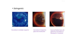
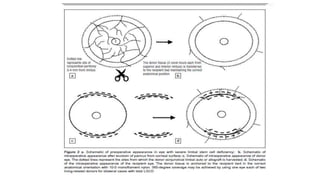
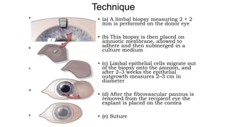
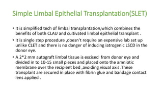
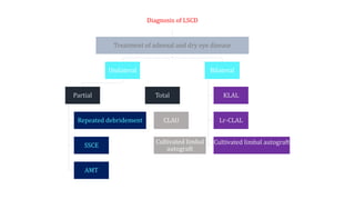
Limbal stem cell deficiency:Current
Management
( TAKEN FROM-Journal of Clinical Ophthalmology & Research) JAN –APRIL 2016 VOL 4 ISSUE 1
Authors:Ikeda Lal,Neha Gupta, Jyoti Purushotham,Virender S.Sangwan
(by DR PAWAN JARWAL ,
RESIDENT DOCTOR ,
SSG HOSPITAL
VADODARA.)
INTRODUCTION
• Stem cells-
• Undifferentiated proliferating cells
• Present in all self-renewing tissues e.g blood, testis and stratified sq. epithelia.
• 0.5%-10% of total cell population
• Properties:
• Long lived, long cell cycle time
• have increased potential for error free proliferation with poor differentiation
• capability to divide in asymmetric manner
RELEVANT ANATOMY
• The corneal epithelium -nonkeratinised, stratified squamous
epithelial cells.
• Thickness ~50 μm
• Limbus-
• CORNEA-stratified, nonkeratinised squamous epithelium
• CONJUNCTIVA-stratified, nonkeratinised columnar vascularizedepithelium
with mucin-secreting goblet cells
• architecture of the limbus - palisade (of Vogt) arrangement.
SITE OF LIMBAL STEM CELL;
• The limbal stem cells probably reside in the basal layer of the palisades of Vogt
• Limbal stem cell- boundary of cornea, conjunctiva and sclera
• One division of each LESC generates a daughter TAC that migrates
centrally across the cornea while the original stem cell remains
within its niche in the basal epithelium of the limbus
• TACs (Transient amplifying cells)- divide rapidly in basal cell layer
• PMCs (Post mitotic cells)- wing cell layer
• TDCs (Terminally differentiated cells)- squamous layer
• The result of this migration and differentiation is that the corneal
epithelium is renewed every 7–10 days in this manner
LESC Proliferation
PROLIFERATION OF LIMBAL STEM CELLS
TAC
STEM CELL
PMC
TDC
STESTEM CELL RESERVOIRM
CORNEAL EPITHELIUM
The XYZ Hypothesis:
• X vector - combination of proliferation and centripetal migration of
the basal epithelial cells.
• Xs - asymmetric division of limbal epithelial stem cells
• one daughter cell that remains in the limbus as a stem cell
• a second daughter cell which migrates out of the limbus into
the basal corneal epithelium - transient amplifying cell (X TA ).
• Th e X TA vector arises through a combination of proliferation and
centripetal migration of the basal TACs.
• Y vector- As the basal epithelial cells divide they give rise to
suprabasal cells that form the stratified layers of the cornea.
• Z vector - shedding of squamous epithelial cells from the surface of
cornea into the tear fi lm.
TYPES OF LIMBAL STEM CELL DEFICIENCY
• According to Etiology:
• Primary
• Secondary
• PRIMARY-
• related to an insufficient stromal microenvironment to support stem cell function
• presumed site of pathophysiology -limbal stromal niche
• Eg.-
• CONGENITAL:
• aniridia, dominantly inherited keratitis, Ectodermal dysplasia
• ACQUIRED:
• neurotrophic (neural and ischaemic) keratopathy and chronic limbitis
• SECONDARY-(more common)-
• ACQUIRED:
• related to external factors that destroy limbal stem cells
• presumed site of pathophysiology – Limbal cells themselves
• Eg.-
• Traumatic- chemical (most common) or thermal
injuries, iatrogenic (multiple surgeries or cryotherapies)
• Stevens-Johnson Syndrome
• ocular cicatricial pemphigoid (OCP)
• contact lens wear
• extensive microbial infection
TYPES
• Iatrogenic:
Secondary to multiple surgeries Secondary to long term
glaucoma medication
Sectorial limbal stem cell
deficiency Secondary to
Mitomycin C treatment
TYPES
• According to extent of involvement:
• Sectorial
• Diffuse
• SECTORIAL (PARTIAL)
• localized deficiency of LESCs in a region of limbus but an intact
population of LESCs in other areas.
• Microscopically:
• columnar keratopathy
• mosaic pattern of stain with impression cytology
TYPES
• DIFFUSE (TOTAL)
• functional loss of the entire LESC population
• conjunctivalization of the entire cornea
TYPES
CLINICAL MANIFESTATIONS
• SYMPTOMS:
• Tearing
• Blepharospasm
• Photophobia
• decreased vision
• recurrent episodes of pain (epithelial
breakdown)
• history of chronic inflammation with
redness
• SIGNS:
• The presence of a conjunctival phenotype on the cornea
(conjunctival overgrowth, conjunctivalization) is central to
the diagnosis of LSCD
• dull and irregular reflex of the corneal epithelium which
varies in thickness and transparency
• an ingrowth of thickened fibrovascular pannus, chronic
keratitis, scarring and calcification.
• Persistent epithelial defects- stippled fluorescein staining
• melting and perforation of the cornea can occur
• Partial stem cell deficiency-
• sectoral ingrowth of conjunctival epithelium from focal areas of SC
deficiency
• clear line of demarcation- often, but not always, visible between
corneal and conjunctival phenotype of cells
• At the line of contact of the two phenotypes, tiny "bud like projections"
of normal corneal epithelium can be seen extending into the
conjunctivalised area
• fluorescein pooling on the conjunctivalised side -because of its relative
thinness
DIAGNOSTIC TOOLS
• Diagnosis is crucial because these patients are poor candidates for
conventional corneal transplantation
• Histologically (impression cytology)
• goblet cells containing conjunctival epithelium on the corneal
surface
• In advanced disease- especially those where keratinisation of the
epithelium occurs (SJS, ocular pemphigoid, Lyell syndrome),
conjunctival goblet cells may be completely absent-not detectable
• Immuno histo-chemically (monoclonal antibodies)
• absence of a cornea-type differentiation (such as the absence of
keratin CK3,12)
• Presence of conjunctival phenotype (CK19)
AVAILABLE TREATMENT OPTIONS
• PARTIAL LIMBAL STEM CELL DEFICIENCY-
• CONSERVATIVE OPTIONS:
• In Acute phase:
• Immunosuppresion-
• Topical steroids
• Cyclosporine
• use of intensive non-preserved lubrication
• bandage contact lenses
• autologous serum eye drops.
• Only the latter is supported by evidence in the literature
Conservative treatment usually provides temporary remission but
the condition tends to deteriorate over time.
• SURGICAL OPTIONS:
• In the acute phase following injury-
• repeated debridement of migrating conjunctival epithelium
(sequential sector conjunctival epitheliectomy (SSCE)- can reduce or
prevent conjunctival ingrowth.
• The use of an amniotic membrane graft has also been reported to be
successful
• TOTAL LIMBAL STEM CELL DEFICIENCY-
to restore a corneal phenotype- ocular surface
reconstruction (OSR) is required
Indicated in-
bilateral blinding ocular surface diseases such as Stevens Johnson
syndrome (SJS), ocular cicatricial pemphigoid (OCP), and severe
chemical/ thermal burns.
SURGICAL OPTIONS
Clinically, the process involves a sequential three-step
approach.
I. Correct any dry eye disease and lid abnormality that is
contributing to ocular surface failure
• correction of
• meibomian gland dysfunction
• Corneal exposure
• Trichiasis
• entropion
• Punctal occlusion
• Repair of symblepharon
• frequent application of preservative-free artificial tears or autologous serum
SURGICAL OPTIONS
II. Remove the conjunctival epithelium from the cornea
and restore a normal stromal environment
• Debridement of abnormal conjunctival epithelium and
subepithelial fi brous tissue
• mechanically -combined tissue peeled off the cornea.
• peritomy and resection of the conjunctival epithelium for up to 4 mm
from the limbus, with application of mitomycin C over the exposed
sclera to reduce recurrence of scarring and subepithelial fibroblastic
proliferation.
SURGICAL OPTIONS
III. Transplant corneal LESCs to re-establish an intact
and transparent epithelium
• conjunctival limbal autograft (CLAU)
• living-related conjunctival limbal allograft (Lr-CLAL)
• Keratolimbal allograft (KLAL)
• Ex- vivo expansion and transplantation of cultured LESCs.
• Simple limbal epithelial transplantion (SLET)
SURGICAL OPTIONS
• CONJUNCTIVAL LIMBAL AUTOGRAFT (CLAU)
• Transfer of autologous limbal tissue from the
unaffected fellow eye to the stem cell deficient eye.
• Imperative to exclude LSCD in the donor tissue
• Optimum amount of limbal tissue.
• Conservative removal to prevent iatrogenic LSCD in donor eye
SURGICAL OPTIONS
SURGICAL OPTIONS
LIVING-RELATED CONJUNCTIVAL LIMBAL ALLOGRAFT TRANSPLANT
(Lr-CLAL)
• In bilateral total LSCD - only potential source of LESCs -
allogenic limbus
• The surgical technique - identical to CLAU
• Amniotic membrane can be used similarly-
• To eliminate the concern of removing LESCs from the
healthy donor eye
• To augment the effect of CLAU in the recipient eye
• Risk - rejection of a Lr- CLAL
• systemic immunosuppression required
SURGICAL OPTIONS
Keratolimbal allograft ( KLAL)
• Cadaveric tissue, the stem cell carrier may be either
conjunctiva or cornea
• Donor Tissue Selection :
• Youngest possible donor with an upper limit of 50 years
• Surgery should be performed within 72 hours
• Systemic Immuno-suppression required
Surgical technique
• Corneo scleral rim (4-5mm wide) of cadaveric eye is taken & central
7.5mm of corneal rim is removed
• Corneo-scleral rim is cut into 2 equal halves
• For 4 crescents, we require both eyes of the cadaver
• By lamellar dissection , posterior half of each lenticule is removed (post
sclera + stroma + Dm + endothelium)
• Cover 360 degrees of recipient with donor tissue
• Suture the edges – patch eye with shield
• Most important limiting factor- Allograft
rejection (despite systemic immunosuppression)
• Signs of allograft rejection-
• Telangiectasia and engorged limbal blood vessels
• Epithelial rejection lines and epithelial breakdown
• Punctate epithelial keratopathy
• Severe limbal inflammation
• Elevated Perilimbal area
SURGICAL OPTIONS
Ex Vivo EXPANSION AND TRANSPLANTATION OF
CULTURED LIMBAL STEM CELLS
• Most exciting and promising technique currently
• Substantially smaller size of the limbal biopsy is
required (although more than one biopsy may be
required to obtain a successful explant or cell
culture)
• Minimizes the risk of precipitating stem cell failure in
the donor eye and provides the option for a second
biopsy if necessary
• Less chances of rejection as only epithelial cells are
transplanted
SURGICAL OPTIONS
• (a) A limbal biopsy measuring 2 × 2
mm is performed on the donor eye
• (b) This biopsy is then placed on
amniotic membrane, allowed to
adhere and then submerged in a
culture medium
• (c) Limbal epithelial cells migrate out
of the biopsy onto the amnion, and
after 2–3 weeks the epithelial
outgrowth measures 2–3 cm in
diameter
• (d) After the fibrovascular pannus is
removed from the recipient eye the
explant is placed on the cornea
• (e) Suture
Technique
Simple Limbal Epithelial Transplantation(SLET)
• It is simplified tech of limbal transplantation,which combines the
benefits of both CLAU and cultivated limbal epithelial transplant .
• It is single step procedure ,doesn’t require an expensive lab set up
unlike CLET and there is no danger of inducing iatrogenic LSCD in the
donor eye.
• A 2*2 mm autograft limbal tissue is excised from donor eye and
divided in to 10-15 small pieces and placed onto the amniotic
membrane over the recipient bed ,avoiding visual axis .These
transplant are secured in place with fibrin glue and bandage contact
lens applied .
Diagnosis of LSCD
Treatment of adnexal and dry eye disease
Unilateral
Partial
Repeated debridement
SSCE
AMT
Total
CLAU
Cultivated limbal
autograft
Bilateral
KLAL
Lr-CLAL
Cultivated limbal autograft
Significance of Limbal Stem Cell Transplantation
• Effective method for limbal stem cell deficiency to recover the integrity of
ocular surface and rebuild limbal barrier
• Successful limbal transplantation can achieve:
• rapid surface healing
• stable ocular surface without recurrent erosions or persistent epithelial defects
• regression of corneal vascularization
• restoration of a smooth and optically improved ocular surface
• resulting in:
• improved visual acuity
• probably, increased success for subsequent keratoplasty.
RECENT ADVANCES
• ALTERNATIVE SOURCES OF AUTOLOGOUS STEM CELLS
• Oral mucosa:
• Potential advantages-
• the cells are autologous- no risk of immune mediated rejection- immuosuppression is not
required.
• oral mucosa -lower stage of differentiation than epidermal keratinocytes- they divide rapidly -
can be maintained in culture for prolonged periods without keratinization.
• Theoretical disadvantage -
• In treatment of autoimmune diseases (such as OCP) is that the oral and ocular mucosa may
both secrete a common basement membrane target antigen
• Co-cultivated limbal and conjunctival epithelium:
• Central area of HAM (human amniotic membrane) has limbal epithelial cells
• Peripheral area- conjunctival epithelial cells
• Advantage- total ocular surface reconstruction simultaneously
• Other autologous sources:
• Hemopoetic stem cells
• Advantage- better long term survival without need of immunosuppression
THANK YOU

Limbal stem cell deficiency

  • 1.
    Limbal stem celldeficiency:Current Management ( TAKEN FROM-Journal of Clinical Ophthalmology & Research) JAN –APRIL 2016 VOL 4 ISSUE 1 Authors:Ikeda Lal,Neha Gupta, Jyoti Purushotham,Virender S.Sangwan (by DR PAWAN JARWAL , RESIDENT DOCTOR , SSG HOSPITAL VADODARA.)
  • 2.
    INTRODUCTION • Stem cells- •Undifferentiated proliferating cells • Present in all self-renewing tissues e.g blood, testis and stratified sq. epithelia. • 0.5%-10% of total cell population • Properties: • Long lived, long cell cycle time • have increased potential for error free proliferation with poor differentiation • capability to divide in asymmetric manner
  • 3.
    RELEVANT ANATOMY • Thecorneal epithelium -nonkeratinised, stratified squamous epithelial cells. • Thickness ~50 μm • Limbus- • CORNEA-stratified, nonkeratinised squamous epithelium • CONJUNCTIVA-stratified, nonkeratinised columnar vascularizedepithelium with mucin-secreting goblet cells • architecture of the limbus - palisade (of Vogt) arrangement.
  • 4.
    SITE OF LIMBALSTEM CELL; • The limbal stem cells probably reside in the basal layer of the palisades of Vogt • Limbal stem cell- boundary of cornea, conjunctiva and sclera
  • 5.
    • One divisionof each LESC generates a daughter TAC that migrates centrally across the cornea while the original stem cell remains within its niche in the basal epithelium of the limbus • TACs (Transient amplifying cells)- divide rapidly in basal cell layer • PMCs (Post mitotic cells)- wing cell layer • TDCs (Terminally differentiated cells)- squamous layer • The result of this migration and differentiation is that the corneal epithelium is renewed every 7–10 days in this manner LESC Proliferation
  • 6.
    PROLIFERATION OF LIMBALSTEM CELLS TAC STEM CELL PMC TDC STESTEM CELL RESERVOIRM CORNEAL EPITHELIUM
  • 7.
    The XYZ Hypothesis: •X vector - combination of proliferation and centripetal migration of the basal epithelial cells. • Xs - asymmetric division of limbal epithelial stem cells • one daughter cell that remains in the limbus as a stem cell • a second daughter cell which migrates out of the limbus into the basal corneal epithelium - transient amplifying cell (X TA ). • Th e X TA vector arises through a combination of proliferation and centripetal migration of the basal TACs. • Y vector- As the basal epithelial cells divide they give rise to suprabasal cells that form the stratified layers of the cornea. • Z vector - shedding of squamous epithelial cells from the surface of cornea into the tear fi lm.
  • 8.
    TYPES OF LIMBALSTEM CELL DEFICIENCY • According to Etiology: • Primary • Secondary • PRIMARY- • related to an insufficient stromal microenvironment to support stem cell function • presumed site of pathophysiology -limbal stromal niche • Eg.- • CONGENITAL: • aniridia, dominantly inherited keratitis, Ectodermal dysplasia • ACQUIRED: • neurotrophic (neural and ischaemic) keratopathy and chronic limbitis
  • 9.
    • SECONDARY-(more common)- •ACQUIRED: • related to external factors that destroy limbal stem cells • presumed site of pathophysiology – Limbal cells themselves • Eg.- • Traumatic- chemical (most common) or thermal injuries, iatrogenic (multiple surgeries or cryotherapies) • Stevens-Johnson Syndrome • ocular cicatricial pemphigoid (OCP) • contact lens wear • extensive microbial infection TYPES
  • 10.
    • Iatrogenic: Secondary tomultiple surgeries Secondary to long term glaucoma medication Sectorial limbal stem cell deficiency Secondary to Mitomycin C treatment TYPES
  • 11.
    • According toextent of involvement: • Sectorial • Diffuse • SECTORIAL (PARTIAL) • localized deficiency of LESCs in a region of limbus but an intact population of LESCs in other areas. • Microscopically: • columnar keratopathy • mosaic pattern of stain with impression cytology TYPES
  • 12.
    • DIFFUSE (TOTAL) •functional loss of the entire LESC population • conjunctivalization of the entire cornea TYPES
  • 13.
    CLINICAL MANIFESTATIONS • SYMPTOMS: •Tearing • Blepharospasm • Photophobia • decreased vision • recurrent episodes of pain (epithelial breakdown) • history of chronic inflammation with redness
  • 14.
    • SIGNS: • Thepresence of a conjunctival phenotype on the cornea (conjunctival overgrowth, conjunctivalization) is central to the diagnosis of LSCD • dull and irregular reflex of the corneal epithelium which varies in thickness and transparency • an ingrowth of thickened fibrovascular pannus, chronic keratitis, scarring and calcification. • Persistent epithelial defects- stippled fluorescein staining • melting and perforation of the cornea can occur
  • 15.
    • Partial stemcell deficiency- • sectoral ingrowth of conjunctival epithelium from focal areas of SC deficiency • clear line of demarcation- often, but not always, visible between corneal and conjunctival phenotype of cells • At the line of contact of the two phenotypes, tiny "bud like projections" of normal corneal epithelium can be seen extending into the conjunctivalised area • fluorescein pooling on the conjunctivalised side -because of its relative thinness
  • 16.
    DIAGNOSTIC TOOLS • Diagnosisis crucial because these patients are poor candidates for conventional corneal transplantation • Histologically (impression cytology) • goblet cells containing conjunctival epithelium on the corneal surface • In advanced disease- especially those where keratinisation of the epithelium occurs (SJS, ocular pemphigoid, Lyell syndrome), conjunctival goblet cells may be completely absent-not detectable • Immuno histo-chemically (monoclonal antibodies) • absence of a cornea-type differentiation (such as the absence of keratin CK3,12) • Presence of conjunctival phenotype (CK19)
  • 17.
    AVAILABLE TREATMENT OPTIONS •PARTIAL LIMBAL STEM CELL DEFICIENCY- • CONSERVATIVE OPTIONS: • In Acute phase: • Immunosuppresion- • Topical steroids • Cyclosporine • use of intensive non-preserved lubrication • bandage contact lenses • autologous serum eye drops. • Only the latter is supported by evidence in the literature Conservative treatment usually provides temporary remission but the condition tends to deteriorate over time.
  • 18.
    • SURGICAL OPTIONS: •In the acute phase following injury- • repeated debridement of migrating conjunctival epithelium (sequential sector conjunctival epitheliectomy (SSCE)- can reduce or prevent conjunctival ingrowth. • The use of an amniotic membrane graft has also been reported to be successful
  • 19.
    • TOTAL LIMBALSTEM CELL DEFICIENCY- to restore a corneal phenotype- ocular surface reconstruction (OSR) is required Indicated in- bilateral blinding ocular surface diseases such as Stevens Johnson syndrome (SJS), ocular cicatricial pemphigoid (OCP), and severe chemical/ thermal burns. SURGICAL OPTIONS
  • 20.
    Clinically, the processinvolves a sequential three-step approach. I. Correct any dry eye disease and lid abnormality that is contributing to ocular surface failure • correction of • meibomian gland dysfunction • Corneal exposure • Trichiasis • entropion • Punctal occlusion • Repair of symblepharon • frequent application of preservative-free artificial tears or autologous serum SURGICAL OPTIONS
  • 21.
    II. Remove theconjunctival epithelium from the cornea and restore a normal stromal environment • Debridement of abnormal conjunctival epithelium and subepithelial fi brous tissue • mechanically -combined tissue peeled off the cornea. • peritomy and resection of the conjunctival epithelium for up to 4 mm from the limbus, with application of mitomycin C over the exposed sclera to reduce recurrence of scarring and subepithelial fibroblastic proliferation. SURGICAL OPTIONS
  • 22.
    III. Transplant cornealLESCs to re-establish an intact and transparent epithelium • conjunctival limbal autograft (CLAU) • living-related conjunctival limbal allograft (Lr-CLAL) • Keratolimbal allograft (KLAL) • Ex- vivo expansion and transplantation of cultured LESCs. • Simple limbal epithelial transplantion (SLET) SURGICAL OPTIONS
  • 23.
    • CONJUNCTIVAL LIMBALAUTOGRAFT (CLAU) • Transfer of autologous limbal tissue from the unaffected fellow eye to the stem cell deficient eye. • Imperative to exclude LSCD in the donor tissue • Optimum amount of limbal tissue. • Conservative removal to prevent iatrogenic LSCD in donor eye SURGICAL OPTIONS
  • 24.
  • 25.
    LIVING-RELATED CONJUNCTIVAL LIMBALALLOGRAFT TRANSPLANT (Lr-CLAL) • In bilateral total LSCD - only potential source of LESCs - allogenic limbus • The surgical technique - identical to CLAU • Amniotic membrane can be used similarly- • To eliminate the concern of removing LESCs from the healthy donor eye • To augment the effect of CLAU in the recipient eye • Risk - rejection of a Lr- CLAL • systemic immunosuppression required SURGICAL OPTIONS
  • 26.
    Keratolimbal allograft (KLAL) • Cadaveric tissue, the stem cell carrier may be either conjunctiva or cornea • Donor Tissue Selection : • Youngest possible donor with an upper limit of 50 years • Surgery should be performed within 72 hours • Systemic Immuno-suppression required
  • 27.
    Surgical technique • Corneoscleral rim (4-5mm wide) of cadaveric eye is taken & central 7.5mm of corneal rim is removed • Corneo-scleral rim is cut into 2 equal halves • For 4 crescents, we require both eyes of the cadaver • By lamellar dissection , posterior half of each lenticule is removed (post sclera + stroma + Dm + endothelium) • Cover 360 degrees of recipient with donor tissue • Suture the edges – patch eye with shield
  • 29.
    • Most importantlimiting factor- Allograft rejection (despite systemic immunosuppression) • Signs of allograft rejection- • Telangiectasia and engorged limbal blood vessels • Epithelial rejection lines and epithelial breakdown • Punctate epithelial keratopathy • Severe limbal inflammation • Elevated Perilimbal area SURGICAL OPTIONS
  • 30.
    Ex Vivo EXPANSIONAND TRANSPLANTATION OF CULTURED LIMBAL STEM CELLS • Most exciting and promising technique currently • Substantially smaller size of the limbal biopsy is required (although more than one biopsy may be required to obtain a successful explant or cell culture) • Minimizes the risk of precipitating stem cell failure in the donor eye and provides the option for a second biopsy if necessary • Less chances of rejection as only epithelial cells are transplanted SURGICAL OPTIONS
  • 31.
    • (a) Alimbal biopsy measuring 2 × 2 mm is performed on the donor eye • (b) This biopsy is then placed on amniotic membrane, allowed to adhere and then submerged in a culture medium • (c) Limbal epithelial cells migrate out of the biopsy onto the amnion, and after 2–3 weeks the epithelial outgrowth measures 2–3 cm in diameter • (d) After the fibrovascular pannus is removed from the recipient eye the explant is placed on the cornea • (e) Suture Technique
  • 32.
    Simple Limbal EpithelialTransplantation(SLET) • It is simplified tech of limbal transplantation,which combines the benefits of both CLAU and cultivated limbal epithelial transplant . • It is single step procedure ,doesn’t require an expensive lab set up unlike CLET and there is no danger of inducing iatrogenic LSCD in the donor eye. • A 2*2 mm autograft limbal tissue is excised from donor eye and divided in to 10-15 small pieces and placed onto the amniotic membrane over the recipient bed ,avoiding visual axis .These transplant are secured in place with fibrin glue and bandage contact lens applied .
  • 33.
    Diagnosis of LSCD Treatmentof adnexal and dry eye disease Unilateral Partial Repeated debridement SSCE AMT Total CLAU Cultivated limbal autograft Bilateral KLAL Lr-CLAL Cultivated limbal autograft
  • 34.
    Significance of LimbalStem Cell Transplantation • Effective method for limbal stem cell deficiency to recover the integrity of ocular surface and rebuild limbal barrier • Successful limbal transplantation can achieve: • rapid surface healing • stable ocular surface without recurrent erosions or persistent epithelial defects • regression of corneal vascularization • restoration of a smooth and optically improved ocular surface • resulting in: • improved visual acuity • probably, increased success for subsequent keratoplasty.
  • 35.
    RECENT ADVANCES • ALTERNATIVESOURCES OF AUTOLOGOUS STEM CELLS • Oral mucosa: • Potential advantages- • the cells are autologous- no risk of immune mediated rejection- immuosuppression is not required. • oral mucosa -lower stage of differentiation than epidermal keratinocytes- they divide rapidly - can be maintained in culture for prolonged periods without keratinization. • Theoretical disadvantage - • In treatment of autoimmune diseases (such as OCP) is that the oral and ocular mucosa may both secrete a common basement membrane target antigen
  • 36.
    • Co-cultivated limbaland conjunctival epithelium: • Central area of HAM (human amniotic membrane) has limbal epithelial cells • Peripheral area- conjunctival epithelial cells • Advantage- total ocular surface reconstruction simultaneously • Other autologous sources: • Hemopoetic stem cells • Advantage- better long term survival without need of immunosuppression
  • 37.