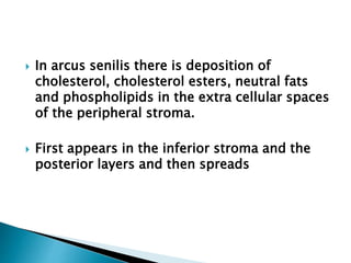
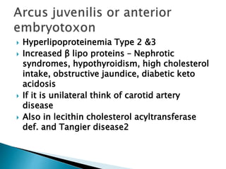
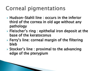
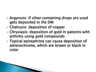
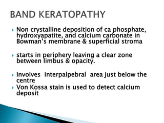
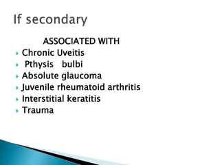
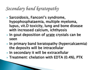
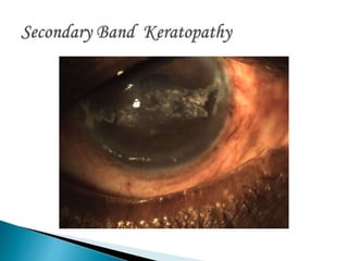
Dr. K. Vasantha M.S., F.R.C.S.
Director RIO Chennai (Rtd)
 Bilateral, symmetrical
 Mostly central cornea
 Non-inflammatory,
hereditary disorders,
little or no
relationship to
systemic or
environmental
factors
 Not accompanied by
vascularisation
 Mostly AD
 Unilateral,asymmetric
 Mostly peripheral
cornea
 D/t aging,trauma,
inflammation or
systemic diseases
 Accompanied by
vascularisation
 Deterioration or
change in a normal
tissue that renders
the tissue less
functional
Corneal degenerations
 Bilateral-type 2
hyper-
lipoproteinemia
 Gray/white/yello
w band
separated from
limbus by a
clear zone of
0.2-0.3 mm
clear zone called
lucid interval of
Vogt. This may
get thinned-
senile furrow
Diffuse central &
sharp peripheral
border
 In arcus senilis there is deposition of
cholesterol, cholesterol esters, neutral fats
and phospholipids in the extra cellular spaces
of the peripheral stroma.
 First appears in the inferior stroma and the
posterior layers and then spreads
 Hyperlipoproteinemia Type 2 &3
 Increased β lipo proteins – Nephrotic
syndromes, hypothyroidism, high cholesterol
intake, obstructive jaundice, diabetic keto
acidosis
 If it is unilateral think of carotid artery
disease
 Also in lecithin cholesterol acyltransferase
def. and Tangier disease2
 Hudson-Stahli line : occurs in the inferior
third of the cornea in old age without any
pathology
 Fleischer’s ring : epithelial iron deposit at the
base of the keratoconus
 Ferry’s line: corneal margin of the filtering
bleb
 Stocker’s line : proximal to the advancing
edge of the pterygium
 Argyrosis: if silver containing drops are used
gets deposited in the DM
 Chalcosis: deposition of copper
 Chrysiasis: deposition of gold in patients with
arthritis using gold compounds
 Topical epinephrine can cause deposition of
adrenochrome, which are brown or black in
color
 Iron deposition at level of
bowman’s layer
 Associated with previous corneal
foreign body
 White crescent like
line along nasal &
temporal limbus
 Type1-seperated
from limbus by a
clear zone
 Type 2-no clear zone
between it & limbus
 HPE: subepithelial
lesion with
hyperelastosis & mild
hyaline degeneration
 Area within interpalpebral fissure is affected.
Centre of band will be slightly inferior to the
centre of cornea
 Swiss-cheese like appearance: clear circular
areas within the band is seen where nerve
endings perforate Bowman’s layer
 Non crystalline deposition of ca phosphate,
hydroxyapatite, and calcium carbonate in
Bowman’s membrane & superficial stroma
 starts in periphery leaving a clear zone
between limbus & opacity.
 Involves interpalpebral area just below the
centre
 Von Kossa stain is used to detect calcium
deposit
ASSOCIATED WITH
 Chronic Uveitis
 Pthysis bulbi
 Absolute glaucoma
 Juvenile rheumatoid arthritis
 Interstitial keratitis
 Trauma
 Sarcoidosis, Fanconi’s syndrome,
hypophosphataemia, multiple myeloma,
lupus, vit.D toxicity, lung and bone disease
with increased calcium, ichthyosis
 In gout deposition of urate crystals can be
seen
 In primary band keratopathy (hypercalcaemia)
the deposits will be intracellular
 In secondary it will be extracellular
 Treatment: chelation with EDTA (0.4%), PTK
 Sometimes in long
standing opacities
calcium deposits will
occur. If the deposit
falls off it disrupts
the epithelium
forming an ulcer
called atheromatous
ulcer
 Unlike band keratopathy it involves deeper
layers of cornea also.
 Seen in grossly distorted globes and
leucomas
 Maybe associated with intraocular bone
formation
 In corneas that had been
inflamed many years
earlier
 E.g.: phlyctenular
keratitis, trachoma,
vernal keratitis etc.,
 Elevated blue-grey,
fibrous nodules in
superficial stroma, just
beneath epithelium
 Base of nodule maybe
surrounded by iron
deposition
 HPE: mound of dense
collagenous tissue
Common characteristics of both Pellucid and
Terrien’s are
 Non inflammatory
 Slowly progressive
 Epithelium and endothelium are normal
 Stromal thinning
 Bowman’s will be disrupted
 Defective vision due to against the rule
astigmatism
 Bilateral, narrow band of thinning in periphery
with normal tissue between limbus &
degenerated cornea in the inferior quadrant
 Typical kissing birds sign with topography
 High astigmatism, keratoconus or keratoglobus
can be seen in other members of the family
 Treatment – C shaped lamellar or full thickness
graft
 Hybrid or scleral lenses
 Very rarely hydrops can occur
 Unilateral or asymmetrically bilateral
 Fine, punctate stromal opacity with a lucid
zone is seen in the superior cornea
 Lucid zone becomes superficially
vascularised and can form a pseudo
pterygium
 The thinning is parallel to limbus
 Lipid deposits are seen in the lower margin
which is steep but no over hanging edge as
in Mooren’s
 Corneal elastosis, Labrador
keratopathy, climatic droplet
keratopathy, Bietti nodular
dystrophy etc.,
 Predisposing factor: UV-
exposure
 Clusters of fine, yellow, gray,
amber or gold colored droplets
are seen beneath epithelium of
cornea
 Begins peripherally & advances
towards the centre
 Dark red
proteinaceous
deposits are seen in
the anterior stroma
replacing BM
 Type 1. bilateral without other ocular
pathologies
 Type 2. Secondary to other ocular pathology
 Type 3. with conjunctival deposits either with
type one or two
 The deposits contain tryptophan, tyrosine
and other sulphur containing amino acids.
 Immunoflorescence techniques show
presence of IgA and IgG
 If the lesion is superficial without
involvement of the Bowman’s, simple
scraping or PTK can be done
 If it is deeper lamellar keratoplasty is done
 Ocular surface disorders like dry eye must be
looked for as it is prevalent in these patients
 Can occur primarily
in normal cornea
 Secondary to other
chronic
inflammations
 Treat the
vascularisation first
 Keratoplasty
 Discrete, tiny, gray/white opacities, scattered
throughout deep corneal stroma-FLOUR
DUST APPEARANCE
 HPE: lipofuscin like material-due to ageing,
wear & tear
 Punctate and filamentous opacities;
appear transparent and glassy on
retro illumination
 Lesions are axial and posteriorly
located
 SENILE FURROW DEGN: in the lucid interval
between an arcus & limbus
 CORNEA GUTTATA: common, bilateral, tiny
dark spots on central endothelium
rarely progress to Fuchs’ dystrophy
 HASSAL-HENLE BODIES/WARTS: localized areas
of nodular thickening in peripheral cornea
 Filamentary keratitis
 K.F. ring
 Seen in severe dry eye – to be treated with
cyclosporine and artificial tear drops
 Superior limbic kerato conjunctivitis
 Mucus strands attached to the cornea are
surrounded by epithelium. These have to be
sometimes removed and anterior stromal
puncture done to prevent recurrence
 Deposition of copper in the Descemet’s
membrane
 Starts in the periphery. So if a patient is
referred to rule out K.F. ring one must see
with a gonioscope also
 Seen in chronic liver damage due to Wilson’s
disease
 In Wilson’s disease it is usually starts at the
age of around ten.
 It is also seen in primary biliary cirrhosis and
familial cholestatic disease
 Diffuse deposits are seen in pulmonary
carcinoma and multiple myeloma
 Bilateral
 Present at birth, with uniform thinning
 Familial association with keratoconus has
been noticed
 Seen in Ehlers- Danlos also
 Spectacle correction will yield reasonable
vision
 Large diameter lamellar or penetrating
keratoplasty can be tried
Corneal degenerations

Corneal degenerations

  • 1.
    Dr. K. VasanthaM.S., F.R.C.S. Director RIO Chennai (Rtd)
  • 2.
     Bilateral, symmetrical Mostly central cornea  Non-inflammatory, hereditary disorders, little or no relationship to systemic or environmental factors  Not accompanied by vascularisation  Mostly AD  Unilateral,asymmetric  Mostly peripheral cornea  D/t aging,trauma, inflammation or systemic diseases  Accompanied by vascularisation  Deterioration or change in a normal tissue that renders the tissue less functional Corneal degenerations
  • 3.
     Bilateral-type 2 hyper- lipoproteinemia Gray/white/yello w band separated from limbus by a clear zone of 0.2-0.3 mm clear zone called lucid interval of Vogt. This may get thinned- senile furrow Diffuse central & sharp peripheral border
  • 4.
     In arcussenilis there is deposition of cholesterol, cholesterol esters, neutral fats and phospholipids in the extra cellular spaces of the peripheral stroma.  First appears in the inferior stroma and the posterior layers and then spreads
  • 5.
     Hyperlipoproteinemia Type2 &3  Increased β lipo proteins – Nephrotic syndromes, hypothyroidism, high cholesterol intake, obstructive jaundice, diabetic keto acidosis  If it is unilateral think of carotid artery disease  Also in lecithin cholesterol acyltransferase def. and Tangier disease2
  • 6.
     Hudson-Stahli line: occurs in the inferior third of the cornea in old age without any pathology  Fleischer’s ring : epithelial iron deposit at the base of the keratoconus  Ferry’s line: corneal margin of the filtering bleb  Stocker’s line : proximal to the advancing edge of the pterygium
  • 7.
     Argyrosis: ifsilver containing drops are used gets deposited in the DM  Chalcosis: deposition of copper  Chrysiasis: deposition of gold in patients with arthritis using gold compounds  Topical epinephrine can cause deposition of adrenochrome, which are brown or black in color
  • 8.
     Iron depositionat level of bowman’s layer  Associated with previous corneal foreign body
  • 9.
     White crescentlike line along nasal & temporal limbus  Type1-seperated from limbus by a clear zone  Type 2-no clear zone between it & limbus  HPE: subepithelial lesion with hyperelastosis & mild hyaline degeneration
  • 10.
     Area withininterpalpebral fissure is affected. Centre of band will be slightly inferior to the centre of cornea  Swiss-cheese like appearance: clear circular areas within the band is seen where nerve endings perforate Bowman’s layer
  • 12.
     Non crystallinedeposition of ca phosphate, hydroxyapatite, and calcium carbonate in Bowman’s membrane & superficial stroma  starts in periphery leaving a clear zone between limbus & opacity.  Involves interpalpebral area just below the centre  Von Kossa stain is used to detect calcium deposit
  • 13.
    ASSOCIATED WITH  ChronicUveitis  Pthysis bulbi  Absolute glaucoma  Juvenile rheumatoid arthritis  Interstitial keratitis  Trauma
  • 14.
     Sarcoidosis, Fanconi’ssyndrome, hypophosphataemia, multiple myeloma, lupus, vit.D toxicity, lung and bone disease with increased calcium, ichthyosis  In gout deposition of urate crystals can be seen  In primary band keratopathy (hypercalcaemia) the deposits will be intracellular  In secondary it will be extracellular  Treatment: chelation with EDTA (0.4%), PTK
  • 16.
     Sometimes inlong standing opacities calcium deposits will occur. If the deposit falls off it disrupts the epithelium forming an ulcer called atheromatous ulcer
  • 17.
     Unlike bandkeratopathy it involves deeper layers of cornea also.  Seen in grossly distorted globes and leucomas  Maybe associated with intraocular bone formation
  • 18.
     In corneasthat had been inflamed many years earlier  E.g.: phlyctenular keratitis, trachoma, vernal keratitis etc.,  Elevated blue-grey, fibrous nodules in superficial stroma, just beneath epithelium  Base of nodule maybe surrounded by iron deposition  HPE: mound of dense collagenous tissue
  • 19.
    Common characteristics ofboth Pellucid and Terrien’s are  Non inflammatory  Slowly progressive  Epithelium and endothelium are normal  Stromal thinning  Bowman’s will be disrupted  Defective vision due to against the rule astigmatism
  • 21.
     Bilateral, narrowband of thinning in periphery with normal tissue between limbus & degenerated cornea in the inferior quadrant  Typical kissing birds sign with topography  High astigmatism, keratoconus or keratoglobus can be seen in other members of the family  Treatment – C shaped lamellar or full thickness graft  Hybrid or scleral lenses  Very rarely hydrops can occur
  • 22.
     Unilateral orasymmetrically bilateral  Fine, punctate stromal opacity with a lucid zone is seen in the superior cornea  Lucid zone becomes superficially vascularised and can form a pseudo pterygium  The thinning is parallel to limbus  Lipid deposits are seen in the lower margin which is steep but no over hanging edge as in Mooren’s
  • 23.
     Corneal elastosis,Labrador keratopathy, climatic droplet keratopathy, Bietti nodular dystrophy etc.,  Predisposing factor: UV- exposure  Clusters of fine, yellow, gray, amber or gold colored droplets are seen beneath epithelium of cornea  Begins peripherally & advances towards the centre  Dark red proteinaceous deposits are seen in the anterior stroma replacing BM
  • 24.
     Type 1.bilateral without other ocular pathologies  Type 2. Secondary to other ocular pathology  Type 3. with conjunctival deposits either with type one or two
  • 25.
     The depositscontain tryptophan, tyrosine and other sulphur containing amino acids.  Immunoflorescence techniques show presence of IgA and IgG
  • 26.
     If thelesion is superficial without involvement of the Bowman’s, simple scraping or PTK can be done  If it is deeper lamellar keratoplasty is done  Ocular surface disorders like dry eye must be looked for as it is prevalent in these patients
  • 27.
     Can occurprimarily in normal cornea  Secondary to other chronic inflammations  Treat the vascularisation first  Keratoplasty
  • 28.
     Discrete, tiny,gray/white opacities, scattered throughout deep corneal stroma-FLOUR DUST APPEARANCE  HPE: lipofuscin like material-due to ageing, wear & tear
  • 29.
     Punctate andfilamentous opacities; appear transparent and glassy on retro illumination  Lesions are axial and posteriorly located
  • 30.
     SENILE FURROWDEGN: in the lucid interval between an arcus & limbus  CORNEA GUTTATA: common, bilateral, tiny dark spots on central endothelium rarely progress to Fuchs’ dystrophy  HASSAL-HENLE BODIES/WARTS: localized areas of nodular thickening in peripheral cornea
  • 31.
  • 33.
     Seen insevere dry eye – to be treated with cyclosporine and artificial tear drops  Superior limbic kerato conjunctivitis  Mucus strands attached to the cornea are surrounded by epithelium. These have to be sometimes removed and anterior stromal puncture done to prevent recurrence
  • 34.
     Deposition ofcopper in the Descemet’s membrane  Starts in the periphery. So if a patient is referred to rule out K.F. ring one must see with a gonioscope also  Seen in chronic liver damage due to Wilson’s disease
  • 35.
     In Wilson’sdisease it is usually starts at the age of around ten.  It is also seen in primary biliary cirrhosis and familial cholestatic disease  Diffuse deposits are seen in pulmonary carcinoma and multiple myeloma
  • 37.
     Bilateral  Presentat birth, with uniform thinning  Familial association with keratoconus has been noticed  Seen in Ehlers- Danlos also  Spectacle correction will yield reasonable vision  Large diameter lamellar or penetrating keratoplasty can be tried