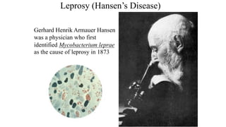
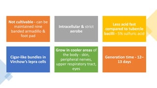
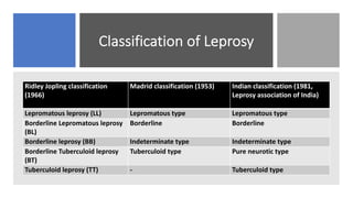
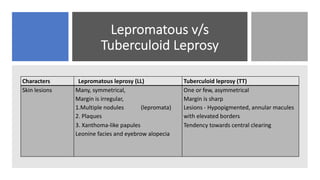
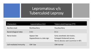
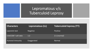
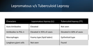
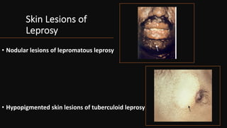
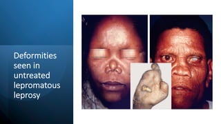
LEPROSY
Leprosy
• A chronic mycobacterial disease
of ancient world.
• First bacterial pathogen of
human to have been described.
Leprosy (Hansen’s Disease)
Gerhard Henrik Armauer Hansen
was a physician who first
identified Mycobacterium leprae
as the cause of leprosy in 1873
MYCOBACTERIUM LEPRAE
History: recognized since Vedic times in India (described
as Kushta Roga in Sushruta Samhita, 600 BC
G. H. Armauer Hansen (1873) - discovered of
lepra bacilli
Shepard (1960) – multiplying lepra bacilli in
footpads of mice kept at a low temperature .
Not cultivable - can be
maintained nine
banded armadillo &
foot pad
Intracellular & strict
aerobe
Less acid fast
compared to tubercle
bacilli - 5% sulfuric acid
Cigar-like bundles in
Virchow’s lepra cells
Grow in cooler areas of
the body - skin,
peripheral nerves,
upper respiratory tract,
eyes
Generation time - 12–
13 days
Classification of Leprosy
Ridley Jopling classification
(1966)
Madrid classification (1953) Indian classification (1981,
Leprosy association of India)
Lepromatous leprosy (LL) Lepromatous type Lepromatous type
Borderline Lepromatous leprosy
(BL)
Borderline Borderline
Borderline leprosy (BB) Indeterminate type Indeterminate type
Borderline Tuberculoid leprosy
(BT)
Tuberculoid type Pure neurotic type
Tuberculoid leprosy (TT) - Tuberculoid type
Lepromatous v/s
Tuberculoid Leprosy
Characters Lepromatous leprosy (LL) Tuberculoid leprosy (TT)
Skin lesions Many, symmetrical,
Margin is irregular,
1.Multiple nodules (lepromata)
2. Plaques
3. Xanthoma-like papules
Leonine facies and eyebrow alopecia
One or few, asymmetrical
Margin is sharp
Lesions - Hypopigmented, annular macules
with elevated borders
Tendency towards central clearing
Lepromatous v/s
Tuberculoid Leprosy
Characters Lepromatous (LL) Tuberculoid leprosy (TT)
Bacillary load Multibacillary Paucibacillary
Bacteriological index 4–6+ 0–1+
Nerve lesion Appear late
Hypoesthesia is a late sign
Early anesthetic skin lesion,
Enlarged thickened nerves,
Nerve abscess seen (common in BT)
Cell mediated immunity CMI low CMI normal
Lepromatous v/s
Tuberculoid Leprosy
Characters Lepromatous (LL) Tuberculoid leprosy (TT)
Lepromin test Negative Positive
CD4/CD8 T cell ratio 1:2 2:1 (normal)
Humoral immunity Exaggerated Normal
Lepromatous v/s Tuberculoid Leprosy
Characters Lepromatous leprosy (LL) Tuberculoid leprosy (TT)
Auto Antibodies Elevated Not seen
Antibodies to PGL-1 Elevated in 95% of cases Elevated in 60% of cases
Macrophages Foamy type (lipid laden) Epithelioid type
Langhans giant cells Not seen Found
Skin Lesions of
Leprosy
• Nodular lesions of lepromatous leprosy
• Hypopigmented skin lesions of tuberculoid leprosy
Other
Categories of
Leprosy
• Characteristics in between tuberculoid and
lepromatous types
• May shift to either TT or LL type with chemotherapy or
alterations in host resistance
Borderline type:
• Early unstable cases with one or two hypopigmented
macules
• definite sensory impairment
• bacteriologically negative
Indeterminate type:
• Neural involvement without any skin lesion
• Bacteriologically negative
Pure neurotic type:
IMMUNITY Humoral antibodies are produced. Minor
role in disease control as bacilli are
intracellular
CMI: vital role in control of the disease
People with low CMI usually develop LL type
of lesions- Lepromin test Negative
People with intact CMI usually develop TT
type lesions – lepromin test positive
- TT patients - release of TH1 specific cytokines (IL2,
interferon γ) à macrophage activation àphagocytose
and kill bacilli
Epidemiology
Source: Multibacillary (LL and
BL) cases
Mode of transmission: .
Aerosols containing M.leprae
Portal of entry - nose or skin
Contact transmission (skin):
• Direct contact from person to
person
• Indirect contact with infected soil,
fomites (clothes, linens)
• Direct dermal inoculation during
tattooing.
Complications – Lepra
Reactions
Characters Lepra Reaction Type I Lepra Reaction Type II
Hypersensitivity
reaction
Type IV (delayed hyper sensitivity) Type III (immune complex mediated)
Seen with Borderline leprosy Lepromatous variety (BL,LL)
Manifests as Inflammation of previous lesions, new skin lesions and
neuritis
Crops of painful erythematous papules which
become nodular
T helper response TH1 predominates TH2 predominates
Other organs Usually not affected Eyes, testes and kidney are affected
Complications
CHARACTERS LEPRA REACTION TYPE I LEPRA REACTION TYPE II
Progresses as If occurs before treatment – Progresses towards
LL (down grading reaction)
If occurs after treatment- Progress towards TT
(reversal reaction)
It usually occurs following the start of
chemotherapy.
Treatment Glucocorticoid Glucocorticoid, thalidomide,
clofazimine and antipyretics
Complications -
Deformities
• About 25% of untreated cases develop deformities
• Due to—nerve injury, disease process, injury
- Face: Leonine facies, sagging face, loss of eyebrow/eye lashes, saddle
nose and corneal opacity and ulcers
- Hands: Claw hand and wrist drop
- Feet: Foot drop, clawing of toes, inversion of foot, and plantar ulcers
Deformities
seen in
untreated
lepromatous
leprosy
Laboratory
Diagnosis
Specimen Collection
• - Six samples – four skin (forehead,
cheek, chin and buttock), one from
ear lobe and nasal mucosa by
nasal blow/scraping
• Slit skin smear
• Nasal scraping
• Biopsy - thickened nerves and
nodular lesions
Microscopy
• Ziehl–Neelsen (5% sulfuric acid)
• Acid fast bacilli
• Singly or in groups (cigar like
bundles)
• Globi in Virchow‘s lepra cells or
foamy cells
• Live bacilli - uniformly stained
with parallel sides
• Dead bacilli - less uniformly
stained, fragmented & granular
Grading
of the
Smear
1–10 bacilli in 100
OIF =1+
1–10 bacilli in 10
OIF = 2+
1–10 bacilli per OIF
= 3+
10–100 bacilli per
OIF = 4+
100–1000 bacilli
per OIF = 5+
>1000 bacilli or
bacilli in clumps
and globi in each
OIF = 6+
Indices
Bacteriological index (BI): Total
number of bacilli (live and dead) per oil
immersion field
Morphological index (MI): Percentage
of uniformly stained bacilli out of the
total number of bacilli counted
- MI is a better marker to monitor the
treatment response
Antibody Detection
•FLA-ABS (Fluorescent leprosy antibody
absorption test):
•ELISA detecting IgM antibodies to PGL-1
(phenolic glycolipid-1) antigen of M.
Leprae
Lepromin test
Lepromin –A Antigen
0.1mL injected
intradermally
Early or Fernandez
reaction: >24–48
hours
Induration
surrounded by
erythema. >10 mm
diameter - positive
DTH, Indicates past
exposure to lepra
bacilli
Late or Mitsuda
reaction: After 21 days
indicates patient’s CMI
is intact
: Nodule >5 mm
àulcerates
WHO Treatment regimens
Criteria Paucibacillary Multibacillary
Treatment
regimen
1)Dapsone (100mg) given daily,
self administered
2)Rifampicin (600mg) given
once a month under
supervision
1)Dapsone (100mg) daily
2)Rifampicin (600mg) once a month
3)Clofazimine–300mg once a month
under supervision, à 50mg daily, self
administered
Duration of
treatment
Up to 6months Upto 1 yr or till smear negative
Follow up Annually till 2 yrs Annually till 5 yrs

Leprosy microbiology

  • 1.
  • 2.
    Leprosy • A chronicmycobacterial disease of ancient world. • First bacterial pathogen of human to have been described.
  • 3.
    Leprosy (Hansen’s Disease) GerhardHenrik Armauer Hansen was a physician who first identified Mycobacterium leprae as the cause of leprosy in 1873
  • 4.
    MYCOBACTERIUM LEPRAE History: recognizedsince Vedic times in India (described as Kushta Roga in Sushruta Samhita, 600 BC G. H. Armauer Hansen (1873) - discovered of lepra bacilli Shepard (1960) – multiplying lepra bacilli in footpads of mice kept at a low temperature .
  • 5.
    Not cultivable -can be maintained nine banded armadillo & foot pad Intracellular & strict aerobe Less acid fast compared to tubercle bacilli - 5% sulfuric acid Cigar-like bundles in Virchow’s lepra cells Grow in cooler areas of the body - skin, peripheral nerves, upper respiratory tract, eyes Generation time - 12– 13 days
  • 6.
    Classification of Leprosy RidleyJopling classification (1966) Madrid classification (1953) Indian classification (1981, Leprosy association of India) Lepromatous leprosy (LL) Lepromatous type Lepromatous type Borderline Lepromatous leprosy (BL) Borderline Borderline Borderline leprosy (BB) Indeterminate type Indeterminate type Borderline Tuberculoid leprosy (BT) Tuberculoid type Pure neurotic type Tuberculoid leprosy (TT) - Tuberculoid type
  • 7.
    Lepromatous v/s Tuberculoid Leprosy CharactersLepromatous leprosy (LL) Tuberculoid leprosy (TT) Skin lesions Many, symmetrical, Margin is irregular, 1.Multiple nodules (lepromata) 2. Plaques 3. Xanthoma-like papules Leonine facies and eyebrow alopecia One or few, asymmetrical Margin is sharp Lesions - Hypopigmented, annular macules with elevated borders Tendency towards central clearing
  • 8.
    Lepromatous v/s Tuberculoid Leprosy CharactersLepromatous (LL) Tuberculoid leprosy (TT) Bacillary load Multibacillary Paucibacillary Bacteriological index 4–6+ 0–1+ Nerve lesion Appear late Hypoesthesia is a late sign Early anesthetic skin lesion, Enlarged thickened nerves, Nerve abscess seen (common in BT) Cell mediated immunity CMI low CMI normal
  • 9.
    Lepromatous v/s Tuberculoid Leprosy CharactersLepromatous (LL) Tuberculoid leprosy (TT) Lepromin test Negative Positive CD4/CD8 T cell ratio 1:2 2:1 (normal) Humoral immunity Exaggerated Normal
  • 10.
    Lepromatous v/s TuberculoidLeprosy Characters Lepromatous leprosy (LL) Tuberculoid leprosy (TT) Auto Antibodies Elevated Not seen Antibodies to PGL-1 Elevated in 95% of cases Elevated in 60% of cases Macrophages Foamy type (lipid laden) Epithelioid type Langhans giant cells Not seen Found
  • 11.
    Skin Lesions of Leprosy •Nodular lesions of lepromatous leprosy • Hypopigmented skin lesions of tuberculoid leprosy
  • 12.
    Other Categories of Leprosy • Characteristicsin between tuberculoid and lepromatous types • May shift to either TT or LL type with chemotherapy or alterations in host resistance Borderline type: • Early unstable cases with one or two hypopigmented macules • definite sensory impairment • bacteriologically negative Indeterminate type: • Neural involvement without any skin lesion • Bacteriologically negative Pure neurotic type:
  • 13.
    IMMUNITY Humoral antibodiesare produced. Minor role in disease control as bacilli are intracellular CMI: vital role in control of the disease People with low CMI usually develop LL type of lesions- Lepromin test Negative People with intact CMI usually develop TT type lesions – lepromin test positive
  • 14.
    - TT patients- release of TH1 specific cytokines (IL2, interferon γ) à macrophage activation àphagocytose and kill bacilli
  • 15.
    Epidemiology Source: Multibacillary (LLand BL) cases Mode of transmission: . Aerosols containing M.leprae Portal of entry - nose or skin Contact transmission (skin): • Direct contact from person to person • Indirect contact with infected soil, fomites (clothes, linens) • Direct dermal inoculation during tattooing.
  • 16.
    Complications – Lepra Reactions CharactersLepra Reaction Type I Lepra Reaction Type II Hypersensitivity reaction Type IV (delayed hyper sensitivity) Type III (immune complex mediated) Seen with Borderline leprosy Lepromatous variety (BL,LL) Manifests as Inflammation of previous lesions, new skin lesions and neuritis Crops of painful erythematous papules which become nodular T helper response TH1 predominates TH2 predominates Other organs Usually not affected Eyes, testes and kidney are affected
  • 17.
    Complications CHARACTERS LEPRA REACTIONTYPE I LEPRA REACTION TYPE II Progresses as If occurs before treatment – Progresses towards LL (down grading reaction) If occurs after treatment- Progress towards TT (reversal reaction) It usually occurs following the start of chemotherapy. Treatment Glucocorticoid Glucocorticoid, thalidomide, clofazimine and antipyretics
  • 18.
    Complications - Deformities • About25% of untreated cases develop deformities • Due to—nerve injury, disease process, injury - Face: Leonine facies, sagging face, loss of eyebrow/eye lashes, saddle nose and corneal opacity and ulcers - Hands: Claw hand and wrist drop - Feet: Foot drop, clawing of toes, inversion of foot, and plantar ulcers
  • 19.
  • 20.
    Laboratory Diagnosis Specimen Collection • -Six samples – four skin (forehead, cheek, chin and buttock), one from ear lobe and nasal mucosa by nasal blow/scraping • Slit skin smear • Nasal scraping • Biopsy - thickened nerves and nodular lesions
  • 21.
    Microscopy • Ziehl–Neelsen (5%sulfuric acid) • Acid fast bacilli • Singly or in groups (cigar like bundles) • Globi in Virchow‘s lepra cells or foamy cells • Live bacilli - uniformly stained with parallel sides • Dead bacilli - less uniformly stained, fragmented & granular
  • 22.
    Grading of the Smear 1–10 bacilliin 100 OIF =1+ 1–10 bacilli in 10 OIF = 2+ 1–10 bacilli per OIF = 3+ 10–100 bacilli per OIF = 4+ 100–1000 bacilli per OIF = 5+ >1000 bacilli or bacilli in clumps and globi in each OIF = 6+
  • 23.
    Indices Bacteriological index (BI):Total number of bacilli (live and dead) per oil immersion field Morphological index (MI): Percentage of uniformly stained bacilli out of the total number of bacilli counted - MI is a better marker to monitor the treatment response
  • 24.
    Antibody Detection •FLA-ABS (Fluorescentleprosy antibody absorption test): •ELISA detecting IgM antibodies to PGL-1 (phenolic glycolipid-1) antigen of M. Leprae
  • 25.
    Lepromin test Lepromin –AAntigen 0.1mL injected intradermally Early or Fernandez reaction: >24–48 hours Induration surrounded by erythema. >10 mm diameter - positive DTH, Indicates past exposure to lepra bacilli Late or Mitsuda reaction: After 21 days indicates patient’s CMI is intact : Nodule >5 mm àulcerates
  • 26.
    WHO Treatment regimens CriteriaPaucibacillary Multibacillary Treatment regimen 1)Dapsone (100mg) given daily, self administered 2)Rifampicin (600mg) given once a month under supervision 1)Dapsone (100mg) daily 2)Rifampicin (600mg) once a month 3)Clofazimine–300mg once a month under supervision, à 50mg daily, self administered Duration of treatment Up to 6months Upto 1 yr or till smear negative Follow up Annually till 2 yrs Annually till 5 yrs