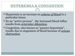
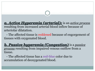
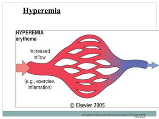
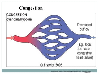
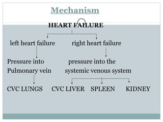
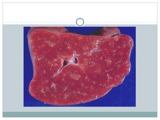
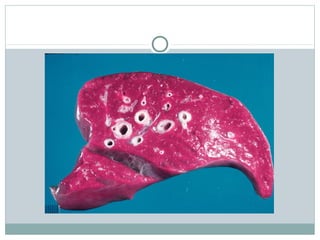
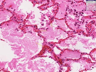
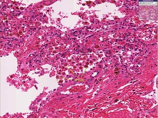
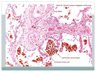
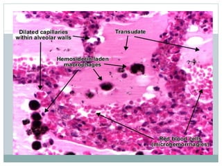
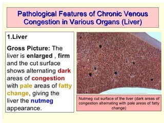
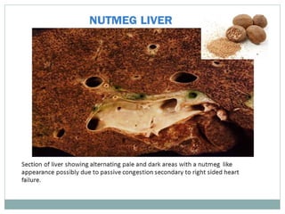
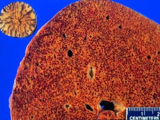
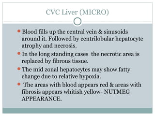
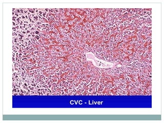
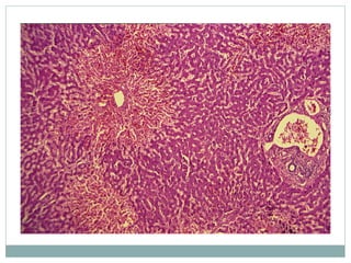
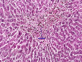
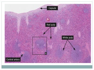
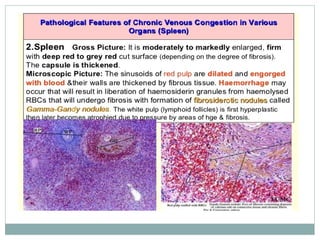
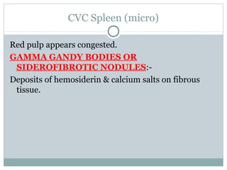
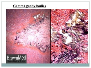
-DR SOWMYA SRINIVAS
CVC LUNG, LIVER AND
SPLEEN
HYPEREMIA & CONGESTION
Hyperemia is an increase in volume of blood in a
particular tissue.
Its an “active process” , the increased blood influx
results from arteriolar dilatation.
Congestion, also known as “passive hyperemia”,
results due to stagnation of blood because of venous
obstruction.
Downloaded from: Robbins & Cotran Pathologic Basis of Disease (on 27 June 2005 07:26 PM)
© 2005 Elsevier
Normal
a. Active Hyperemia (arterial):a. Active Hyperemia (arterial): is an active process
resulting from increased arterial blood inflow because of
arteriolar dilatation.
- The affected tissue is reddened because of engorgement of
tissues with oxygenated blood.
b. Passive hyperemia (Congestion)b. Passive hyperemia (Congestion) is a passive
process resulting from impaired venous outflow from a
tissue.
- The affected tissue has a red-blue color due to
accumulation of deoxygenated blood.
ACTIVE HYPEREMIA
Types:
1. Physiological:
- Hyperemia in skeletal muscles during exercise ,in the gut
following a meal.
2. Pathological: e.g. in acute inflammation.
Downloaded from: Robbins & Cotran Pathologic Basis of Disease (on 27 June 2005 07:26 PM)
© 2005 Elsevier
Hyperemia
PASSIVE HYPEREMIA OR
CONGESTION
Definition:
- Increase in venous blood in an organ as result of
obstruction of venous outflow.
- The veins, venules & capillaries in the organ become
passively dilated (passive hyperemia).
Types:
a. Localized
b. Generalized
Downloaded from: Robbins & Cotran Pathologic Basis of Disease (on 27 June 2005 07:26 PM)
© 2005 Elsevier
Congestion
Examples:-
Hyperemia:-
1. Inflammation
2. Blushing – adrenergic stimulation
3. Exercise – increased blood flow to the muscle.
Congestion:-
Obstruction of veins due to thrombi or backward
pressure due to heart failure.
Color of hyperemic & congested tissue:-
Hyperemic tissue contains increased amounts of
oxygenated blood & therefore the tissue appears
bright red.
Congested tissue contains increased amounts of
deoxygenated blood & appears blue.
Hyperemic tissue is warm, while congested blood is
cold & clammy.
Chronic venous congestion (CVC):-
In CVC there is long standing accumulation of
deoxygenated blood & hence there is damage to the
tissue.
Mechanism
HEART FAILURE
left heart failure right heart failure
Pressure into pressure into the
Pulmonary vein systemic venous system
CVC LUNGS CVC LIVER SPLEEN KIDNEY
CVC Lung
Causes:-
Left heart failure
Gross :-
The lungs are heavy. Lungs appear brown- BROWN
INDURATION OF LUNGS.
CVC Lungs
Micro:-
Rupture of congested vessel results in edema &
hemorrhage.
The alveolar septa appears thickened and fibrotic.
The alveoli are dilated and contain edema fluid and
RBC’s and macrophages.
Lysis of RBC’s releases hemosiderin pigment which
is taken up by macrophages in the alveoli – HEART
FAILURE CELLS.
CVC Liver
Causes:-
1. Right heart failure
2. Occlusion of inferior vena cava or portal vein.
GROSS APPEARANCE:-
NUTMEG APPEARANCE – Alternate areas of red &
yellow .
CVC Liver (MICRO)
Blood fills up the central vein & sinusoids
around it. Followed by centrilobular hepatocyte
atrophy and necrosis.
In the long standing cases the necrotic area is
replaced by fibrous tissue.
The mid zonal hepatocytes may show fatty
change due to relative hypoxia.
 The areas with blood appears red & areas with
fibrosis appears whitish yellow- NUTMEG
APPEARANCE.
CVC Spleen
Causes:-
1. Right heart failure
2. Portal hypertension
GROSS:-
Spleen is enlarged & congested.
CVC Spleen (micro)
Red pulp appears congested.
GAMMA GANDY BODIES OR
SIDEROFIBROTIC NODULES:-
Deposits of hemosiderin & calcium salts on fibrous
tissue.
Gamma gandy bodies
THANK YOU

Cvc lung, liver and spleen

  • 1.
    -DR SOWMYA SRINIVAS CVCLUNG, LIVER AND SPLEEN
  • 2.
    HYPEREMIA & CONGESTION Hyperemiais an increase in volume of blood in a particular tissue. Its an “active process” , the increased blood influx results from arteriolar dilatation. Congestion, also known as “passive hyperemia”, results due to stagnation of blood because of venous obstruction.
  • 3.
    Downloaded from: Robbins& Cotran Pathologic Basis of Disease (on 27 June 2005 07:26 PM) © 2005 Elsevier Normal
  • 4.
    a. Active Hyperemia(arterial):a. Active Hyperemia (arterial): is an active process resulting from increased arterial blood inflow because of arteriolar dilatation. - The affected tissue is reddened because of engorgement of tissues with oxygenated blood. b. Passive hyperemia (Congestion)b. Passive hyperemia (Congestion) is a passive process resulting from impaired venous outflow from a tissue. - The affected tissue has a red-blue color due to accumulation of deoxygenated blood.
  • 5.
    ACTIVE HYPEREMIA Types: 1. Physiological: -Hyperemia in skeletal muscles during exercise ,in the gut following a meal. 2. Pathological: e.g. in acute inflammation.
  • 6.
    Downloaded from: Robbins& Cotran Pathologic Basis of Disease (on 27 June 2005 07:26 PM) © 2005 Elsevier Hyperemia
  • 7.
    PASSIVE HYPEREMIA OR CONGESTION Definition: -Increase in venous blood in an organ as result of obstruction of venous outflow. - The veins, venules & capillaries in the organ become passively dilated (passive hyperemia). Types: a. Localized b. Generalized
  • 8.
    Downloaded from: Robbins& Cotran Pathologic Basis of Disease (on 27 June 2005 07:26 PM) © 2005 Elsevier Congestion
  • 9.
    Examples:- Hyperemia:- 1. Inflammation 2. Blushing– adrenergic stimulation 3. Exercise – increased blood flow to the muscle. Congestion:- Obstruction of veins due to thrombi or backward pressure due to heart failure.
  • 10.
    Color of hyperemic& congested tissue:- Hyperemic tissue contains increased amounts of oxygenated blood & therefore the tissue appears bright red. Congested tissue contains increased amounts of deoxygenated blood & appears blue. Hyperemic tissue is warm, while congested blood is cold & clammy.
  • 11.
    Chronic venous congestion(CVC):- In CVC there is long standing accumulation of deoxygenated blood & hence there is damage to the tissue.
  • 12.
    Mechanism HEART FAILURE left heartfailure right heart failure Pressure into pressure into the Pulmonary vein systemic venous system CVC LUNGS CVC LIVER SPLEEN KIDNEY
  • 13.
    CVC Lung Causes:- Left heartfailure Gross :- The lungs are heavy. Lungs appear brown- BROWN INDURATION OF LUNGS.
  • 16.
    CVC Lungs Micro:- Rupture ofcongested vessel results in edema & hemorrhage. The alveolar septa appears thickened and fibrotic. The alveoli are dilated and contain edema fluid and RBC’s and macrophages. Lysis of RBC’s releases hemosiderin pigment which is taken up by macrophages in the alveoli – HEART FAILURE CELLS.
  • 21.
    CVC Liver Causes:- 1. Rightheart failure 2. Occlusion of inferior vena cava or portal vein. GROSS APPEARANCE:- NUTMEG APPEARANCE – Alternate areas of red & yellow .
  • 26.
    CVC Liver (MICRO) Bloodfills up the central vein & sinusoids around it. Followed by centrilobular hepatocyte atrophy and necrosis. In the long standing cases the necrotic area is replaced by fibrous tissue. The mid zonal hepatocytes may show fatty change due to relative hypoxia.  The areas with blood appears red & areas with fibrosis appears whitish yellow- NUTMEG APPEARANCE.
  • 31.
    CVC Spleen Causes:- 1. Rightheart failure 2. Portal hypertension GROSS:- Spleen is enlarged & congested.
  • 34.
    CVC Spleen (micro) Redpulp appears congested. GAMMA GANDY BODIES OR SIDEROFIBROTIC NODULES:- Deposits of hemosiderin & calcium salts on fibrous tissue.
  • 35.
  • 36.

Editor's Notes

  • #4 Figure 4-3 Hyperemia versus congestion. In both cases there is an increased volume and pressure of blood in a given tissue with associated capillary dilation and a potential for fluid extravasation.
  • #7 In hyperemia, increased inflow leads to engorgement with oxygenated blood, resulting in erythema.
  • #9 In congestion, diminished outflow leads to a capillary bed swollen with deoxygenated venous blood and resulting in cyanosis.
  • #18 View of pulmonary congestion and edema. Often caused by an increase in hydrostatic pressure, a protein poor transudate seeps into interstitial and alveolar spaces. Note the engorged alveolar wall capillaries. If capillaries rupture, RBCs will escape into the alveolar space
  • #19 Heart failure cells are hemosiderin laden macrophages. Blood escapes into the alveolar space because chronic congestion causes the thin walled alveolar capillaries to burst. Note the thickening of the alveolar septae. This is caused by chronic pulmonary congestion and edema. Note the thickening of the alveolar septae. This is caused by chronic pulmonary congestion and edema.
  • #26 Nutmeg liver compared with actual nutmeg. Courtesy of Dr. Ed Friedlander
  • #31 This view shows a close up of hemorrhagic central necrosis. Necrotic cells in the central area have been removed (cell dropout) and been replaced by cellular debris and hemorrhge. There is evidence that the passive congestion and hemorrhage is chronic, as many of the RBCs have been degraded into hemosiderin.