Download as PDF, PPTX


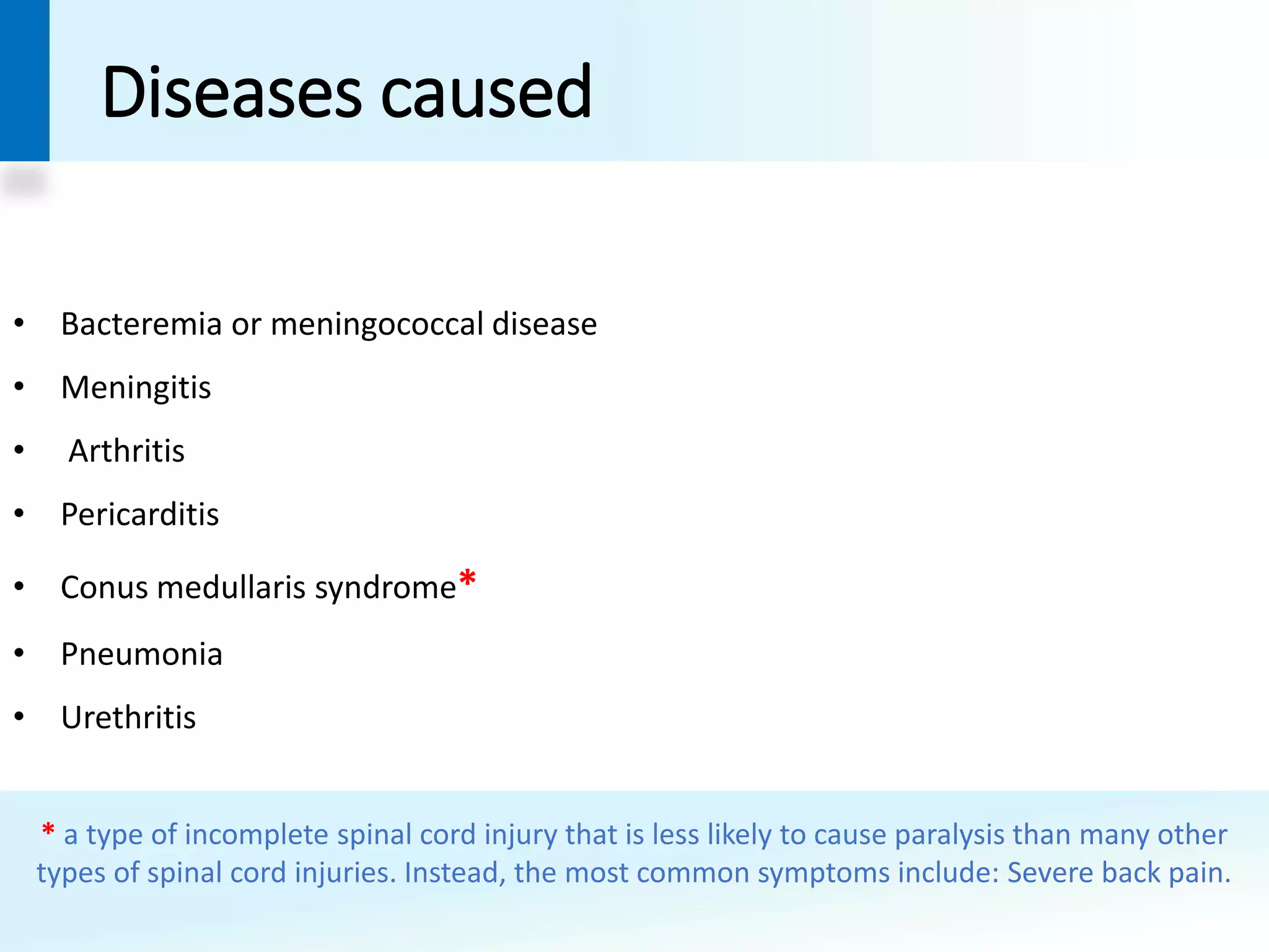

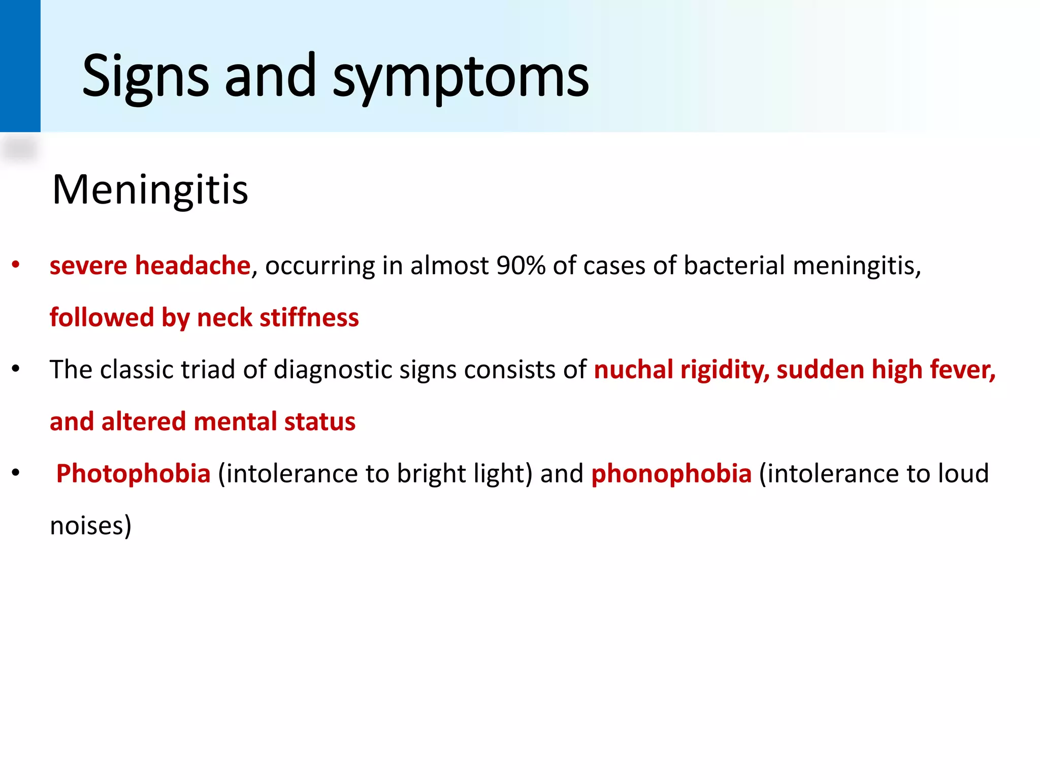

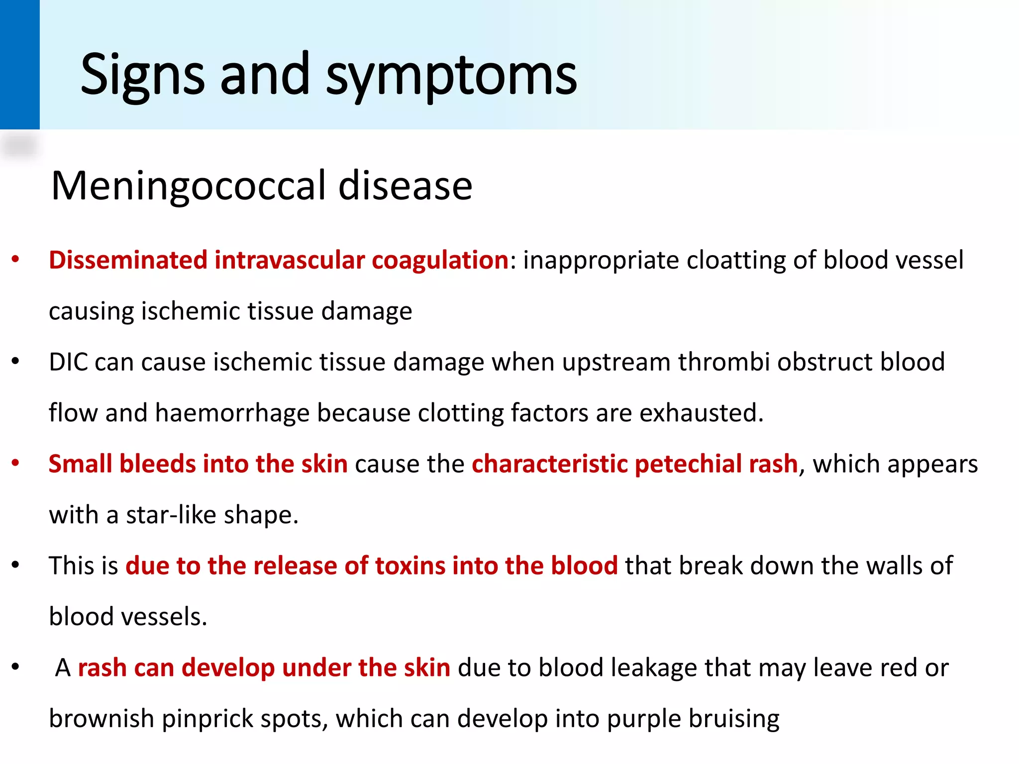


Neisseria meningitidis, a gram-negative bacterium, is primarily responsible for meningococcal diseases, including meningitis and meningococcemia. It frequently colonizes the nasopharynx of humans, leading to severe infections, particularly in sub-Saharan Africa, where epidemics occur cyclically. Diagnosis involves detecting the bacteria in blood and cerebrospinal fluid, with treatment requiring immediate hospitalization and administration of antibiotics.


