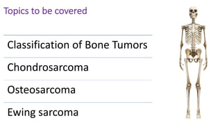
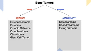
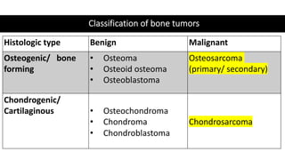
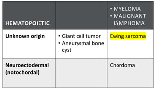
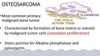
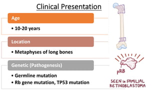
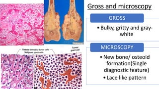
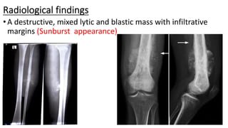
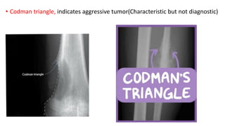
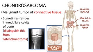
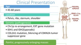
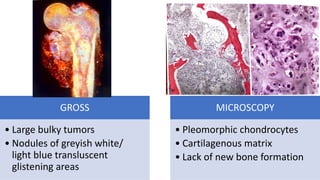
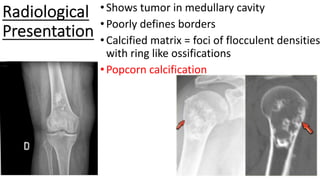
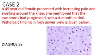
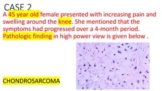
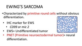
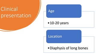
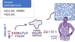
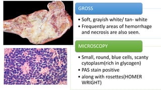
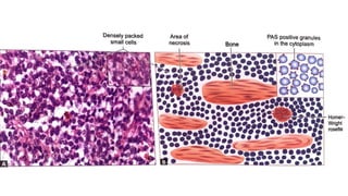
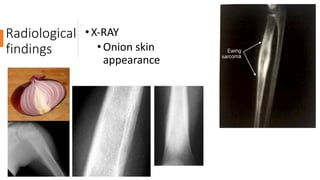
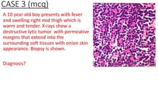
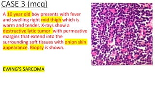
Malignant
bone
tumors
Topics to be covered
Classification of Bone Tumors
Chondrosarcoma
Osteosarcoma
Ewing sarcoma
Classification of bone tumors
Histologic type Benign Malignant
Osteogenic/ bone
forming
• Osteoma
• Osteoid osteoma
• Osteoblastoma
Osteosarcoma
(primary/ secondary)
Chondrogenic/
Cartilaginous • Osteochondroma
• Chondroma
• Chondroblastoma
Chondrosarcoma
HEMATOPOIETIC
• MYELOMA
• MALIGNANT
LYMPHOMA
Unknown origin • Giant cell tumor
• Aneurysmal bone
cyst
Ewing sarcoma
Neuroectodermal
(notochordal)
Chordoma
OSTEOSARCOMA
•Most common primary
malignant bone tumor
• Characterized by formation of bone matrix or osteoid
by malignant tumor cells (osteoblast proliferation)
• Stains positive for Alkaline phosphatase and
osteonectin
Clinical Presentation
• 10-20 years
Age
• Metaphyses of long bones
Location
• Germline mutation
• Rb gene mutation, TP53 mutation
Genetic (Pathogenesis)
Gross and microscopy
•Bulky, gritty and gray-
white
GROSS
•New bone/ osteoid
formation(Single
diagnostic feature)
•Lace like pattern
MICROSCOPY
Radiological findings
•A destructive, mixed lytic and blastic mass with infiltrative
margins (Sunburst appearance)
• Codman triangle, indicates aggressive tumor(Characteristic but not diagnostic)
CASE 1(mcq)
A 20 year old man presented with progressive swelling of
lower end of femur. X-ray shows a characteristic codmans
triangle and CT shows osteoblastic metastasis. Which of
the following is associated with a high risk of the given
clinical condition?
A. Translocation (12:16)
B. Germline mutation of Rb
C. Translocation (x;18)
D. 1p rearrangements
CHONDROSARCOMA
•Malignant tumor of connective tissue
• Sometimes resides
in medullary cavity
of bone
(distinguish this
from
osteochondroma)
Clinical Presentation
• 45-60 years
Age
• Pelvis, ribs, sternum, shoulder
Location
• Chr1p re-arrangement, EXT gene mutation
• IDH1 and IDH2(sporadic)
• COL2A1 mutation, Silencing of CDKN2A tumor
suppressor gene
Genetic (pathogenesis)
Painful, progressively enlarging masses
GROSS
• Large bulky tumors
• Nodules of greyish white/
light blue transluscent
glistening areas
MICROSCOPY
• Pleomorphic chondrocytes
• Cartilagenous matrix
• Lack of new bone formation
Radiological
Presentation
•Shows tumor in medullary cavity
•Poorly defines borders
•Calcified matrix = foci of flocculent densities
with ring like ossifications
•Popcorn calcification
CASE 2
A 45 year old female presented with increasing pain and
swelling around the knee. She mentioned that the
symptoms had progressed over a 4-month period.
Pathologic finding in high power view is given below .
DIAGNOSIS?
CASE 2
A 45 year old female presented with increasing pain and
swelling around the knee. She mentioned that the
symptoms had progressed over a 4-month period.
Pathologic finding in high power view is given below .
CHONDROSARCOMA
EWING’S SARCOMA
•Characterized by primitive round cells without obvious
differentiation.
• IHC marker for EWS
• CD99 or mic 2
• EWS= Undifferentiated tumor
• PNET (Primitive neuroectodermal tumor)= neural
differentiation.
Clinical
presentation
•10-20 years
Age
•Diaphysis of long bones
Location
•t(11:22) EWSR1
•t(21:22)
Genetic
(pathogenesis)
• Soft, grayish white/ tan- white
• Frequently areas of hemorrhage
and necrosis are also seen.
GROSS
• Small, round, blue cells, scanty
cytoplasm(rich in glycogen)
• PAS stain positive
• along with rosettes(HOMER
WRIGHT)
MICROSCOPY
Radiological
findings
• X-RAY
•Onion skin
appearance
CASE 3 (mcq)
A 10 year old boy presents with fever
and swelling right mid thigh which is
warm and tender. X-rays show a
destructive lytic tumor with permeative
margins that extend into the
surrounding soft tissues with onion skin
appearance. Biopsy is shown.
Diagnosis?
CASE 3 (mcq)
A 10 year old boy presents with fever
and swelling right mid thigh which is
warm and tender. X-rays show a
destructive lytic tumor with permeative
margins that extend into the
surrounding soft tissues with onion skin
appearance. Biopsy is shown.
EWING’S SARCOMA
Thank you

Malignant bone tumors

  • 1.
  • 2.
    Topics to becovered Classification of Bone Tumors Chondrosarcoma Osteosarcoma Ewing sarcoma
  • 4.
    Classification of bonetumors Histologic type Benign Malignant Osteogenic/ bone forming • Osteoma • Osteoid osteoma • Osteoblastoma Osteosarcoma (primary/ secondary) Chondrogenic/ Cartilaginous • Osteochondroma • Chondroma • Chondroblastoma Chondrosarcoma
  • 5.
    HEMATOPOIETIC • MYELOMA • MALIGNANT LYMPHOMA Unknownorigin • Giant cell tumor • Aneurysmal bone cyst Ewing sarcoma Neuroectodermal (notochordal) Chordoma
  • 6.
    OSTEOSARCOMA •Most common primary malignantbone tumor • Characterized by formation of bone matrix or osteoid by malignant tumor cells (osteoblast proliferation) • Stains positive for Alkaline phosphatase and osteonectin
  • 7.
    Clinical Presentation • 10-20years Age • Metaphyses of long bones Location • Germline mutation • Rb gene mutation, TP53 mutation Genetic (Pathogenesis)
  • 8.
    Gross and microscopy •Bulky,gritty and gray- white GROSS •New bone/ osteoid formation(Single diagnostic feature) •Lace like pattern MICROSCOPY
  • 9.
    Radiological findings •A destructive,mixed lytic and blastic mass with infiltrative margins (Sunburst appearance)
  • 10.
    • Codman triangle,indicates aggressive tumor(Characteristic but not diagnostic)
  • 11.
    CASE 1(mcq) A 20year old man presented with progressive swelling of lower end of femur. X-ray shows a characteristic codmans triangle and CT shows osteoblastic metastasis. Which of the following is associated with a high risk of the given clinical condition? A. Translocation (12:16) B. Germline mutation of Rb C. Translocation (x;18) D. 1p rearrangements
  • 12.
    CASE 1(mcq) A 20year old man presented with progressive swelling of lower end of femur. X-ray shows a characteristic codmans triangle and CT shows osteoblastic metastasis. Which of the following is associated with a high risk of the given clinical condition? A. Translocation (12:16) B. Germline mutation of Rb C. Translocation (x;18) D. 1p rearrangements
  • 13.
    CHONDROSARCOMA •Malignant tumor ofconnective tissue • Sometimes resides in medullary cavity of bone (distinguish this from osteochondroma)
  • 14.
    Clinical Presentation • 45-60years Age • Pelvis, ribs, sternum, shoulder Location • Chr1p re-arrangement, EXT gene mutation • IDH1 and IDH2(sporadic) • COL2A1 mutation, Silencing of CDKN2A tumor suppressor gene Genetic (pathogenesis) Painful, progressively enlarging masses
  • 15.
    GROSS • Large bulkytumors • Nodules of greyish white/ light blue transluscent glistening areas MICROSCOPY • Pleomorphic chondrocytes • Cartilagenous matrix • Lack of new bone formation
  • 16.
    Radiological Presentation •Shows tumor inmedullary cavity •Poorly defines borders •Calcified matrix = foci of flocculent densities with ring like ossifications •Popcorn calcification
  • 17.
    CASE 2 A 45year old female presented with increasing pain and swelling around the knee. She mentioned that the symptoms had progressed over a 4-month period. Pathologic finding in high power view is given below . DIAGNOSIS?
  • 18.
    CASE 2 A 45year old female presented with increasing pain and swelling around the knee. She mentioned that the symptoms had progressed over a 4-month period. Pathologic finding in high power view is given below . CHONDROSARCOMA
  • 19.
    EWING’S SARCOMA •Characterized byprimitive round cells without obvious differentiation. • IHC marker for EWS • CD99 or mic 2 • EWS= Undifferentiated tumor • PNET (Primitive neuroectodermal tumor)= neural differentiation.
  • 20.
  • 21.
  • 22.
    • Soft, grayishwhite/ tan- white • Frequently areas of hemorrhage and necrosis are also seen. GROSS • Small, round, blue cells, scanty cytoplasm(rich in glycogen) • PAS stain positive • along with rosettes(HOMER WRIGHT) MICROSCOPY
  • 24.
  • 25.
    CASE 3 (mcq) A10 year old boy presents with fever and swelling right mid thigh which is warm and tender. X-rays show a destructive lytic tumor with permeative margins that extend into the surrounding soft tissues with onion skin appearance. Biopsy is shown. Diagnosis?
  • 26.
    CASE 3 (mcq) A10 year old boy presents with fever and swelling right mid thigh which is warm and tender. X-rays show a destructive lytic tumor with permeative margins that extend into the surrounding soft tissues with onion skin appearance. Biopsy is shown. EWING’S SARCOMA
  • 27.