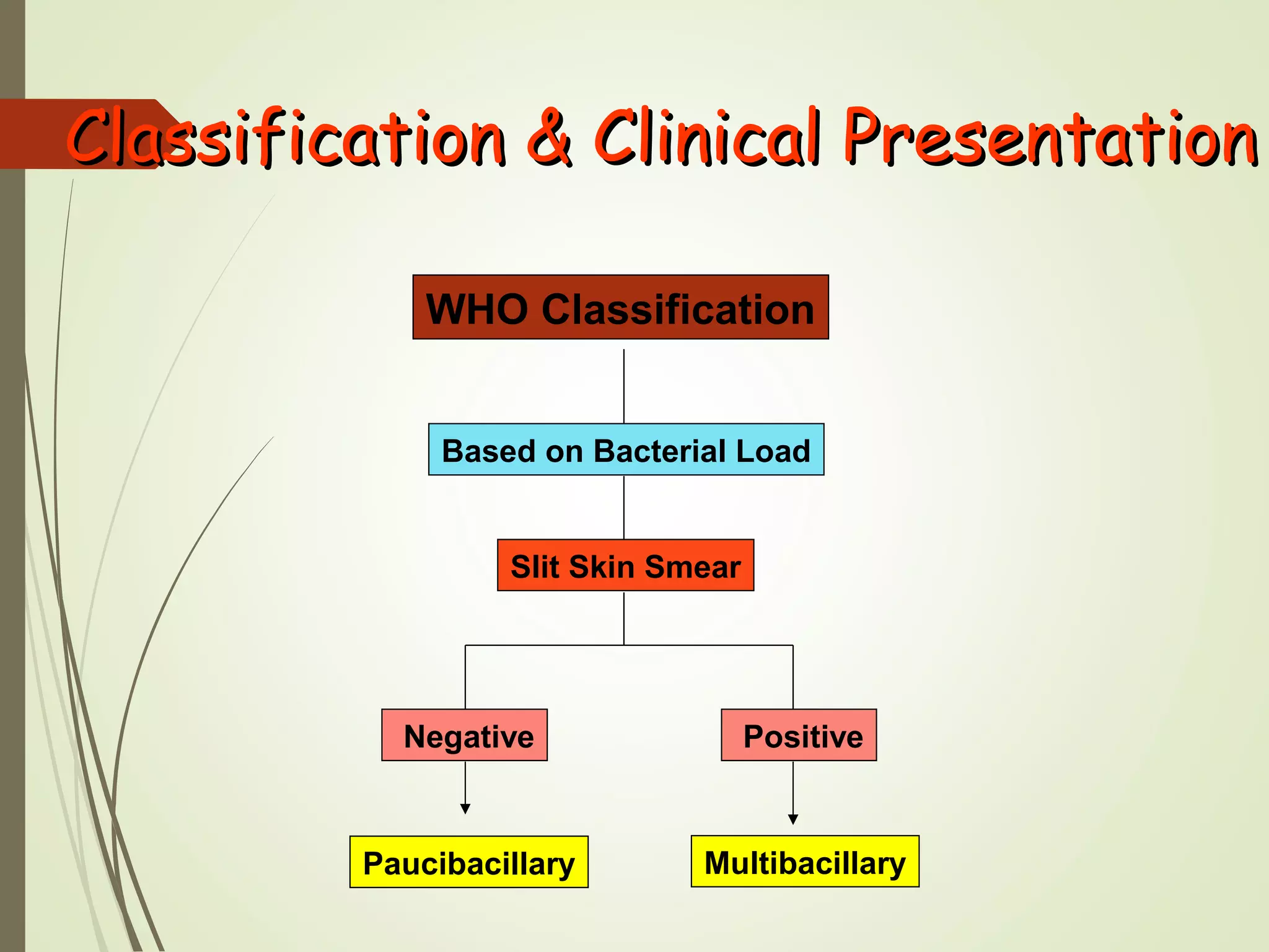
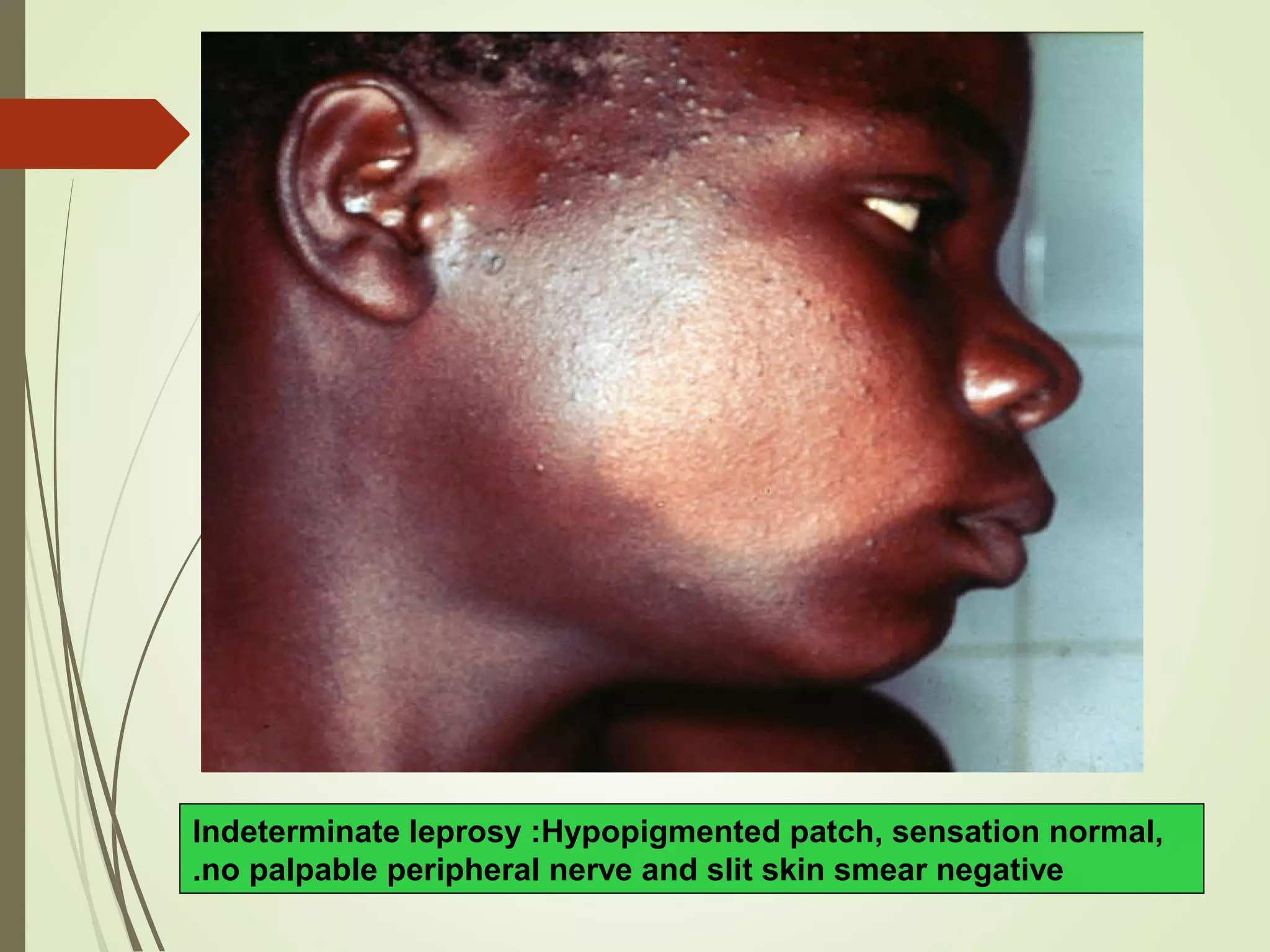
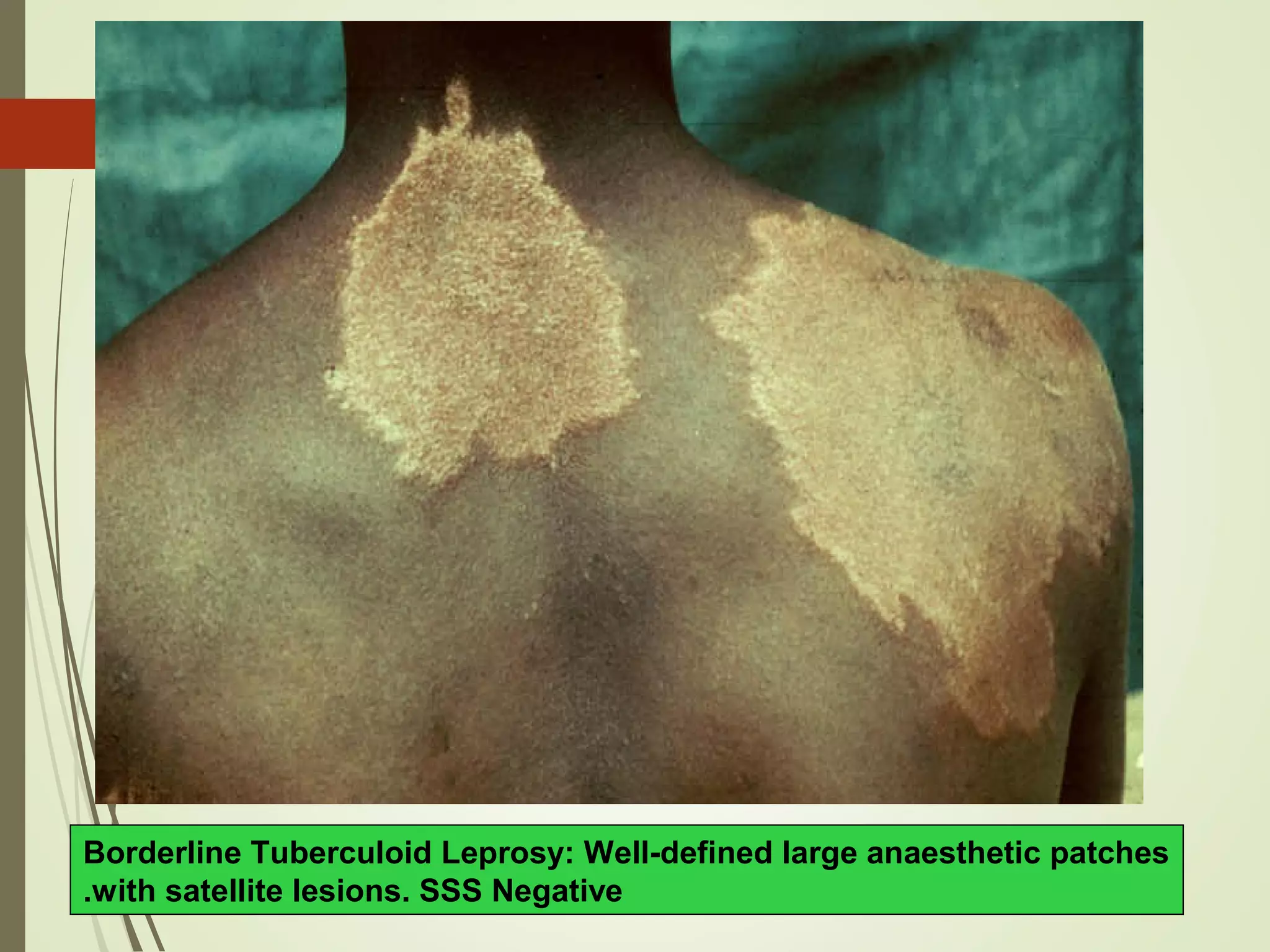
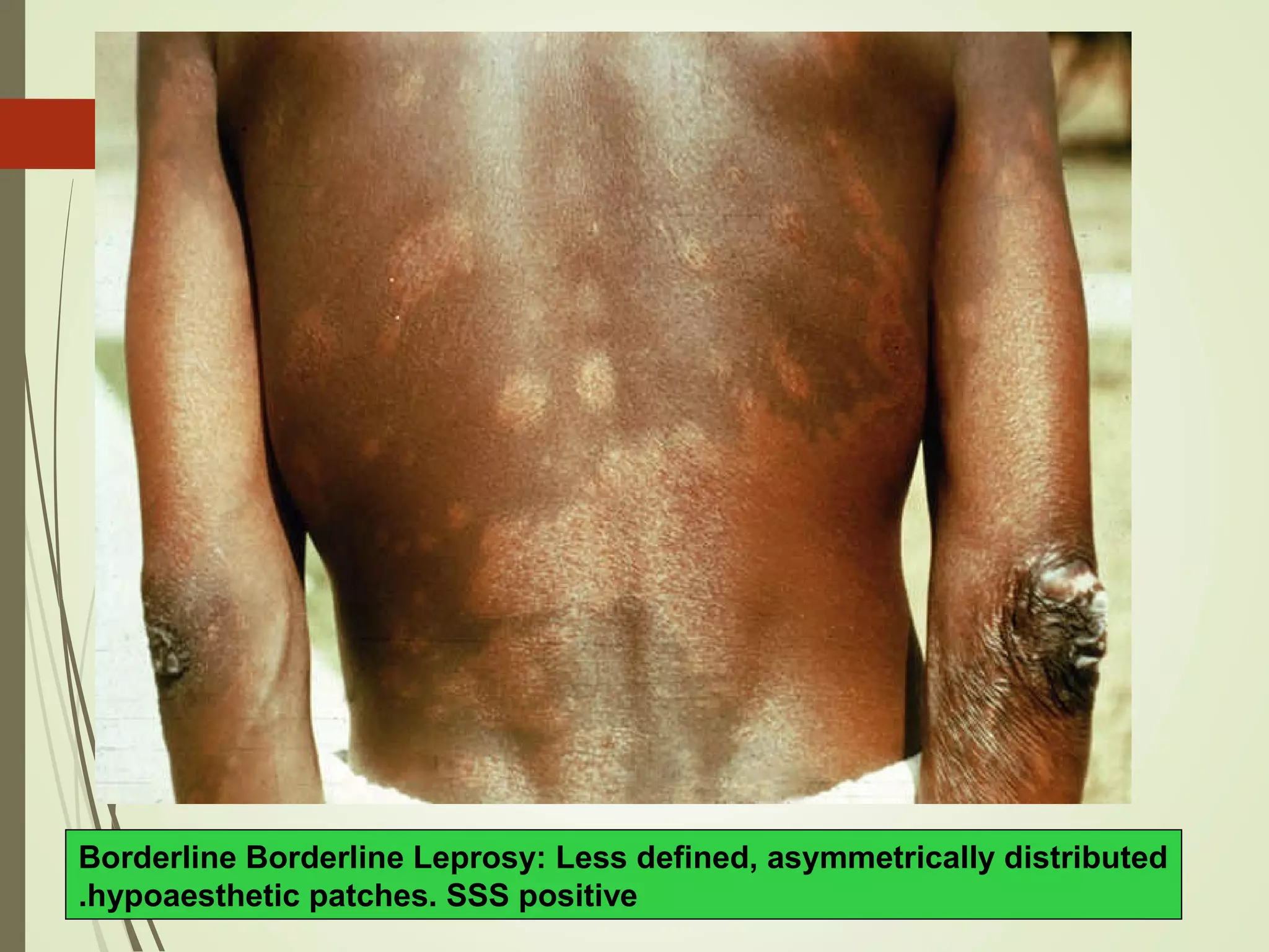
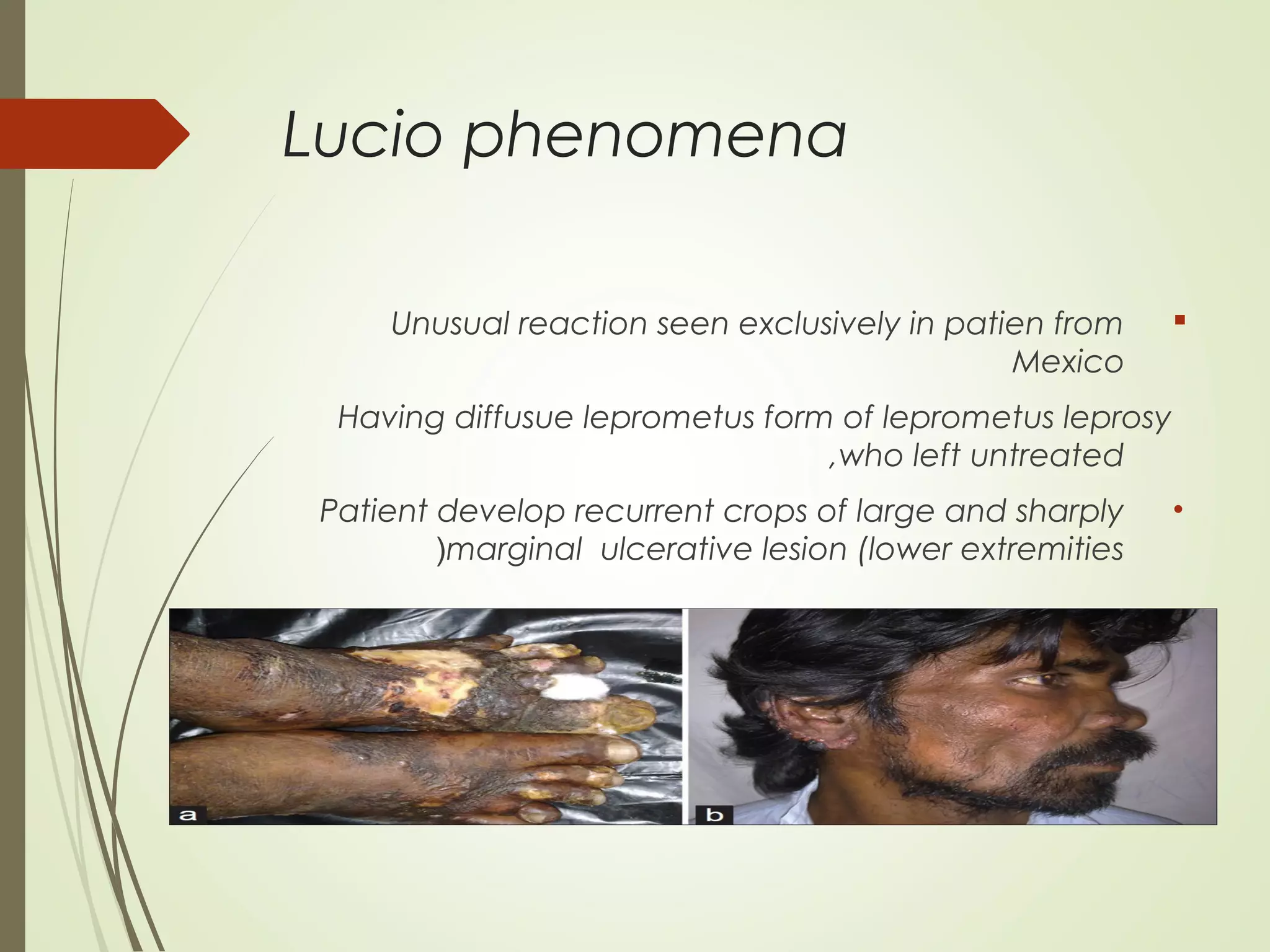
LEPROSY
Hansen`s Disease
:
- :
«
:
49)
it is the oldest disease in history and
still worldwide sociomedical and
economic problem, chronic
infectious diseaseas it can lead to
deformities and can leave a crippled
patient burdens on his family and his
society .
It principally affects the skin and
peripheral nerves.
Leprosy (Hansen’s disease(
Epidemiology
 It is endemic in tropical and subtropical countries .
It is common and endemic in certain area in upper
Egypt e .g Assiut & Giza
In lower Egypt e.g Sharkeia &
Gharbeia .
M. leprae is discovered by Hansen from Norway in 1873
Aetiology
-Mycobacterium leprae .
-discovered by Hansen in Norway 1873
-It is can not be cultured in artificial media .
- it can be growen in mouse footpad and armadillo .
BACTERILOGY (cont.(
 . M . Leprae are straight or slightly curved rod like bacteria
 They look like M. tuberculosis stained with Ziehl – Neelsan
stain . But are less acid fast and alcohol fast .
bacteriological index is the density of M leprae in
smear it includes living and dead
bacilli .
morphological index.
is percentage of living bacilli it indicates
if patient is infectious
or not and response to treatment
Mode of transmission
 droplet infection is main mode of infection
There is personal susceptibility which decides if a contact will be
infected or not
The majority of human beings have no susceptibiity
 Contact through the skin (rare).
 Arthropod-born infection (rare).
 Through placenta and milk.
Leprosy is not STD or directly inherited.
Predisposing or risk factors
Age : in all age groups .Peak of onset is 10-20 years Children
are more susceptible
Sex : male to female ratio in adult was 2 : 1 last years it is
going to be equal .
1.Residence in an endemic area.
2.Having a blood relative with leprosy.
3.Poverty (malnutrition).
4.Contact with affected armadillo.
pathogenesis
Classification
Based on the clinical, ibacteriologic,immunologic
and histopathologicfeatures
leprosy is classified into main
types
1.Paucibacillaryexample: (Tuberculoidleprosy) (TL) (with
scanty or absent bacilli) -Skin lesions, loss of sensation.
2.Multibacillary(Border line) (with numerous bacilli)---numerous
skin lesions, loss of sensation, can go to
3.Multibacillary(lepromatousleprosy) (LL).Nodules and
plaques, thickened dermis, loss of sensation, neuronal damage,
nasal congestion, epistaxis.
Classification & Clinical Presentation
Jopling Classification
Based on Host Immunity
TT BL LL
BT BB BL
Classification & Clinical PresentationClassification & Clinical Presentation
WHO Classification
Based on Bacterial Load
Paucibacillary Multibacillary
Slit Skin Smear
PositiveNegative
LEPROSY
Paucibacillary (PB( Multibacillary (MB(
Indeterminate Leprosy (IL(
Tuberculoid Leprosy (TL(
Borderline Tuberculoid (BT(
Borderline Borderline (BB(
Borderline Lepromatous(BL(
Lepromatous Leprosy (LL(
CLINICAL PICTURE
Indeterminate Leprosy
Tuberculoid Leprosy
Borderline Leprosy
BT BB BL
Lepromatous Leprosy
Indeterminate Leprosy (IL(
 Usually single (multiple) macule / patche.
 Hypopigmented or faintly erythematous.
 Sensation normal but sometimes imparied.
 The peripheral nerves normal.
 Slit skin smear negative.
Indeterminate leprosy :Hypopigmented patch, sensation normal,
no palpable peripheral nerve and slit skin smear negative.
Tuberculoid Leprosy (TL(

Usually single but may be few (<5).
 Hypopigmented / erythematous plaque.
 Well defined borders.
 Sensation markedly imparied.
 Enlarged peripheral nerve.
 Slit skin smear negative
Tuberculoid leprosy: Two hypopigmented patches, hypoasthetic
well defined borders, palpable peripheral nerve and SSS negative.
Tuberculoid Leprosy: Annular, erythematous, anasthetic patch with
well defined and raised borders and SSS Negative.
Borderline Leprosy (BL)
(BT,BB,BL)
 Few / many asymmetrical patches.
 Partly well-defined borders.
 Sensory impairments range from slight to marked.
 Slit skin smear usually positive.
 P. nerves asymmetrically enlarged.
 Annular plaque in leprosy are most commonly border line with characteristic
punched out internal border and sloping outer border(swiss cheese
appearance)
Borderline Tuberculoid Leprosy: Well-defined large anaesthetic patches
with satellite lesions. SSS Negative.
Borderline Borderline Leprosy: Less defined, asymmetrically distributed
hypoaesthetic patches. SSS positive.
Borderline Lepromatous Leprosy: Numerous, hypoaesthetic almost
symmetrically distributed patches . SSS positive.
Lepromatous Leprosy (LL(
 Very numerous ill defined lesions.
(macules, patches, papules,and nodules).
 Symmetrically distributed allover the body
 Loss of eyebrows and eyelashes.
 Leonina facies.
 No sensory impairments in lesions .
 Peripheral nerves symmetrically enlarged.
 Slit skin smear always positive.
Lepromatous Leprosy: Leonine Face
Mucous membrane
involvement
Very common
Usually epistaxis
Dryness ,rhinorrhea and
obstruction of Nose
Nerve involvement
Bilateral symmetrical
thickening
of all nerves
Nerve fibrosis is very late in
lepromatous leprosy, so loss of
sensation is also very late.
Bilateral Glove and stocking
anasthesia
is present in all advanced cases
of lepromatous leprosy.
Diagnosis of Leprosy
1. Clinical Examination.
2. Slit Skin Smear.
3. Skin Biopsy.
1.Clinical examination:
What are the cardinal skin signs of leprosy?
1. Hypopigmented or erythematus patch / plaque
2. Complete / partial loss of sensation.
3. Thickening of peripheral nerves.
1.History
Familial or extrafamilial contact
Parasthesia
Numbness
Epistaxis
2.Clinical examination
I.1(E, Temperature & painxamine all modalities of
sensation in the skin lesion “Touch”
II.2(Examine peripheral nerves for Tenderness,
consistency and size:
III.Head: Supraorbital
IV.Neck: Great auricular n.
V.Elbows: Ulnar n.
VI.Wrists: Superficial radial cut. And median n.
VII.Popliteal fossa: Common peroneal n.
VIII.They may be cord-like, beaded, thinned or may show
abscess formation.
3.Investigations
)1(Nicotinic acid test:
“Professor Dr. El-Arini”
15mg nicotinic acid IM injection Hypopigmented
macule becomes erythematous within 10-15 mins.
*Value:
Quick office test for early detection of hypopigmented
lesions of leprosy.
)2(Bacteriological examination:
Nasal smear
Not recommended)?(:
Painful for the patient & may reveal +ve
results in contacts
On treatment, lepra bacilli disappear from
nasal mucus earlier than from skin.
It’s very difficult to prove the bacilli ara M.
Leprae
2.Slit Skin Smear
 Simple and valuable test.
 It is needed for diagnosis.
 Monitor the progress of the treatment.
Slit Skin Smear (method(.
 Pinch the site tight.
 Incise.
 Scrape & collect material
 Smear on a slide.
 Air dry & fix.
 Stain (Z-N method)
Slit Skin Smear (site(.
 Ear lobe.
 Forehead.
 Gluteal region.
 Active edge of patch.
Slit Skin Smear (Reporting the smear(.
Bacteriological indexBacteriological index
00––no bacilli in 100 fieldsno bacilli in 100 fields
11+:+:1-101-10bacilli in 100 fieldsbacilli in 100 fields
22+:+:1-101-10bacilli in 10 fieldsbacilli in 10 fields
33+:+:1-101-10bacilli in 1 fieldbacilli in 1 field
44+:+:10-10010-100bacilli in 1 fieldbacilli in 1 field
55+:+:100-1000100-1000in 1 fieldin 1 field
66+: <+: <10001000bacilli field (globibacilli field (globi(.(.
Morphological indexMorphological index
The percentage of living bacilli to
the total number of bacilli in the
smear.
3.Skin biopsy
- TO verify the diagnosis & classification
- The biopsy must be deep enough to include dermis
It’s a sudden change in the clinical picture of the disease because
of conflict between the bacilli and the immune system of the host.
What are the precipitating factors ?
1.1. Effective treatment.Effective treatment.
2.2. Intercurrent infection.Intercurrent infection.
3.3. Physical stress.Physical stress.
4.4. Surgical operation.Surgical operation.
5.5. Pregnancy.Pregnancy.
6.6. Sometimes spontaneously.Sometimes spontaneously.
LEPROSY REACTIONLEPROSY REACTION
TYPES OF LEPRA REACTIONS
Type I
•Change in host CMI
•Seen in borderlines
•Skin and nerve lesions
Type II
•Antigen antibody
•Seen in LL & BL leprosy
•Skin, nerve & systemic
involvement
Type I Lepra Reaction
(Reversal Reaction(
 Seen in BT, BB & BL.
 Sudden onset.
 Eythematous & odematous changes in old lesions.
 Appearing of new lesions.
 Tenderness & swelling of peripheral nerves.
Treatment of type I Reaction:
1. Continue MDT.
2. NSAID.
3. Systemic corticosteroid.
Type II Lepra Reaction (ENL(
 Acute onset of constitutional symptoms.
 Appearance of ENL-like skin lesions.
 Visceral manifestations includes :-
Iridocyclitis, hepato-splenomegaly, epididmo-
orchitis, nephritis, pleuritis, lymphadenitis &
neuritis.
Treatment of type II Reaction:
1. Continue MDT.
2. NSAID
3. Thalidoamide ( clofazimine, corticosteroid )
Lucio phenomena
Unusual reaction seen exclusively in patien from
Mexico
Having diffusue leprometus form of leprometus leprosy
,who left untreated
•Patient develop recurrent crops of large and sharply
marginal ulcerative lesion (lower extremities(
COMLICATIONS
OF
LEPROSY
COMLICATIONS OF LEPROSY
1. Reactions.
2. Complications of peripheral nerves.
3. Complications of eyes
4. Complication of bones
COMPLICATIONS
OF
PERIPHERAL NERVES
Peripheral nerves
Sensory Motor Autonomic
Hypoaestesia / anaestesia Muscle paralysis Lack of sweating & sebum
Ulcers Ulnar nerve Claw hand
Radial nerve Wrist drop
Lt. popliteal Foot drop
Post. tibial Claw toes
Facial lagophthalmous
Dry skin
Cracked skin
Ulcers
COMPLICATIONS
OF
EYE
Involvment of the ophthalmic division of the (5th
.) trigeminal nerve
Corneal sensation imparment
Patients ignore injuries
keratitis, conjunctivitis and ulcers
Involvment of zygomatic & temporal braches of the (7th
.) facial nerve.
Lagophthalmos
Unable to close the eye (unbliking stare)
Complications Of Bones
Bone damage in Leprosy is confined to bones of hand , feet &
skull.
In the skull two pathognomonic changes occurs
1-Atrophy of anterior nasal spine.
Nasal collapse
2-Atrophy of maxillary alveolar process.
Loss of upper central incisors
These two skull changes known as “facies leprosa”
TREATMENT
LEPROSY IS A CURABLE DISEASE
 Leprosy treatment is simple, & the drugs are
supplied in backs
 All you have to do is decide which course of
treatment the patient needs and make sure that
he take it regularly.
Drugs used in Leprosy treatment
What are the three commonly used drugs?
1. Dapson.
2. Rifampicine.
3. Clofazimine.
The combination of these three drugs is
known as Multi Drug Therapy (MDT)
 Rifampicin is highly bactericidal 99.999% of bacilli will be killed within 3
monthly doses.
 Dapsone & clofazimine are weekly bactericidal, but in combination
will
kill 99.999% of bacilli within 3 months.
 MDT (Chemotherapy) renders Leprosy patients non-infectious.
MDT for PB leprosy
6 months
Monthly dose
Rifampicin 600mg
Dapsone 100 mg
Daily dose
Dapsone 100 mg
Multidrug Therapy (MDT) for Paucibacillary Leprosy (PB)
MDT for MB leprosy
24 months
Monthly dose
Rifampicin 600mg
Clofazimine 300 mg
Dapsone 100 mg
Daily dose
Dapsone 100mg
Clofazimine 50 mg
Multidrug Therapy (MDT) for Multibacillary Leprosy (MB)
Multi Drug TherapyMulti Drug Therapy
24 months
6 months
“
”
Prophylaxis
1-BCG Vaccine : it has protective effect as it can
help until a vaccine becomes available.
3-small dose of Dapsone can be used as
prophylactic for contacts at risk , though it is
liable to give chance for dapsone resistance . But
now it is the only available prophylactic measure
for contacts at risk.
2- Armadillo Derived Vaccine (heat killed M.lepra) + BCG and
vaccines derived from cultivable mycobacteria are now under trials .
thanks

Leprosy by tanta university student

  • 1.
  • 2.
  • 3.
    it is theoldest disease in history and still worldwide sociomedical and economic problem, chronic infectious diseaseas it can lead to deformities and can leave a crippled patient burdens on his family and his society . It principally affects the skin and peripheral nerves. Leprosy (Hansen’s disease(
  • 4.
    Epidemiology  It isendemic in tropical and subtropical countries . It is common and endemic in certain area in upper Egypt e .g Assiut & Giza In lower Egypt e.g Sharkeia & Gharbeia .
  • 5.
    M. leprae isdiscovered by Hansen from Norway in 1873 Aetiology -Mycobacterium leprae . -discovered by Hansen in Norway 1873 -It is can not be cultured in artificial media . - it can be growen in mouse footpad and armadillo .
  • 6.
    BACTERILOGY (cont.(  .M . Leprae are straight or slightly curved rod like bacteria  They look like M. tuberculosis stained with Ziehl – Neelsan stain . But are less acid fast and alcohol fast . bacteriological index is the density of M leprae in smear it includes living and dead bacilli . morphological index. is percentage of living bacilli it indicates if patient is infectious or not and response to treatment
  • 7.
    Mode of transmission droplet infection is main mode of infection There is personal susceptibility which decides if a contact will be infected or not The majority of human beings have no susceptibiity  Contact through the skin (rare).  Arthropod-born infection (rare).  Through placenta and milk. Leprosy is not STD or directly inherited.
  • 8.
    Predisposing or riskfactors Age : in all age groups .Peak of onset is 10-20 years Children are more susceptible Sex : male to female ratio in adult was 2 : 1 last years it is going to be equal . 1.Residence in an endemic area. 2.Having a blood relative with leprosy. 3.Poverty (malnutrition). 4.Contact with affected armadillo.
  • 9.
  • 10.
    Classification Based on theclinical, ibacteriologic,immunologic and histopathologicfeatures leprosy is classified into main types 1.Paucibacillaryexample: (Tuberculoidleprosy) (TL) (with scanty or absent bacilli) -Skin lesions, loss of sensation. 2.Multibacillary(Border line) (with numerous bacilli)---numerous skin lesions, loss of sensation, can go to 3.Multibacillary(lepromatousleprosy) (LL).Nodules and plaques, thickened dermis, loss of sensation, neuronal damage, nasal congestion, epistaxis.
  • 11.
    Classification & ClinicalPresentation Jopling Classification Based on Host Immunity TT BL LL BT BB BL
  • 12.
    Classification & ClinicalPresentationClassification & Clinical Presentation WHO Classification Based on Bacterial Load Paucibacillary Multibacillary Slit Skin Smear PositiveNegative
  • 13.
    LEPROSY Paucibacillary (PB( Multibacillary(MB( Indeterminate Leprosy (IL( Tuberculoid Leprosy (TL( Borderline Tuberculoid (BT( Borderline Borderline (BB( Borderline Lepromatous(BL( Lepromatous Leprosy (LL(
  • 14.
    CLINICAL PICTURE Indeterminate Leprosy TuberculoidLeprosy Borderline Leprosy BT BB BL Lepromatous Leprosy
  • 15.
    Indeterminate Leprosy (IL( Usually single (multiple) macule / patche.  Hypopigmented or faintly erythematous.  Sensation normal but sometimes imparied.  The peripheral nerves normal.  Slit skin smear negative.
  • 16.
    Indeterminate leprosy :Hypopigmentedpatch, sensation normal, no palpable peripheral nerve and slit skin smear negative.
  • 17.
    Tuberculoid Leprosy (TL(  Usuallysingle but may be few (<5).  Hypopigmented / erythematous plaque.  Well defined borders.  Sensation markedly imparied.  Enlarged peripheral nerve.  Slit skin smear negative
  • 18.
    Tuberculoid leprosy: Twohypopigmented patches, hypoasthetic well defined borders, palpable peripheral nerve and SSS negative.
  • 19.
    Tuberculoid Leprosy: Annular,erythematous, anasthetic patch with well defined and raised borders and SSS Negative.
  • 21.
    Borderline Leprosy (BL) (BT,BB,BL) Few / many asymmetrical patches.  Partly well-defined borders.  Sensory impairments range from slight to marked.  Slit skin smear usually positive.  P. nerves asymmetrically enlarged.  Annular plaque in leprosy are most commonly border line with characteristic punched out internal border and sloping outer border(swiss cheese appearance)
  • 22.
    Borderline Tuberculoid Leprosy:Well-defined large anaesthetic patches with satellite lesions. SSS Negative.
  • 23.
    Borderline Borderline Leprosy:Less defined, asymmetrically distributed hypoaesthetic patches. SSS positive.
  • 24.
    Borderline Lepromatous Leprosy:Numerous, hypoaesthetic almost symmetrically distributed patches . SSS positive.
  • 25.
    Lepromatous Leprosy (LL( Very numerous ill defined lesions. (macules, patches, papules,and nodules).  Symmetrically distributed allover the body  Loss of eyebrows and eyelashes.  Leonina facies.  No sensory impairments in lesions .  Peripheral nerves symmetrically enlarged.  Slit skin smear always positive.
  • 28.
  • 29.
    Mucous membrane involvement Very common Usuallyepistaxis Dryness ,rhinorrhea and obstruction of Nose Nerve involvement Bilateral symmetrical thickening of all nerves Nerve fibrosis is very late in lepromatous leprosy, so loss of sensation is also very late. Bilateral Glove and stocking anasthesia is present in all advanced cases of lepromatous leprosy.
  • 30.
    Diagnosis of Leprosy 1.Clinical Examination. 2. Slit Skin Smear. 3. Skin Biopsy.
  • 31.
    1.Clinical examination: What arethe cardinal skin signs of leprosy? 1. Hypopigmented or erythematus patch / plaque 2. Complete / partial loss of sensation. 3. Thickening of peripheral nerves.
  • 32.
    1.History Familial or extrafamilialcontact Parasthesia Numbness Epistaxis
  • 33.
    2.Clinical examination I.1(E, Temperature& painxamine all modalities of sensation in the skin lesion “Touch” II.2(Examine peripheral nerves for Tenderness, consistency and size: III.Head: Supraorbital IV.Neck: Great auricular n. V.Elbows: Ulnar n. VI.Wrists: Superficial radial cut. And median n. VII.Popliteal fossa: Common peroneal n. VIII.They may be cord-like, beaded, thinned or may show abscess formation.
  • 34.
    3.Investigations )1(Nicotinic acid test: “ProfessorDr. El-Arini” 15mg nicotinic acid IM injection Hypopigmented macule becomes erythematous within 10-15 mins. *Value: Quick office test for early detection of hypopigmented lesions of leprosy.
  • 35.
    )2(Bacteriological examination: Nasal smear Notrecommended)?(: Painful for the patient & may reveal +ve results in contacts On treatment, lepra bacilli disappear from nasal mucus earlier than from skin. It’s very difficult to prove the bacilli ara M. Leprae
  • 36.
    2.Slit Skin Smear Simple and valuable test.  It is needed for diagnosis.  Monitor the progress of the treatment.
  • 37.
    Slit Skin Smear(method(.  Pinch the site tight.  Incise.  Scrape & collect material  Smear on a slide.  Air dry & fix.  Stain (Z-N method)
  • 38.
    Slit Skin Smear(site(.  Ear lobe.  Forehead.  Gluteal region.  Active edge of patch.
  • 39.
    Slit Skin Smear(Reporting the smear(. Bacteriological indexBacteriological index 00––no bacilli in 100 fieldsno bacilli in 100 fields 11+:+:1-101-10bacilli in 100 fieldsbacilli in 100 fields 22+:+:1-101-10bacilli in 10 fieldsbacilli in 10 fields 33+:+:1-101-10bacilli in 1 fieldbacilli in 1 field 44+:+:10-10010-100bacilli in 1 fieldbacilli in 1 field 55+:+:100-1000100-1000in 1 fieldin 1 field 66+: <+: <10001000bacilli field (globibacilli field (globi(.(. Morphological indexMorphological index The percentage of living bacilli to the total number of bacilli in the smear.
  • 40.
    3.Skin biopsy - TOverify the diagnosis & classification - The biopsy must be deep enough to include dermis
  • 45.
    It’s a suddenchange in the clinical picture of the disease because of conflict between the bacilli and the immune system of the host. What are the precipitating factors ? 1.1. Effective treatment.Effective treatment. 2.2. Intercurrent infection.Intercurrent infection. 3.3. Physical stress.Physical stress. 4.4. Surgical operation.Surgical operation. 5.5. Pregnancy.Pregnancy. 6.6. Sometimes spontaneously.Sometimes spontaneously. LEPROSY REACTIONLEPROSY REACTION
  • 46.
    TYPES OF LEPRAREACTIONS Type I •Change in host CMI •Seen in borderlines •Skin and nerve lesions Type II •Antigen antibody •Seen in LL & BL leprosy •Skin, nerve & systemic involvement
  • 47.
    Type I LepraReaction (Reversal Reaction(  Seen in BT, BB & BL.  Sudden onset.  Eythematous & odematous changes in old lesions.  Appearing of new lesions.  Tenderness & swelling of peripheral nerves. Treatment of type I Reaction: 1. Continue MDT. 2. NSAID. 3. Systemic corticosteroid.
  • 48.
    Type II LepraReaction (ENL(  Acute onset of constitutional symptoms.  Appearance of ENL-like skin lesions.  Visceral manifestations includes :- Iridocyclitis, hepato-splenomegaly, epididmo- orchitis, nephritis, pleuritis, lymphadenitis & neuritis. Treatment of type II Reaction: 1. Continue MDT. 2. NSAID 3. Thalidoamide ( clofazimine, corticosteroid )
  • 49.
    Lucio phenomena Unusual reactionseen exclusively in patien from Mexico Having diffusue leprometus form of leprometus leprosy ,who left untreated •Patient develop recurrent crops of large and sharply marginal ulcerative lesion (lower extremities(
  • 50.
  • 51.
    COMLICATIONS OF LEPROSY 1.Reactions. 2. Complications of peripheral nerves. 3. Complications of eyes 4. Complication of bones
  • 52.
  • 53.
    Peripheral nerves Sensory MotorAutonomic Hypoaestesia / anaestesia Muscle paralysis Lack of sweating & sebum Ulcers Ulnar nerve Claw hand Radial nerve Wrist drop Lt. popliteal Foot drop Post. tibial Claw toes Facial lagophthalmous Dry skin Cracked skin Ulcers
  • 57.
  • 58.
    Involvment of theophthalmic division of the (5th .) trigeminal nerve Corneal sensation imparment Patients ignore injuries keratitis, conjunctivitis and ulcers Involvment of zygomatic & temporal braches of the (7th .) facial nerve. Lagophthalmos Unable to close the eye (unbliking stare)
  • 61.
    Complications Of Bones Bonedamage in Leprosy is confined to bones of hand , feet & skull.
  • 62.
    In the skulltwo pathognomonic changes occurs 1-Atrophy of anterior nasal spine. Nasal collapse 2-Atrophy of maxillary alveolar process. Loss of upper central incisors These two skull changes known as “facies leprosa”
  • 65.
  • 66.
    LEPROSY IS ACURABLE DISEASE  Leprosy treatment is simple, & the drugs are supplied in backs  All you have to do is decide which course of treatment the patient needs and make sure that he take it regularly.
  • 67.
    Drugs used inLeprosy treatment What are the three commonly used drugs? 1. Dapson. 2. Rifampicine. 3. Clofazimine. The combination of these three drugs is known as Multi Drug Therapy (MDT)
  • 68.
     Rifampicin ishighly bactericidal 99.999% of bacilli will be killed within 3 monthly doses.  Dapsone & clofazimine are weekly bactericidal, but in combination will kill 99.999% of bacilli within 3 months.  MDT (Chemotherapy) renders Leprosy patients non-infectious.
  • 69.
    MDT for PBleprosy 6 months Monthly dose Rifampicin 600mg Dapsone 100 mg Daily dose Dapsone 100 mg
  • 70.
    Multidrug Therapy (MDT)for Paucibacillary Leprosy (PB)
  • 71.
    MDT for MBleprosy 24 months Monthly dose Rifampicin 600mg Clofazimine 300 mg Dapsone 100 mg Daily dose Dapsone 100mg Clofazimine 50 mg
  • 72.
    Multidrug Therapy (MDT)for Multibacillary Leprosy (MB)
  • 73.
    Multi Drug TherapyMultiDrug Therapy 24 months 6 months
  • 74.
    “ ” Prophylaxis 1-BCG Vaccine :it has protective effect as it can help until a vaccine becomes available. 3-small dose of Dapsone can be used as prophylactic for contacts at risk , though it is liable to give chance for dapsone resistance . But now it is the only available prophylactic measure for contacts at risk. 2- Armadillo Derived Vaccine (heat killed M.lepra) + BCG and vaccines derived from cultivable mycobacteria are now under trials .
  • 75.