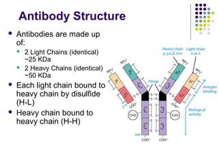
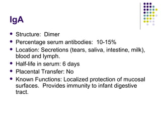
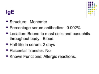
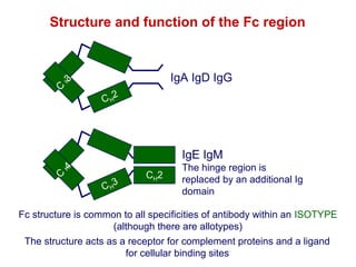
Lecture 5 
Protein Structure and Function II
Antibodies 
 Proteins that recognize and bind to a 
particular antigen with very high specificity. 
 Belong to a group of serum proteins called 
immunoglobulins (Igs). 
 Each antibody has at least two identical sites 
that bind antigen: Antigen binding sites.
Antibody Structure 
 Antibodies are made up 
of: 
 2 Light Chains (identical) 
~25 KDa 
 2 Heavy Chains (identical) 
~50 KDa 
 Each light chain bound to 
heavy chain by disulfide 
(H-L) 
 Heavy chain bound to 
heavy chain (H-H)
Fb 
CH2 
CH3 
Fv 
Fv 
Fv 
Fb 
Fv 
Fb 
Hinge 
Elbow 
CH2 
CH3 
Fv 
Flexibility and 
motion of 
immunoglobulins
Hinge 
CH1 
CH2 
CH3 
VH1 
CL VL
 First 100 a/a of amino terminal vary of both H 
and L chain are variable 
 Referred to as VL , VH, CH And CL 
 CDR (Complementarity Determining 
Regions) are what bind Ag 
 Remaining regions are very similar within 
same class
CDR Are Hypevariable
Non-covalent forces in 
antibody - antigen interactions 
Electrostatic forces Attraction between opposite charges 
Hydrogen bonds Hydrogens shared between electronegative atoms 
Van der Waal’s forces Fluctuations in electron clouds around molecules 
oppositely polarise neighbouring atoms 
Hydrophobic forces Hydrophobic groups pack together to exclude 
water (involves Van der Waal’s forces)
Five classes of antibodies: 
 IgG 
 IgM 
 IgA 
 IgD 
 IgE
Sequencing Of Heavy Chains 
 Sequencing Of Several Immunoglobulins Revealed 
 Five Basic Sequence Patterns 
 a,g, d, e, m 
 IgA, IgG, IgD, IgE and IgM 
 The Above Classes Are Called Isotype 
 Each class can have either k or l light chains 
 Minor Differences Led To Sub-classes For IgA and IgG 
 IgA1, IgGA2 and IgG1, IgG2, IgG3, IgG4
IgG 
 Structure: Monomer 
 Percentage serum antibodies: 80% 
 Location: Blood, lymph, intestine 
 Half-life in serum: 23 days 
 Placental Transfer: Yes 
 Known Functions: Enhances phagocytosis, 
neutralizes toxins and viruses, protects fetus 
and newborn.
IgM 
 Structure: Pentamer 
 Percentage serum antibodies: 5-10% 
 Location: Blood, lymph, B cell surface (monomer) 
 Half-life in serum: 5 days 
 Placental Transfer: No 
 Known Functions: First antibodies produced during 
an infection.
IgA 
 Structure: Dimer 
 Percentage serum antibodies: 10-15% 
 Location: Secretions (tears, saliva, intestine, milk), 
blood and lymph. 
 Half-life in serum: 6 days 
 Placental Transfer: No 
 Known Functions: Localized protection of mucosal 
surfaces. Provides immunity to infant digestive 
tract.
IgD 
 Structure: Monomer 
 Percentage serum antibodies: 0.2% 
 Location: B-cell surface, blood, and lymph 
 Half-life in serum: 3 days 
 Placental Transfer: No 
 Known Functions: In serum function is 
unknown. On B cell surface, initiate immune 
response.
IgE 
 Structure: Monomer 
 Percentage serum antibodies: 0.002% 
 Location: Bound to mast cells and basophils 
throughout body. Blood. 
 Half-life in serum: 2 days 
 Placental Transfer: No 
 Known Functions: Allergic reactions.
Antibody Structure 
 Repeating Domains of ~110 a/a 
 Intrachain disulfide bonds within each domain 
 Heavy chains 
 1 VH and either 3 or 4 CH (CH1, CH2, CH3, CH4) 
 Light chains 
 1 VL and 1 CL 
 Hinge Region 
 Rich in proline residues (flexible) 
 Hinge found in IgG, IgA and IgD 
 Proline residues are target for proteolytic digestion (papain and 
pepsin) 
 Rich in cysteine residues (disulfide bonds) 
 IgM and IgE lack hinge region 
 They instead have extra CH4 Domain
Enzymatic Digestion Of Antibodies 
 Digestion With Papain Yields 
 3 Fragments 
 2 identical Fab and 1 Fc 
 Fab Because Fragment That is Antigen Binding 
 Fc Because Found To Crystallize In Cold Storage 
 Pepsin Digestion 
 F(ab`)2 
 No Fc Recovery, Digested Entirely 
Mercaptoethanol Reduction (Eliminates 
Disulfide Bonds) And Alkylation Showed
Why do antibodies need an Fc region? 
The (Fab)2 fragment can - 
•Detect antigen 
•Precipitate antigen 
•Block the active sites of toxins or pathogen-associated 
molecules 
•Block interactions between host and pathogen-associated 
molecules 
but can not activate 
•Inflammatory and effector functions associated with cells 
•Inflammatory and effector functions of complement 
•The trafficking of antigens into the antigen processing 
pathways
Structure and function of the Fc region 
C H3 
CH2 
IgA IgD IgG 
C H4 
CH3 
CH2 
IgE IgM 
The hinge region is 
replaced by an additional Ig 
domain 
Fc structure is common to all specificities of antibody within an ISOTYPE 
(although there are allotypes) 
The structure acts as a receptor for complement proteins and a ligand 
for cellular binding sites
Monoclonal Antibodies 
 Immunize Animal With Antigen 
 Multiple Clones Are Generated, Good For In Vivo 
 For Clinical Diagnosis, Research, One Clone 
That Reacts To Single Epitope Is Preferred 
 Solution By Kohler and Milstein 
 Fuse A Myeloma Cell (Cancerous) With A Normal 
Plasma Cells 
 Resulting Clones Can Be Cultured Indefinitely 
 Produces An Antibody Recognizing One Epitope
Fc Receptors (FcR) Functions 
 To Transport Abs Across Membranes 
 Secretion of IgA Across Epithelium into lumen 
 Transport of maternal Abs Across Placenta (IgG) 
 Many Cell Types Use FcR 
 Ex. Mast Cells, Macrophages, Neutrophils, B, T, NK 
 Poly IgR 
 Transport of IgA across epithelium 
 FcRN 
 Transport of maternal IgG to fetus
IgA Antibody Transport Across Cell 
(Transcytosis)
Protein section review 
 Proteins are polymers composed of L-amino 
acids. Amino acids are 20 in number
Protein section review 
 Amino acids possess two functional groups 
namely carboxyl (-COOH) and amino (-NH2). 
 In the physiological system, they exist as 
zwitterions.
 The structure of protein is divided into four 
levels of organization. 
 The tertiary and quaternary structures ol 
protein are stabilized by non-covalent bonds 
such as hydrogen bonds, hydrophobic 
interactions, ionic bonds etc.
Protein section review 
 The determination of primary structure of a 
protein: chemical and enzymatic methods are 
employed.
Sample question 
 A heptapeptide (2 M, D, R, K, F, G) was isolated from the 
urine of a three-toed sloth. 
 Reaction of the heptapeptide with FDNB gave DNP-M. 
 Limited proteolysis with carboxypeptidase indicated that M was the first amino 
acid released 
 Cyanogen bromide (CNBr) reaction with the heptapeptide released one 
equivalent of free homoserine lactone 
 Chymotrypsin digestion of the heptapeptide yielded a pentapeptide and a 
dipeptide. Reaction of the pentapeptide with dansyl-Cl gave dansyl-M. 
 Trypsin digestion yielded two M-containing tripeptides and free R. 
 Digestion of the heptapeptide with pepsin gave a tetrapeptide (containing M, R, K 
and D) and a tripeptide (M, F, and G). 
 DNFB & carboxypeptidase (Identify N- and C-terminal residues) 
 CNBr acts only on methionine residues 
 CNBr cleaves peptide chains on the carboxy side of M residues and converts them 
into homoserine lactone 
 Trypsin - cleavage on the C-side of Lys, Arg 
 Chymotrypsin - C-side of Phe, Tyr, Trp 
 Clostripain - like trypsin, but attacks Arg Pepsin hydrolyzes peptide chains on the N-side 
of aromatic AA
Answer 
 N-terminal AA is M 
 C-terminal AA is M 
 CNBr cleaves peptide chains on the carboxy side of M residues 
and converts them into homoserine lactone) - M-?-?-?-?-?-M 
 Chymotrypsin (carboxy side of aromatic AA's) - pentapeptide 
- M-?-?-?-F - dipeptide - ?-M 
 Trypsin digestion - since we know that M is at both the C-terminal 
and the N-terminal, this places K as the 3rd AA 
residue and R as the 4th - M-?-K-R-?-?-M 
 Pepsin digestion - tetrapeptide (M,R,K & D) - sequence must be 
- M-D-K-R - tripeptide (M,F & G) - sequence must be - F-G-M 
 The sequence of the heptapeptide must be: M-D-K-R-F-G-M
Sample questions 
 The imino acid found in protein structure 
 (a) Arginine (b) Proline (c) Histidine (d) Lysin 
 The bonds in protein structure that are not 
broken on denaturation. 
 (a) Hydrogen bonds (b) Peptide bonds (c) 
lonic bond (d) Disulfide bonds

Lecture 5

  • 1.
    Lecture 5 ProteinStructure and Function II
  • 2.
    Antibodies  Proteinsthat recognize and bind to a particular antigen with very high specificity.  Belong to a group of serum proteins called immunoglobulins (Igs).  Each antibody has at least two identical sites that bind antigen: Antigen binding sites.
  • 3.
    Antibody Structure Antibodies are made up of:  2 Light Chains (identical) ~25 KDa  2 Heavy Chains (identical) ~50 KDa  Each light chain bound to heavy chain by disulfide (H-L)  Heavy chain bound to heavy chain (H-H)
  • 5.
    Fb CH2 CH3 Fv Fv Fv Fb Fv Fb Hinge Elbow CH2 CH3 Fv Flexibility and motion of immunoglobulins
  • 6.
    Hinge CH1 CH2 CH3 VH1 CL VL
  • 7.
     First 100a/a of amino terminal vary of both H and L chain are variable  Referred to as VL , VH, CH And CL  CDR (Complementarity Determining Regions) are what bind Ag  Remaining regions are very similar within same class
  • 8.
  • 9.
    Non-covalent forces in antibody - antigen interactions Electrostatic forces Attraction between opposite charges Hydrogen bonds Hydrogens shared between electronegative atoms Van der Waal’s forces Fluctuations in electron clouds around molecules oppositely polarise neighbouring atoms Hydrophobic forces Hydrophobic groups pack together to exclude water (involves Van der Waal’s forces)
  • 10.
    Five classes ofantibodies:  IgG  IgM  IgA  IgD  IgE
  • 11.
    Sequencing Of HeavyChains  Sequencing Of Several Immunoglobulins Revealed  Five Basic Sequence Patterns  a,g, d, e, m  IgA, IgG, IgD, IgE and IgM  The Above Classes Are Called Isotype  Each class can have either k or l light chains  Minor Differences Led To Sub-classes For IgA and IgG  IgA1, IgGA2 and IgG1, IgG2, IgG3, IgG4
  • 12.
    IgG  Structure:Monomer  Percentage serum antibodies: 80%  Location: Blood, lymph, intestine  Half-life in serum: 23 days  Placental Transfer: Yes  Known Functions: Enhances phagocytosis, neutralizes toxins and viruses, protects fetus and newborn.
  • 13.
    IgM  Structure:Pentamer  Percentage serum antibodies: 5-10%  Location: Blood, lymph, B cell surface (monomer)  Half-life in serum: 5 days  Placental Transfer: No  Known Functions: First antibodies produced during an infection.
  • 14.
    IgA  Structure:Dimer  Percentage serum antibodies: 10-15%  Location: Secretions (tears, saliva, intestine, milk), blood and lymph.  Half-life in serum: 6 days  Placental Transfer: No  Known Functions: Localized protection of mucosal surfaces. Provides immunity to infant digestive tract.
  • 15.
    IgD  Structure:Monomer  Percentage serum antibodies: 0.2%  Location: B-cell surface, blood, and lymph  Half-life in serum: 3 days  Placental Transfer: No  Known Functions: In serum function is unknown. On B cell surface, initiate immune response.
  • 16.
    IgE  Structure:Monomer  Percentage serum antibodies: 0.002%  Location: Bound to mast cells and basophils throughout body. Blood.  Half-life in serum: 2 days  Placental Transfer: No  Known Functions: Allergic reactions.
  • 17.
    Antibody Structure Repeating Domains of ~110 a/a  Intrachain disulfide bonds within each domain  Heavy chains  1 VH and either 3 or 4 CH (CH1, CH2, CH3, CH4)  Light chains  1 VL and 1 CL  Hinge Region  Rich in proline residues (flexible)  Hinge found in IgG, IgA and IgD  Proline residues are target for proteolytic digestion (papain and pepsin)  Rich in cysteine residues (disulfide bonds)  IgM and IgE lack hinge region  They instead have extra CH4 Domain
  • 18.
    Enzymatic Digestion OfAntibodies  Digestion With Papain Yields  3 Fragments  2 identical Fab and 1 Fc  Fab Because Fragment That is Antigen Binding  Fc Because Found To Crystallize In Cold Storage  Pepsin Digestion  F(ab`)2  No Fc Recovery, Digested Entirely Mercaptoethanol Reduction (Eliminates Disulfide Bonds) And Alkylation Showed
  • 20.
    Why do antibodiesneed an Fc region? The (Fab)2 fragment can - •Detect antigen •Precipitate antigen •Block the active sites of toxins or pathogen-associated molecules •Block interactions between host and pathogen-associated molecules but can not activate •Inflammatory and effector functions associated with cells •Inflammatory and effector functions of complement •The trafficking of antigens into the antigen processing pathways
  • 21.
    Structure and functionof the Fc region C H3 CH2 IgA IgD IgG C H4 CH3 CH2 IgE IgM The hinge region is replaced by an additional Ig domain Fc structure is common to all specificities of antibody within an ISOTYPE (although there are allotypes) The structure acts as a receptor for complement proteins and a ligand for cellular binding sites
  • 23.
    Monoclonal Antibodies Immunize Animal With Antigen  Multiple Clones Are Generated, Good For In Vivo  For Clinical Diagnosis, Research, One Clone That Reacts To Single Epitope Is Preferred  Solution By Kohler and Milstein  Fuse A Myeloma Cell (Cancerous) With A Normal Plasma Cells  Resulting Clones Can Be Cultured Indefinitely  Produces An Antibody Recognizing One Epitope
  • 24.
    Fc Receptors (FcR)Functions  To Transport Abs Across Membranes  Secretion of IgA Across Epithelium into lumen  Transport of maternal Abs Across Placenta (IgG)  Many Cell Types Use FcR  Ex. Mast Cells, Macrophages, Neutrophils, B, T, NK  Poly IgR  Transport of IgA across epithelium  FcRN  Transport of maternal IgG to fetus
  • 25.
    IgA Antibody TransportAcross Cell (Transcytosis)
  • 26.
    Protein section review  Proteins are polymers composed of L-amino acids. Amino acids are 20 in number
  • 27.
    Protein section review  Amino acids possess two functional groups namely carboxyl (-COOH) and amino (-NH2).  In the physiological system, they exist as zwitterions.
  • 28.
     The structureof protein is divided into four levels of organization.  The tertiary and quaternary structures ol protein are stabilized by non-covalent bonds such as hydrogen bonds, hydrophobic interactions, ionic bonds etc.
  • 29.
    Protein section review  The determination of primary structure of a protein: chemical and enzymatic methods are employed.
  • 30.
    Sample question A heptapeptide (2 M, D, R, K, F, G) was isolated from the urine of a three-toed sloth.  Reaction of the heptapeptide with FDNB gave DNP-M.  Limited proteolysis with carboxypeptidase indicated that M was the first amino acid released  Cyanogen bromide (CNBr) reaction with the heptapeptide released one equivalent of free homoserine lactone  Chymotrypsin digestion of the heptapeptide yielded a pentapeptide and a dipeptide. Reaction of the pentapeptide with dansyl-Cl gave dansyl-M.  Trypsin digestion yielded two M-containing tripeptides and free R.  Digestion of the heptapeptide with pepsin gave a tetrapeptide (containing M, R, K and D) and a tripeptide (M, F, and G).  DNFB & carboxypeptidase (Identify N- and C-terminal residues)  CNBr acts only on methionine residues  CNBr cleaves peptide chains on the carboxy side of M residues and converts them into homoserine lactone  Trypsin - cleavage on the C-side of Lys, Arg  Chymotrypsin - C-side of Phe, Tyr, Trp  Clostripain - like trypsin, but attacks Arg Pepsin hydrolyzes peptide chains on the N-side of aromatic AA
  • 31.
    Answer  N-terminalAA is M  C-terminal AA is M  CNBr cleaves peptide chains on the carboxy side of M residues and converts them into homoserine lactone) - M-?-?-?-?-?-M  Chymotrypsin (carboxy side of aromatic AA's) - pentapeptide - M-?-?-?-F - dipeptide - ?-M  Trypsin digestion - since we know that M is at both the C-terminal and the N-terminal, this places K as the 3rd AA residue and R as the 4th - M-?-K-R-?-?-M  Pepsin digestion - tetrapeptide (M,R,K & D) - sequence must be - M-D-K-R - tripeptide (M,F & G) - sequence must be - F-G-M  The sequence of the heptapeptide must be: M-D-K-R-F-G-M
  • 32.
    Sample questions The imino acid found in protein structure  (a) Arginine (b) Proline (c) Histidine (d) Lysin  The bonds in protein structure that are not broken on denaturation.  (a) Hydrogen bonds (b) Peptide bonds (c) lonic bond (d) Disulfide bonds