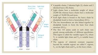
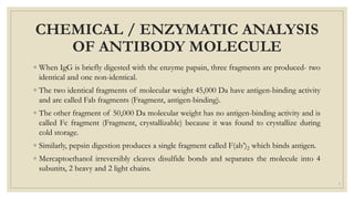
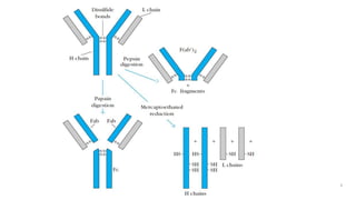
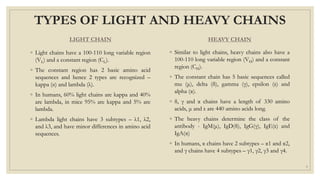
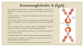
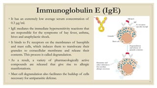
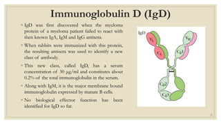
IMMUNOGLOBULIN
STRUCTURE, FUNCTION AND TYPES
Aman Kumar Jaiswar, 422MB00173, I M.Sc. Applied Microbiology
1
INTRODUCTION
◦ Antibodies are antigen binding proteins present on B-cell membrane
and secreted by plasma cells.
◦ Antibodies circulate in the blood, where they participate in humoral
immunity.
◦ They act by searching out and neutralizing antigens or marking them
for elimination.
◦ All antibodies share structural features, bind to antigen, and
participate in a limited number of effector functions.
2
ELECTROPHORETIC STUDY OF
IMMUNE SERA
◦ Carried out by A. Tiselius and E. A. Kabat in 1939.
◦ They immunized rabbits with the protein ovalbumin and then divided the immunized rabbits’ serum
into two aliquots.
◦ Electrophoresis of one serum aliquot revealed four peaks corresponding to albumin and the alpha (α),
beta (β), and gamma (γ) globulins.
◦ The other serum aliquot was reacted with ovalbumin, and the precipitate that formed was removed.
◦ The remaining serum proteins, which did not react with the antigen, were then electrophoresed.
◦ On comparison, significant drop in γ-globulin peak in the aliquot that had been reacted in the antigen.
◦ Thus, the γ -globulin fraction was identified as containing serum antibodies, which were called
immunoglobulins to distinguish them from any other proteins in the same fraction.
3
Blue line: electrophoretic pattern
of untreated antiserum
Black line: pattern of antiserum
that was incubated with OVA
4
BASIC STRUCTURE
OF AN ANTIBODY
5
 4 peptide chains, 2 identical light (L) chains and 2
identical heavy (H) chains.
 Light chains have a molecular weight of about
25,000 Da whereas heavy chains have a molecular
weight of 50,000 Da and more.
 Each light chain is bound to the heavy chain by
disulphide bond to form a heterodimer (H-L).
 The two heterodimers are also linked together by
disulphide bonds to form the basic four chain
structure (H-L)2.
 The first 110 amino acids in each chain vary
greatly among antibodies of different specificities.
This region is called the variable region (V), where
VL is variable light chain and VH is variable heavy
chain.
 The regions of relatively constant sequence
beyond the variable regions are called C regions,
CL on the light chain and CH on the heavy chain.
6
CHEMICAL / ENZYMATIC ANALYSIS
OF ANTIBODY MOLECULE
◦ When IgG is briefly digested with the enzyme papain, three fragments are produced- two
identical and one non-identical.
◦ The two identical fragments of molecular weight 45,000 Da have antigen-binding activity
and are called Fab fragments (Fragment, antigen-binding).
◦ The other fragment of 50,000 Da molecular weight has no antigen-binding activity and is
called Fc fragment (Fragment, crystallizable) because it was found to crystallize during
cold storage.
◦ Similarly, pepsin digestion produces a single fragment called F(ab’)2 which binds antigen.
◦ Mercaptoethanol irreversibly cleaves disulfide bonds and separates the molecule into 4
subunits, 2 heavy and 2 light chains.
7
8
TYPES OF LIGHT AND HEAVY CHAINS
LIGHT CHAIN
◦ Light chains have a 100-110 long variable region
(VL) and a constant region (CL).
◦ The constant region has 2 basic amino acid
sequences and hence 2 types are recognized –
kappa (κ) and lambda (λ).
◦ In humans, 60% light chains are kappa and 40%
are lambda, in mice 95% are kappa and 5% are
lambda.
◦ Lambda light chains have 3 subtypes – λ1, λ2,
and λ3, and have minor differences in amino acid
sequences.
HEAVY CHAIN
◦ Similar to light chains, heavy chains also have a
100-110 long variable region (VH) and a constant
region (CH).
◦ The constant chain has 5 basic sequences called
mu (μ), delta (δ), gamma (γ), epsilon (ε) and
alpha (α).
◦ δ, γ and α chains have a length of 330 amino
acids, μ and ε are 440 amino acids long.
◦ The heavy chains determine the class of the
antibody - IgM(μ), IgD(δ), IgG(γ), IgE(ε) and
IgA(α)
◦ In humans, α chains have 2 subtypes – α1 and α2,
and γ chains have 4 subtypes – γ1, γ2, γ3 and γ4.
9
OTHER CHARACTERISTICS
◦ Both heavy and light chains are folded into domains, homologous units of 110 amino acids
with an intrachain disulphide bond that forms a loop of about 60 amino acids.
◦ Light chains have 1 constant (CL) and 1 variable (VL) domain. Heavy chains have 1 variable
(VH) and 3-4 constant domains (CH1, CH2, CH3 and CH4).
◦ The loops present in VL and VH chains show maximum variability and are called
hypervariable regions or complementarity determining regions (CDRs) and are the antigen
binding sites.
◦ The remainder of VL and VH show less variability and are called framework regions (FRs).
◦ δ, γ and α have an extended peptide sequence between CH1 and CH2 called hinge region
which is rich in proline and gives IgD, IgG and IgA flexibility.
10
11
FUNCTIONS
Apart from antigen-binding, which is carried out by variable regions of the antibody,
there are several other effector functions that are carried out by the constant region.
These include opsonization, complement activation, antibody-dependent cell-mediated
cytotoxicity and transcytosis.
12
FUNCTIONS
OPSONIZATION
◦ It is the promotion of phagocytosis of
antigens by macrophages and neutrophils.
◦ Macrophages and neutrophils contain Fc
receptors that bind to the Fc region of
immunoglobulins.
◦ When Fc receptors bind to the Fc regions in
an antigen-antibody complex, it initiates
binding of the complex to the phagocyte
membrane as well as phagocytosis.
◦ The complex is then destructed using
enzymes or oxidizing agents.
COMPLEMENT ACTIVATION
◦ IgM and IgG can activate a collection of
serum glycoproteins called complements
that can perforate cell membranes.
◦ C3b binds non-specifically to antigen-
antibody complexes which also acts as a
receptor for macrophages and promotes
phagocytosis.
◦ The collaboration between antibody and the
complement system is important for the
inactivation and removal of antigens and
the killing of pathogens.
13
14
OPSONIZATION COMPLEMENT ACTIVATION
FUNCTIONS
ANTIBODY-DEPENDENT CELL-
MEDIATED CYTOXICITY
◦ Some other cells such as Natural Killer
(NK) cells also have Fc receptors and
hence can bind to antigen-antibody
complexes.
◦ This leads to cytotoxic activities of the
effector cell towards target cell by a
process called antibody-dependent cell-
mediated cytotoxicity.
◦ Here, antibody acts as a receptor that
enables the attacking cell to recognize
and kill the target cell.
TRANSCYTOSIS
◦ The delivery of antibodies to mucosal
surfaces as well as breast milk across
epithelial layers is called transcytosis.
◦ It greatly depends on the properties of
the constant region.
◦ Examples for immunoglobulins capable
of transcytosis include IgA, IgM and
IgG.
◦ IgG can also cross maternal placenta to
confer passive immunity to the fetus.
15
16
ANTIBODY-DEPENDENT CELL-MEDIATED CYTOTOXICITY (ADCC)
ANTIBODY CLASSES
There are five major classes of antibodies that differ in the
amino acid sequences in their heavy chain constant regions.
These confer class-specific structural and functional properties.
17
Immunoglobulin G (IgG)
◦ It is the most abundant in serum, and constitutes about 80% of total
serum immunoglobulin.
◦ IgG molecule consists of 2 γ heavy chains and 2 κ/λ light chains.
◦ There are 4 subclasses distinguished by differences in γ chain
sequences – IgG1, IgG2, IgG3 and IgG4.
◦ The major differences between these subclasses are the size of the
hinge region and number and position of interchain disulphide bonds
between the heavy chains.
◦ These differences affect the biological properties as given below:
◦ 1. IgG1, IgG3 and IgG4 are involved in transplacental movement and
passive immunization.
◦ 2. IgG3 is the most effective complement activator followed by IgG1
and IgG2. IgG4 does not activate complement.
◦ 3. IgG1 and IgG3 have high affinity to Fc receptors and thus mediate
opsonization compared to IgG4 and IgG2 that have low affinity.
18
Immunoglobulin M (IgM)
◦ IgM accounts for 5-10% of total serum immunoglobulin with an average
serum concentration of 1.5 mg/ml.
◦ It had a molecular weight of about 1,80,000 Da.
◦ It is secreted as a pentamer with 5 monomer units held together with
disulphide bonds that link their heavy chains.
◦ Each pentamer contains an additional Fc-linked polypeptide called J
(joining) chain, which is bound to 2 of the 10 μ chains by disulphide
bonds.
◦ J chain is required for pentamer formation and is added just before the
secretion of the IgM molecule.
◦ It is the first immunoglobulin produced in response to an antigen and
the first immunoglobulin produced by a neonate.
◦ It is a good agglutinin due to its large size and is also more efficient than
IgG in activating complement.
◦ It plays an important accessory role as secretory immunoglobulin and is
present is mucosal tissue.
19
Immunoglobulin A (IgA)
◦ IgA constitutes about 10-15% of total immunoglobulin in serum, and is
predominant in external secretions such as breast milk, saliva, tears and
mucus.
◦ It exists primarily as a monomer, but polymeric forms are also seen, all
containing a J-chain.
◦ Secretory IgA consists of a dimer, a J-chain and a polypeptide chain called
secretory component, which is receptor-derived from mucosal epithelial
cells.
◦ Secretory component has a molecular weight of 70,000 Da and contains 5
domains that bind to Fc region of the IgA by disulphide bonds.
◦ It is the most secreted antibody, and about 5-15g of IgA is secreted into
mucous secretions daily.
◦ Its functions include cross-linking of large antigens with multiple epitopes.
◦ It also prevents attachment of pathogens to mucosal membranes, thus
inhibiting infection and colonization.
◦ It is present in breast milk and provides passive immunity to the neonate.
20
Immunoglobulin E (IgE)
◦ It has an extremely low average serum concentration of
0.3 μg/ml.
◦ IgE mediates the immediate hypersensitivity reactions that
are responsible for the symptoms of hay fever, asthma,
hives and anaphylactic shock.
◦ It binds to Fc receptors on the membranes of basophils
and mast cells, which induces them to translocate their
granules to extracellular membrane and release their
contents. This process is called degranulation.
◦ As a result, a variety of pharmacologically active
compounds are released that give rise to allergic
manifestations.
◦ Mast cell degranulation also facilitates the buildup of cells
necessary for antiparasitic defense.
21
Immunoglobulin D (IgD)
◦ IgD was first discovered when the myeloma
protein of a myeloma patient failed to react with
then known IgA, IgM and IgG antisera.
◦ When rabbits were immunized with this protein,
the resulting antisera was used to identify a new
class of antibody.
◦ This new class, called IgD, has a serum
concentration of 30 μg/ml and constitutes about
0.2% of the total immunoglobulin in the serum.
◦ Along with IgM, it is the major membrane bound
immunoglobulin expressed by mature B cells.
◦ No biological effector function has been
identified for IgD so far.
22

Immunoglobulin.pptx

  • 1.
    IMMUNOGLOBULIN STRUCTURE, FUNCTION ANDTYPES Aman Kumar Jaiswar, 422MB00173, I M.Sc. Applied Microbiology 1
  • 2.
    INTRODUCTION ◦ Antibodies areantigen binding proteins present on B-cell membrane and secreted by plasma cells. ◦ Antibodies circulate in the blood, where they participate in humoral immunity. ◦ They act by searching out and neutralizing antigens or marking them for elimination. ◦ All antibodies share structural features, bind to antigen, and participate in a limited number of effector functions. 2
  • 3.
    ELECTROPHORETIC STUDY OF IMMUNESERA ◦ Carried out by A. Tiselius and E. A. Kabat in 1939. ◦ They immunized rabbits with the protein ovalbumin and then divided the immunized rabbits’ serum into two aliquots. ◦ Electrophoresis of one serum aliquot revealed four peaks corresponding to albumin and the alpha (α), beta (β), and gamma (γ) globulins. ◦ The other serum aliquot was reacted with ovalbumin, and the precipitate that formed was removed. ◦ The remaining serum proteins, which did not react with the antigen, were then electrophoresed. ◦ On comparison, significant drop in γ-globulin peak in the aliquot that had been reacted in the antigen. ◦ Thus, the γ -globulin fraction was identified as containing serum antibodies, which were called immunoglobulins to distinguish them from any other proteins in the same fraction. 3
  • 4.
    Blue line: electrophoreticpattern of untreated antiserum Black line: pattern of antiserum that was incubated with OVA 4
  • 5.
  • 6.
     4 peptidechains, 2 identical light (L) chains and 2 identical heavy (H) chains.  Light chains have a molecular weight of about 25,000 Da whereas heavy chains have a molecular weight of 50,000 Da and more.  Each light chain is bound to the heavy chain by disulphide bond to form a heterodimer (H-L).  The two heterodimers are also linked together by disulphide bonds to form the basic four chain structure (H-L)2.  The first 110 amino acids in each chain vary greatly among antibodies of different specificities. This region is called the variable region (V), where VL is variable light chain and VH is variable heavy chain.  The regions of relatively constant sequence beyond the variable regions are called C regions, CL on the light chain and CH on the heavy chain. 6
  • 7.
    CHEMICAL / ENZYMATICANALYSIS OF ANTIBODY MOLECULE ◦ When IgG is briefly digested with the enzyme papain, three fragments are produced- two identical and one non-identical. ◦ The two identical fragments of molecular weight 45,000 Da have antigen-binding activity and are called Fab fragments (Fragment, antigen-binding). ◦ The other fragment of 50,000 Da molecular weight has no antigen-binding activity and is called Fc fragment (Fragment, crystallizable) because it was found to crystallize during cold storage. ◦ Similarly, pepsin digestion produces a single fragment called F(ab’)2 which binds antigen. ◦ Mercaptoethanol irreversibly cleaves disulfide bonds and separates the molecule into 4 subunits, 2 heavy and 2 light chains. 7
  • 8.
  • 9.
    TYPES OF LIGHTAND HEAVY CHAINS LIGHT CHAIN ◦ Light chains have a 100-110 long variable region (VL) and a constant region (CL). ◦ The constant region has 2 basic amino acid sequences and hence 2 types are recognized – kappa (κ) and lambda (λ). ◦ In humans, 60% light chains are kappa and 40% are lambda, in mice 95% are kappa and 5% are lambda. ◦ Lambda light chains have 3 subtypes – λ1, λ2, and λ3, and have minor differences in amino acid sequences. HEAVY CHAIN ◦ Similar to light chains, heavy chains also have a 100-110 long variable region (VH) and a constant region (CH). ◦ The constant chain has 5 basic sequences called mu (μ), delta (δ), gamma (γ), epsilon (ε) and alpha (α). ◦ δ, γ and α chains have a length of 330 amino acids, μ and ε are 440 amino acids long. ◦ The heavy chains determine the class of the antibody - IgM(μ), IgD(δ), IgG(γ), IgE(ε) and IgA(α) ◦ In humans, α chains have 2 subtypes – α1 and α2, and γ chains have 4 subtypes – γ1, γ2, γ3 and γ4. 9
  • 10.
    OTHER CHARACTERISTICS ◦ Bothheavy and light chains are folded into domains, homologous units of 110 amino acids with an intrachain disulphide bond that forms a loop of about 60 amino acids. ◦ Light chains have 1 constant (CL) and 1 variable (VL) domain. Heavy chains have 1 variable (VH) and 3-4 constant domains (CH1, CH2, CH3 and CH4). ◦ The loops present in VL and VH chains show maximum variability and are called hypervariable regions or complementarity determining regions (CDRs) and are the antigen binding sites. ◦ The remainder of VL and VH show less variability and are called framework regions (FRs). ◦ δ, γ and α have an extended peptide sequence between CH1 and CH2 called hinge region which is rich in proline and gives IgD, IgG and IgA flexibility. 10
  • 11.
  • 12.
    FUNCTIONS Apart from antigen-binding,which is carried out by variable regions of the antibody, there are several other effector functions that are carried out by the constant region. These include opsonization, complement activation, antibody-dependent cell-mediated cytotoxicity and transcytosis. 12
  • 13.
    FUNCTIONS OPSONIZATION ◦ It isthe promotion of phagocytosis of antigens by macrophages and neutrophils. ◦ Macrophages and neutrophils contain Fc receptors that bind to the Fc region of immunoglobulins. ◦ When Fc receptors bind to the Fc regions in an antigen-antibody complex, it initiates binding of the complex to the phagocyte membrane as well as phagocytosis. ◦ The complex is then destructed using enzymes or oxidizing agents. COMPLEMENT ACTIVATION ◦ IgM and IgG can activate a collection of serum glycoproteins called complements that can perforate cell membranes. ◦ C3b binds non-specifically to antigen- antibody complexes which also acts as a receptor for macrophages and promotes phagocytosis. ◦ The collaboration between antibody and the complement system is important for the inactivation and removal of antigens and the killing of pathogens. 13
  • 14.
  • 15.
    FUNCTIONS ANTIBODY-DEPENDENT CELL- MEDIATED CYTOXICITY ◦Some other cells such as Natural Killer (NK) cells also have Fc receptors and hence can bind to antigen-antibody complexes. ◦ This leads to cytotoxic activities of the effector cell towards target cell by a process called antibody-dependent cell- mediated cytotoxicity. ◦ Here, antibody acts as a receptor that enables the attacking cell to recognize and kill the target cell. TRANSCYTOSIS ◦ The delivery of antibodies to mucosal surfaces as well as breast milk across epithelial layers is called transcytosis. ◦ It greatly depends on the properties of the constant region. ◦ Examples for immunoglobulins capable of transcytosis include IgA, IgM and IgG. ◦ IgG can also cross maternal placenta to confer passive immunity to the fetus. 15
  • 16.
  • 17.
    ANTIBODY CLASSES There arefive major classes of antibodies that differ in the amino acid sequences in their heavy chain constant regions. These confer class-specific structural and functional properties. 17
  • 18.
    Immunoglobulin G (IgG) ◦It is the most abundant in serum, and constitutes about 80% of total serum immunoglobulin. ◦ IgG molecule consists of 2 γ heavy chains and 2 κ/λ light chains. ◦ There are 4 subclasses distinguished by differences in γ chain sequences – IgG1, IgG2, IgG3 and IgG4. ◦ The major differences between these subclasses are the size of the hinge region and number and position of interchain disulphide bonds between the heavy chains. ◦ These differences affect the biological properties as given below: ◦ 1. IgG1, IgG3 and IgG4 are involved in transplacental movement and passive immunization. ◦ 2. IgG3 is the most effective complement activator followed by IgG1 and IgG2. IgG4 does not activate complement. ◦ 3. IgG1 and IgG3 have high affinity to Fc receptors and thus mediate opsonization compared to IgG4 and IgG2 that have low affinity. 18
  • 19.
    Immunoglobulin M (IgM) ◦IgM accounts for 5-10% of total serum immunoglobulin with an average serum concentration of 1.5 mg/ml. ◦ It had a molecular weight of about 1,80,000 Da. ◦ It is secreted as a pentamer with 5 monomer units held together with disulphide bonds that link their heavy chains. ◦ Each pentamer contains an additional Fc-linked polypeptide called J (joining) chain, which is bound to 2 of the 10 μ chains by disulphide bonds. ◦ J chain is required for pentamer formation and is added just before the secretion of the IgM molecule. ◦ It is the first immunoglobulin produced in response to an antigen and the first immunoglobulin produced by a neonate. ◦ It is a good agglutinin due to its large size and is also more efficient than IgG in activating complement. ◦ It plays an important accessory role as secretory immunoglobulin and is present is mucosal tissue. 19
  • 20.
    Immunoglobulin A (IgA) ◦IgA constitutes about 10-15% of total immunoglobulin in serum, and is predominant in external secretions such as breast milk, saliva, tears and mucus. ◦ It exists primarily as a monomer, but polymeric forms are also seen, all containing a J-chain. ◦ Secretory IgA consists of a dimer, a J-chain and a polypeptide chain called secretory component, which is receptor-derived from mucosal epithelial cells. ◦ Secretory component has a molecular weight of 70,000 Da and contains 5 domains that bind to Fc region of the IgA by disulphide bonds. ◦ It is the most secreted antibody, and about 5-15g of IgA is secreted into mucous secretions daily. ◦ Its functions include cross-linking of large antigens with multiple epitopes. ◦ It also prevents attachment of pathogens to mucosal membranes, thus inhibiting infection and colonization. ◦ It is present in breast milk and provides passive immunity to the neonate. 20
  • 21.
    Immunoglobulin E (IgE) ◦It has an extremely low average serum concentration of 0.3 μg/ml. ◦ IgE mediates the immediate hypersensitivity reactions that are responsible for the symptoms of hay fever, asthma, hives and anaphylactic shock. ◦ It binds to Fc receptors on the membranes of basophils and mast cells, which induces them to translocate their granules to extracellular membrane and release their contents. This process is called degranulation. ◦ As a result, a variety of pharmacologically active compounds are released that give rise to allergic manifestations. ◦ Mast cell degranulation also facilitates the buildup of cells necessary for antiparasitic defense. 21
  • 22.
    Immunoglobulin D (IgD) ◦IgD was first discovered when the myeloma protein of a myeloma patient failed to react with then known IgA, IgM and IgG antisera. ◦ When rabbits were immunized with this protein, the resulting antisera was used to identify a new class of antibody. ◦ This new class, called IgD, has a serum concentration of 30 μg/ml and constitutes about 0.2% of the total immunoglobulin in the serum. ◦ Along with IgM, it is the major membrane bound immunoglobulin expressed by mature B cells. ◦ No biological effector function has been identified for IgD so far. 22