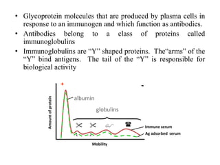
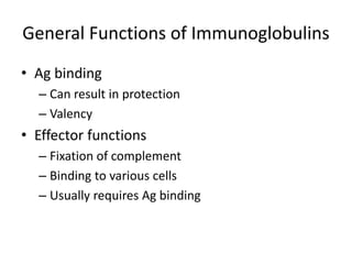
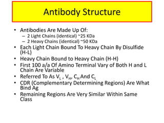
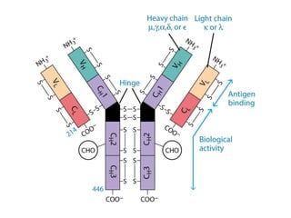
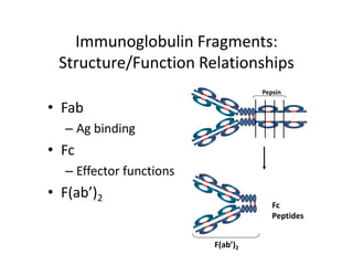
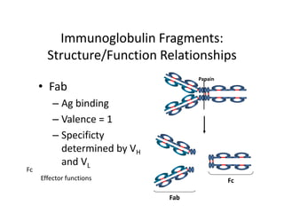
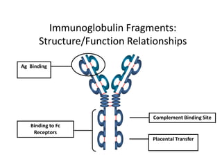
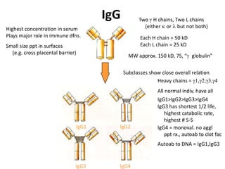
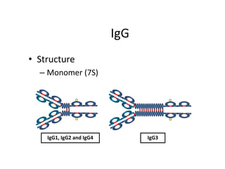
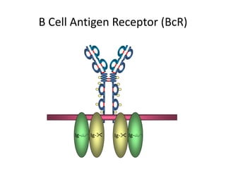
Antibodies: Structure And
Function
• Glycoprotein molecules that are produced by plasma cells in
response to an immunogen and which function as antibodies.
• Antibodies belong to a class of proteins called
immunoglobulins
• Immunoglobulins are “Y” shaped proteins. The“arms” of the
“Y” bind antigens. The tail of the “Y” is responsible for
biological activity
Immune serum
Ag adsorbed serum

1

2
 
+ -
albumin
globulins
Mobility
Amountofprotein
General Functions of Immunoglobulins
• Ag binding
– Can result in protection
– Valency
• Effector functions
– Fixation of complement
– Binding to various cells
– Usually requires Ag binding
Basic Immunoglobulin Structure
• Immunoglobulins - heterogeneous
• Myeloma proteins - homogeneous
immunoglobulins
Antibody Structure
• Antibodies Are Made Up Of:
– 2 Light Chains (identical) ~25 KDa
– 2 Heavy Chains (identical) ~50 KDa
• Each Light Chain Bound To Heavy Chain By Disulfide
(H-L)
• Heavy Chain Bound to Heavy Chain (H-H)
• First 100 a/a Of Amino Terminal Vary of Both H and L
Chain Are Variable
• Referred To As VL , VH, CH And CL
• CDR (Complementary Determining Regions) Are What
Bind Ag
• Remaining Regions Are Very Similar Within Same
Class
• Repeating Domains of ~110 a/a
– Intrachain disulfide bonds within each domain
• Heavy chains
– 1 VH and either 3 or 4 CH (CH1, CH2, CH3, CH4)
• Light chains
– 1 VL and 1 CL
• Hinge Region
– Rich in proline residues (flexible)
– Hinge found in IgG, IgA and IgD
– Proline residues are target for proteolytic digestion (papain and
pepsin)
– Rich in cysteine residues (disulfide bonds)
– IgM and IgE lack hinge region
– They instead have extra CH4 Domain
Antibody Structure
• Digestion With Papain Yields
– 3 Fragments
– 2 identical Fab and 1 Fc
– Fab Because Fragment That is Antigen Binding
– Fc Because Found To Crystallize In Cold Storage
• Pepsin Digestion
– F(ab`)2
– No Fc Recovery, Digested Entirely
• Mercaptoethanol Reduction (Eliminates Disulfide Bonds)
And Alkylation Showed
Enzymatic Digestion Of Antibodies
• Sequencing Of Several Immunoglobulins Revealed
– 100-110 Amino Terminus, Highly Variable (V)
– Five Basic Sequence Patterns
– , , , , 
– IgA, IgD, IgG, IgE and IgM
– The Above Classes Are Called Isotype
– Each class can have either  or  light chains
– Minor differences Led To Sub-classes For IgA and IgG
– IgA1, IgGA2 and IgG1, IgG2, IgG3, IgG4
Sequencing Of Heavy Chains
Characteristics of hinge region
•Immunoglobulins (with possible exception of IgM & IgE)
contain hinge between
• CH1 & CH2
•No homology between AA sequence of hinge & heavy chains
•AA sequence differs with different classes
•Comprised of many cysteine and proline residues
•Cysteine involved in formation of interchain disulfide bonds
•Proline prevents folding in a globular structure, allowing
flexibility between
two Fab arms of the Y-shaped antibody; allows open & close to
accommodate
binding to two epitopes; because it is open, it can be cleaved by
proteases
(e.g. papain) to generate the Fab & Fc fragments
Immunoglobulin Fragments:
Structure/Function Relationships
• Fab
– Ag binding
• Fc
– Effector functions
• F(ab’)2
Pepsin
Fc
Peptides
F(ab’)2
Immunoglobulin Fragments:
Structure/Function Relationships
• Fab
– Ag binding
– Valence = 1
– Specificty
determined by VH
and VL
Papain
Fc
Fab
Fc
Effector functions
Immunoglobulin Fragments:
Structure/Function Relationships
Ag Binding
Complement Binding Site
Placental Transfer
Binding to Fc
Receptors
IgG
IgG1 IgG2
IgG3 IgG4
Subclasses show close overall relation
Heavy chains = 1,2,3,4
Highest concentration in serum
Plays major role in immune dfns.
Small size ppt in surfaces
(e.g. cross placental barrier)
All normal indiv. have all
IgG1>IgG2>IgG3>IgG4
IgG3 has shortest 1/2 life,
highest catabolic rate,
highest # S-S
IgG4 = monoval. no aggl
ppt rx., autoab to clot fac
Autoab to DNA = IgG1,IgG3
Two  H chains, Two L chains
(either  or  but not both)
Each H chain = 50 kD
Each L chain = 25 kD
MW approx. 150 kD, 7S, “ globulin”
IgG
• Structure
– Monomer (7S)
IgG1, IgG2 and IgG4 IgG3
IgG
• Structure
• Properties
– Major serum Ig
– Major Ig in extravascular spaces
– The only antibody to cross the placental
– Fixes complement
– Binds to Fc receptors
• Phagocytes - opsonization
• NK cells – ADCC
– Binds to SPA
IgM
• Structure
• Properties
– 3rd highest serum Ig
– First Ig made by fetus and B
cells
– Produced early in the
primary response
– The most efficient Ig
– Fixes complement
Tail
Piece
Agglutinating Ig
Binds to Fc receptors
B cell surface Ig
IgM
• Structure
– Pentamer (19S)
composed 5
H2L2 units plus one
molecule of
J chain
– Extra domain (CH4)
– J chain
CH4
J Chain
Structural features of IgM
Pentamer (5)
First Ig produced following immun.
Macroglobulin (M)
900 kD, 19S
SSS S
SS
J chain Doesn’t have hinge region
has additional H domain
Has a J chain (one of 2 Ig isotypes)
15 kD
IgD
• Structure
– Monomer
– Tail piece
Tail Piece
IgD
• Structure
• Properties
– 4th highest serum Ig
– B cell surface Ig
– Does not bind complement
Structural features of IgD
Primarily a B cell antigen receptor
Long exposed hinge region
170kD, 7S, migrates as fast 
No interchain S-S bridges in H chains
Readily denatured
IgA
• Properties
–2nd highest serum Ig
–Major secretory Ig ( saliva, tears,
respiratory, intestinal, and genital
tract secretions.)
–Does not fix complement unless
aggregated
–Binds to Fc receptors on some cells
IgA
• Structure
–Serum - monomer
–Secretions (sIgA)
–Dimer (11S), sIgA molecule consists of two
H2L2 units plus one molecule each of J chain
and secretory component(SC or SP)
J ChainSecretory Piece
IgE
• Structure
– Monomer
– Extra domain (CH4)
CH4
Structural features of IgE
Sometimes called reaginic ag
Mediates allergies (Type I
hypersensitivies)
190 kDa, 8S, migrates as fast 
Contains an extra domain (CH4)
which binds to mast cells
& basophils
May remain attached for long time
when ag reappears, cross links
IgE on mast cell surface, release
mast-cell granules & signs of
anaphylaxis
IgE
• Structure
• Properties
– Least common serum Ig
– Allergic reactions
– Parasitic infections
– Does not fix complement
Fixation of C1 by IgG and IgM Abs
No activation Activation
B Cell Antigen Receptor (BcR)
Ig-Ig- Ig-Ig-
L & H chain folding to yield 3 CDR in each
chain to form walls of ag binding groove

Imunoglobulins-Basics

  • 1.
  • 2.
    • Glycoprotein moleculesthat are produced by plasma cells in response to an immunogen and which function as antibodies. • Antibodies belong to a class of proteins called immunoglobulins • Immunoglobulins are “Y” shaped proteins. The“arms” of the “Y” bind antigens. The tail of the “Y” is responsible for biological activity Immune serum Ag adsorbed serum  1  2   + - albumin globulins Mobility Amountofprotein
  • 3.
    General Functions ofImmunoglobulins • Ag binding – Can result in protection – Valency • Effector functions – Fixation of complement – Binding to various cells – Usually requires Ag binding
  • 4.
    Basic Immunoglobulin Structure •Immunoglobulins - heterogeneous • Myeloma proteins - homogeneous immunoglobulins
  • 5.
    Antibody Structure • AntibodiesAre Made Up Of: – 2 Light Chains (identical) ~25 KDa – 2 Heavy Chains (identical) ~50 KDa • Each Light Chain Bound To Heavy Chain By Disulfide (H-L) • Heavy Chain Bound to Heavy Chain (H-H) • First 100 a/a Of Amino Terminal Vary of Both H and L Chain Are Variable • Referred To As VL , VH, CH And CL • CDR (Complementary Determining Regions) Are What Bind Ag • Remaining Regions Are Very Similar Within Same Class
  • 7.
    • Repeating Domainsof ~110 a/a – Intrachain disulfide bonds within each domain • Heavy chains – 1 VH and either 3 or 4 CH (CH1, CH2, CH3, CH4) • Light chains – 1 VL and 1 CL • Hinge Region – Rich in proline residues (flexible) – Hinge found in IgG, IgA and IgD – Proline residues are target for proteolytic digestion (papain and pepsin) – Rich in cysteine residues (disulfide bonds) – IgM and IgE lack hinge region – They instead have extra CH4 Domain Antibody Structure
  • 9.
    • Digestion WithPapain Yields – 3 Fragments – 2 identical Fab and 1 Fc – Fab Because Fragment That is Antigen Binding – Fc Because Found To Crystallize In Cold Storage • Pepsin Digestion – F(ab`)2 – No Fc Recovery, Digested Entirely • Mercaptoethanol Reduction (Eliminates Disulfide Bonds) And Alkylation Showed Enzymatic Digestion Of Antibodies
  • 11.
    • Sequencing OfSeveral Immunoglobulins Revealed – 100-110 Amino Terminus, Highly Variable (V) – Five Basic Sequence Patterns – , , , ,  – IgA, IgD, IgG, IgE and IgM – The Above Classes Are Called Isotype – Each class can have either  or  light chains – Minor differences Led To Sub-classes For IgA and IgG – IgA1, IgGA2 and IgG1, IgG2, IgG3, IgG4 Sequencing Of Heavy Chains
  • 12.
    Characteristics of hingeregion •Immunoglobulins (with possible exception of IgM & IgE) contain hinge between • CH1 & CH2 •No homology between AA sequence of hinge & heavy chains •AA sequence differs with different classes •Comprised of many cysteine and proline residues •Cysteine involved in formation of interchain disulfide bonds •Proline prevents folding in a globular structure, allowing flexibility between two Fab arms of the Y-shaped antibody; allows open & close to accommodate binding to two epitopes; because it is open, it can be cleaved by proteases (e.g. papain) to generate the Fab & Fc fragments
  • 13.
    Immunoglobulin Fragments: Structure/Function Relationships •Fab – Ag binding • Fc – Effector functions • F(ab’)2 Pepsin Fc Peptides F(ab’)2
  • 14.
    Immunoglobulin Fragments: Structure/Function Relationships •Fab – Ag binding – Valence = 1 – Specificty determined by VH and VL Papain Fc Fab Fc Effector functions
  • 15.
    Immunoglobulin Fragments: Structure/Function Relationships AgBinding Complement Binding Site Placental Transfer Binding to Fc Receptors
  • 16.
    IgG IgG1 IgG2 IgG3 IgG4 Subclassesshow close overall relation Heavy chains = 1,2,3,4 Highest concentration in serum Plays major role in immune dfns. Small size ppt in surfaces (e.g. cross placental barrier) All normal indiv. have all IgG1>IgG2>IgG3>IgG4 IgG3 has shortest 1/2 life, highest catabolic rate, highest # S-S IgG4 = monoval. no aggl ppt rx., autoab to clot fac Autoab to DNA = IgG1,IgG3 Two  H chains, Two L chains (either  or  but not both) Each H chain = 50 kD Each L chain = 25 kD MW approx. 150 kD, 7S, “ globulin”
  • 17.
    IgG • Structure – Monomer(7S) IgG1, IgG2 and IgG4 IgG3
  • 18.
    IgG • Structure • Properties –Major serum Ig – Major Ig in extravascular spaces – The only antibody to cross the placental – Fixes complement – Binds to Fc receptors • Phagocytes - opsonization • NK cells – ADCC – Binds to SPA
  • 19.
    IgM • Structure • Properties –3rd highest serum Ig – First Ig made by fetus and B cells – Produced early in the primary response – The most efficient Ig – Fixes complement Tail Piece Agglutinating Ig Binds to Fc receptors B cell surface Ig
  • 20.
    IgM • Structure – Pentamer(19S) composed 5 H2L2 units plus one molecule of J chain – Extra domain (CH4) – J chain CH4 J Chain
  • 21.
    Structural features ofIgM Pentamer (5) First Ig produced following immun. Macroglobulin (M) 900 kD, 19S SSS S SS J chain Doesn’t have hinge region has additional H domain Has a J chain (one of 2 Ig isotypes) 15 kD
  • 22.
    IgD • Structure – Monomer –Tail piece Tail Piece
  • 23.
    IgD • Structure • Properties –4th highest serum Ig – B cell surface Ig – Does not bind complement
  • 24.
    Structural features ofIgD Primarily a B cell antigen receptor Long exposed hinge region 170kD, 7S, migrates as fast  No interchain S-S bridges in H chains Readily denatured
  • 26.
    IgA • Properties –2nd highestserum Ig –Major secretory Ig ( saliva, tears, respiratory, intestinal, and genital tract secretions.) –Does not fix complement unless aggregated –Binds to Fc receptors on some cells
  • 27.
    IgA • Structure –Serum -monomer –Secretions (sIgA) –Dimer (11S), sIgA molecule consists of two H2L2 units plus one molecule each of J chain and secretory component(SC or SP) J ChainSecretory Piece
  • 28.
    IgE • Structure – Monomer –Extra domain (CH4) CH4
  • 29.
    Structural features ofIgE Sometimes called reaginic ag Mediates allergies (Type I hypersensitivies) 190 kDa, 8S, migrates as fast  Contains an extra domain (CH4) which binds to mast cells & basophils May remain attached for long time when ag reappears, cross links IgE on mast cell surface, release mast-cell granules & signs of anaphylaxis
  • 30.
    IgE • Structure • Properties –Least common serum Ig – Allergic reactions – Parasitic infections – Does not fix complement
  • 31.
    Fixation of C1by IgG and IgM Abs No activation Activation
  • 32.
    B Cell AntigenReceptor (BcR) Ig-Ig- Ig-Ig-
  • 33.
    L & Hchain folding to yield 3 CDR in each chain to form walls of ag binding groove