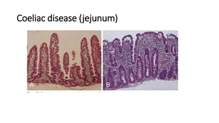
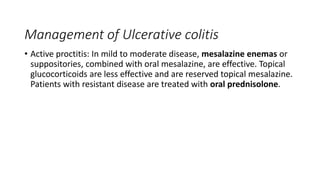
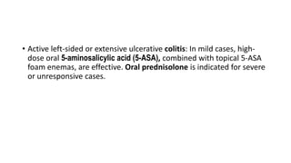
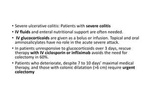
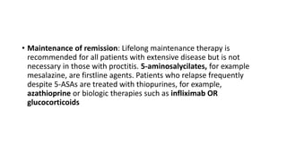
Coeliac disease
By Dr. Hadeer Fathy
• Coeliac disease is an immunologically mediated inflammatory
disorder of the small bowel occurring in genetically susceptible
individuals. It is caused by intolerance to wheat gluten and similar
proteins in rye, barley and oats. It can result in malabsorption and
responds to a gluten-free diet.
• Coeliac disease is associated with other HLA-linked autoimmune
disorders and with certain other diseases.
• individuals at higher risk of developing coeliac disease, e.g. people
with type 1 diabetes,.
Clinical features
• • Infants: failure to thrive, malabsorption.
• • Older children: delayed growth and puberty, malnutrition, mild
abdominal distension.
• • Adults: presents in third or fourth decade; 2:1 female
predominance. malabsorption in some; others present with
tiredness, weight loss, iron or folate deficiency.
• Oral ulceration, dyspepsia and bloating.
Investigations
• Duodenal biopsy: The mucosa may appear macroscopically normal,
but multiple biopsies should be taken.
• Villous atrophy is characteristic but other causes should also be
considered.
Coeliac disease (jejunum)
Antibodies
• These are a valuable screening tool in patients with suggestive
symptoms but are not a substitute for duodenal biopsy.
• IgA anti-endomysial antibodies are detectable in most untreated
cases and are sensitive and specific.
Others
• There is microcytic or macrocytic anemia from iron or folate
deficiency.
• . Calcium, magnesium, total protein, albumin and vitamin D are
reduced
Complications
• There is an increased risk of malignancy, particularly of
enteropathyassociated T-cell lymphoma, small bowel carcinoma and
squamous carcinoma of the oesophagus.
• Ulcerative jejunoileitis may occur; fever, pain, obstruction or
perforation may then occure., and some patients require surgical
resection and parenteral nutrition.
• Osteoporosis and osteomalacia occur but are less common in
patients who adhere strictly to a gluten-free diet
Management
• • Lifelong gluten-free diet aids mucosal healing.
• • Correction of deficiencies of iron, folate, calcium and vitamin D
symptoms, weight and nutrition.
Inflammatory bowel diseases (IBD)
• Ulcerative colitis and Crohn’s disease are chronic inflammatory bowel
diseases that relapse and remit over years. The diseases have many
similarities
• ulcerative colitis only involves the colona and rectum, whereas
Crohn’s disease can involve any part of the GI tract.
• Both diseases present most commonly in the second and third
decades.
• Ulcerative colitis has a prevalence of 100 to 200 per 100 000,
compared with 50 to 100 per 100 000 for Crohn’s disease.
Pathogenesis
• It is thought that IBD develops because genetically susceptible
individuals mount an abnormal inflammatory response to
environmental triggers, such as intestinal bacteria.
• There is emerging evidence that microbial dysbiosis, the virome and
the mycobiome (fungal species) may be important in the
development of IBD
Clinical features
Ulcerative colitis
• The cardinal symptoms are rectal bleeding with passage of mucus
and bloody diarrhoea. Findings vary with the site and activity of the
disease
• Proctitis: Rectal bleeding and mucus discharge, sometimes with
tenesmus. Some pass frequent, small-volume fluid stools, while
others are constipated. Constitutional symptoms do not occur.
Extensive colitis
• Bloody diarrhoea with mucus, anorexia, malaise, weight loss and
abdominal pain. The patient is toxic, with fever, tachycardia and
peritoneal inflammation.
• The first attack is usually the most severe and is followed by relapses
and remissions; a few have unremitting symptoms.
• Stress, intercurrent infection, gastroenteritis, antibiotics or NSAIDs
may provoke relapse.
Disease severity
Clinical features of Crohn’s disease
• The major symptoms are abdominal pain, diarrhoea and weight loss.
• Crohn’s colitis: Presents exactly like ulcerative colitis, with bloody
diarrhoea, mucus, lethargy, malaise, anorexia and weight loss.
Rectal sparing and perianal disease suggest Crohn’s disease
rather than ulcerative colitis.
• Ileal Crohn’s disease: Presents with abdominal pain caused by
subacute intestinal obstruction, an inflammatory mass, intra-
abdominal abscess or acute obstruction. Diarrhoea is watery
without blood or mucus.
• Many patients present with both small bowel and colonic disease. A
few have isolated perianal disease,
• vomiting from jejunal strictures or severe oral ulceration
• Weight loss, anaemia, glossitis and angular stomatitis. Weight loss is
as a result of anorexia or malabsorption with fat, protein or vitamin
deficiencies.
• • Abdominal tenderness, most marked over the inflamed area.
• • Abdominal mass as a result of matted loops of thickened bowel or an
intra-abdominal abscess.
• • Perianal skin tags, fissures or fistulae in at least 50% of patients.
• Differences between
ulcerative colitis and
crohn’s disease
Complications in IBD
Life-threatening colonic inflammation:
This occurs in both ulcerative colitis and Crohn’s disease.
In extreme cases, the colon dilates (toxic megacolon), and bacterial
toxins cross the diseased mucosa into the circulation. This occurs most
commonly during the first attack of colitis and is associated with the
severity indicators
• an emergency usually requiring colectomy. If an AXR shows the
transverse colon is dilated to more than 6 cm, there is a high risk of
perforation, although perforation can also occur in the absence of
toxic megacolon.
• Haemorrhage: Due to erosion of a major artery, occurs rarely.
• Fistulae: These occur only in Crohn’s disease. Enteroenteric fistulae
cause diarrhoea and malabsorption.
• Enterovesical fistulation causes recurrent urinary infections and
pneumaturia.
• Enterovaginal fistula causes faeculent vaginal discharge. Fistulation
from the bowel may also cause or ischiorectal abscesses, and fissures
Cancer
• Extensive, long-lasting colitis increases the risk of cancer. The
cumulative risk for ulcerative colitis reaches 20% after 30 years but is
lower for Crohn’s colitis. Small bowel adenocarcinoma occasionally
complicates long-standing small bowel Crohn’s disease. Patients with
chronic colitis should start surveillance colonoscopy 10 years after
diagnosis, with targeted biopsy of areas showing abnormal dye
staining (pancolonic chromo-endoscopy).
• Those with high-grade dysplasia should be considered for
panproctocolectomy to prevent cancer.
Sysytemic complications of IBD
Investigations
• CBC: May show anaemia from bleeding or malabsorption of iron, folic acid or
vitamin B12.
• Serum albumin is low because of protein-losing enteropathy or poor nutrition.
• ESR: Raised in exacerbations or abscess.
• CRP: Helpful in monitoring Crohn’s disease activity.
• Faecal calprotectin: Sensitive; useful to distinguish from irritable bowel syndrome
and for monitoring activity.
• Stool cultures: Help to exclude superimposed enteric infection in exacerbations.
• Blood cultures: Advisable in febrile patients with known colitis or Crohn’s disease
Endoscopy
• Ileocolonoscopy should be performed in those with diarrhoea and raised
inflammatory markers. In ulcerative colitis, there is loss of vascular pattern,
granularity, friability and ulceration. In Crohn’s disease, patchy
inflammation is seen with discrete, deep ulcers, perianal disease or rectal
sparing. Biopsies are taken to define disease extent and to seek dysplasia
in long-standing colitis.
• In ulcerative colitis, the abnormalities are confluent and most severe in
the distal colon and rectum. Stricture formation does not occur in the
absence of a carcinoma. In Crohn’s colitis, the endoscopic abnormalities
are patchy, with intervening normal mucosa, and ulcers and strictures are
common.
• Enteroscopy and upper GI endoscopy may be required for complete
evaluation of Crohn’s disease
Radiology
• Barium enema can show ulcers or strictures but is less sensitive than
colonoscopy. If colonoscopy is incomplete, CT colonogram is
preferred. Small bowel imaging is essential for staging Crohn’s
disease, and MRI enterography has replaced barium follow-through,
as it can also show extra-intestinal and pelvic manifestations. It can
also distinguish inflammatory from fibrotic strictures; the former
respond to antiinflammatory treatment, but the latter require surgery
or balloon dilatation.
• Abdominal X-ray is useful in active disease to show dilatation of the
colon, mucosal oedema or evidence of perforation. In small bowel
Crohn’s disease, there may be intestinal obstruction or displacement
of bowel loops by a mass.
Management
• Multidisciplinary management by physicians, surgeons, radiologists
and dietitians is advantageous. Ulcerative colitis and Crohn’s disease
are lifelong conditions, and counsellors and patient support groups
have important roles. The key aims are to:
• Treat acute attacks
.• Prevent bowel INFLAMMATION and relapses.
• Detect carcinoma early.
• Select patients for surgery.
Management of Ulcerative colitis
• Active proctitis: In mild to moderate disease, mesalazine enemas or
suppositories, combined with oral mesalazine, are effective. Topical
glucocorticoids are less effective and are reserved topical mesalazine.
Patients with resistant disease are treated with oral prednisolone.
• Active left-sided or extensive ulcerative colitis: In mild cases, high-
dose oral 5-aminosalicylic acid (5-ASA), combined with topical 5-ASA
foam enemas, are effective. Oral prednisolone is indicated for severe
or unresponsive cases.
• Severe ulcerative colitis: Patients with severe colitis
• IV fluids and enteral nutritional support are often needed.
• IV glucocorticoids are given as a bolus or infusion. Topical and oral
aminosalicylates have no role in the acute severe attack.
• In patients unresponsive to glucocorticoids over 3 days, rescue
therapy with IV ciclosporin or infliximab avoids the need for
colectomy in 60%.
• Patients who deteriorate, despite 7 to 10 days’ maximal medical
therapy, and those with colonic dilatation (>6 cm) require urgent
colectomy
• Maintenance of remission: Lifelong maintenance therapy is
recommended for all patients with extensive disease but is not
necessary in those with proctitis. 5-aminosalycilates, for example
mesalazine, are firstline agents. Patients who relapse frequently
despite 5-ASAs are treated with thiopurines, for example,
azathioprine or biologic therapies such as infliximab OR
glucocorticoids
Management of Crohn’s disease
• Crohn’s is a progressive disease with fistula and stricture formation if
suboptimally managed. The goal is induction of remission, then
maintenance with minimum glucocorticoid use.
• Colitis or ileal disease is treated with oral prednisolone. Patients on
glucocorticoids should also receive calcium and vitamin D. Nutritional
therapy using polymeric or elemental diets can induce remission
without glucocorticoids and is a useful option in children and in
extensive ileal disease
• Severe colonic disease is treated with IV glucocorticoids. Severe ileal
or panenteric disease requires anti-TNF therapy (infliximab or
adalimubab).
• These are used to induce remission, provided abscess and
perforation have been excluded.
• Maintenance of remission: A thiopurine (azathioprine or
mercaptopurine) or methotrexate is widely used for maintenance.
Patients with unresponsive disease are managed with both
immunomodulating agents and anti-TNF therapy.
• Smoking cessation is important, as continued smoking predicts
relapse
• Fistulating and perianal disease: The site of fistulation is defined
using imaging, usually pelvic MRI. Examination under anaesthetic and
surgical
• intervention are usually required, and nutritional support is also
frequently necessary. For simple perianal disease, antibiotics;
metronidazole and/or ciprofloxacin can aid healing. Thiopurines are
given in chronic disease. Anti-TNF therapy helps to heal
enterocutaneous fistulae and perianal disease.
Surgical treatment in IBD
• Ulcerative colitis Up to 60% of patients with extensive ulcerative
colitis eventually require surgery.
• Indications include impaired quality of life, failure of medical
therapy, fulminant colitis, cancer or severe dysplasia.
Panproctocolectomy with ileostomy or proctocolectomy with ileal–
anal pouch anastomosis cures the patient. Before surgery, patients
must be counselled both by staff and by patients who have had
surgery.
• Crohn’s disease The indications for surgery are similar to those for
ulcerative colitis. Operations are often necessary to deal with fistulae,
abscesses and perianal disease, or to relieve small or large bowel
obstruction.
• In contrast to ulcerative colitis, surgery is not curative, and disease
recurrence is the rule, so a conservative approach is used.
• Those with extensive colitis require total colectomy, but ileal– anal
pouch formation should be avoided because of the high risk of
recurrence within the pouch with fistulae, abscess formation and
pouch failure.
Thank you

a lecture about intestinal disorders.pptx

  • 1.
  • 2.
    • Coeliac diseaseis an immunologically mediated inflammatory disorder of the small bowel occurring in genetically susceptible individuals. It is caused by intolerance to wheat gluten and similar proteins in rye, barley and oats. It can result in malabsorption and responds to a gluten-free diet.
  • 3.
    • Coeliac diseaseis associated with other HLA-linked autoimmune disorders and with certain other diseases. • individuals at higher risk of developing coeliac disease, e.g. people with type 1 diabetes,.
  • 4.
    Clinical features • •Infants: failure to thrive, malabsorption. • • Older children: delayed growth and puberty, malnutrition, mild abdominal distension. • • Adults: presents in third or fourth decade; 2:1 female predominance. malabsorption in some; others present with tiredness, weight loss, iron or folate deficiency. • Oral ulceration, dyspepsia and bloating.
  • 5.
    Investigations • Duodenal biopsy:The mucosa may appear macroscopically normal, but multiple biopsies should be taken. • Villous atrophy is characteristic but other causes should also be considered.
  • 6.
  • 7.
    Antibodies • These area valuable screening tool in patients with suggestive symptoms but are not a substitute for duodenal biopsy. • IgA anti-endomysial antibodies are detectable in most untreated cases and are sensitive and specific.
  • 8.
    Others • There ismicrocytic or macrocytic anemia from iron or folate deficiency. • . Calcium, magnesium, total protein, albumin and vitamin D are reduced
  • 9.
    Complications • There isan increased risk of malignancy, particularly of enteropathyassociated T-cell lymphoma, small bowel carcinoma and squamous carcinoma of the oesophagus. • Ulcerative jejunoileitis may occur; fever, pain, obstruction or perforation may then occure., and some patients require surgical resection and parenteral nutrition. • Osteoporosis and osteomalacia occur but are less common in patients who adhere strictly to a gluten-free diet
  • 10.
    Management • • Lifelonggluten-free diet aids mucosal healing. • • Correction of deficiencies of iron, folate, calcium and vitamin D symptoms, weight and nutrition.
  • 11.
  • 12.
    Inflammatory bowel diseases(IBD) • Ulcerative colitis and Crohn’s disease are chronic inflammatory bowel diseases that relapse and remit over years. The diseases have many similarities • ulcerative colitis only involves the colona and rectum, whereas Crohn’s disease can involve any part of the GI tract.
  • 13.
    • Both diseasespresent most commonly in the second and third decades. • Ulcerative colitis has a prevalence of 100 to 200 per 100 000, compared with 50 to 100 per 100 000 for Crohn’s disease.
  • 14.
    Pathogenesis • It isthought that IBD develops because genetically susceptible individuals mount an abnormal inflammatory response to environmental triggers, such as intestinal bacteria. • There is emerging evidence that microbial dysbiosis, the virome and the mycobiome (fungal species) may be important in the development of IBD
  • 15.
    Clinical features Ulcerative colitis •The cardinal symptoms are rectal bleeding with passage of mucus and bloody diarrhoea. Findings vary with the site and activity of the disease • Proctitis: Rectal bleeding and mucus discharge, sometimes with tenesmus. Some pass frequent, small-volume fluid stools, while others are constipated. Constitutional symptoms do not occur.
  • 16.
    Extensive colitis • Bloodydiarrhoea with mucus, anorexia, malaise, weight loss and abdominal pain. The patient is toxic, with fever, tachycardia and peritoneal inflammation. • The first attack is usually the most severe and is followed by relapses and remissions; a few have unremitting symptoms. • Stress, intercurrent infection, gastroenteritis, antibiotics or NSAIDs may provoke relapse.
  • 17.
  • 18.
    Clinical features ofCrohn’s disease • The major symptoms are abdominal pain, diarrhoea and weight loss. • Crohn’s colitis: Presents exactly like ulcerative colitis, with bloody diarrhoea, mucus, lethargy, malaise, anorexia and weight loss. Rectal sparing and perianal disease suggest Crohn’s disease rather than ulcerative colitis. • Ileal Crohn’s disease: Presents with abdominal pain caused by subacute intestinal obstruction, an inflammatory mass, intra- abdominal abscess or acute obstruction. Diarrhoea is watery without blood or mucus.
  • 19.
    • Many patientspresent with both small bowel and colonic disease. A few have isolated perianal disease, • vomiting from jejunal strictures or severe oral ulceration • Weight loss, anaemia, glossitis and angular stomatitis. Weight loss is as a result of anorexia or malabsorption with fat, protein or vitamin deficiencies. • • Abdominal tenderness, most marked over the inflamed area. • • Abdominal mass as a result of matted loops of thickened bowel or an intra-abdominal abscess. • • Perianal skin tags, fissures or fistulae in at least 50% of patients.
  • 20.
    • Differences between ulcerativecolitis and crohn’s disease
  • 21.
    Complications in IBD Life-threateningcolonic inflammation: This occurs in both ulcerative colitis and Crohn’s disease. In extreme cases, the colon dilates (toxic megacolon), and bacterial toxins cross the diseased mucosa into the circulation. This occurs most commonly during the first attack of colitis and is associated with the severity indicators • an emergency usually requiring colectomy. If an AXR shows the transverse colon is dilated to more than 6 cm, there is a high risk of perforation, although perforation can also occur in the absence of toxic megacolon.
  • 23.
    • Haemorrhage: Dueto erosion of a major artery, occurs rarely. • Fistulae: These occur only in Crohn’s disease. Enteroenteric fistulae cause diarrhoea and malabsorption. • Enterovesical fistulation causes recurrent urinary infections and pneumaturia. • Enterovaginal fistula causes faeculent vaginal discharge. Fistulation from the bowel may also cause or ischiorectal abscesses, and fissures
  • 24.
    Cancer • Extensive, long-lastingcolitis increases the risk of cancer. The cumulative risk for ulcerative colitis reaches 20% after 30 years but is lower for Crohn’s colitis. Small bowel adenocarcinoma occasionally complicates long-standing small bowel Crohn’s disease. Patients with chronic colitis should start surveillance colonoscopy 10 years after diagnosis, with targeted biopsy of areas showing abnormal dye staining (pancolonic chromo-endoscopy). • Those with high-grade dysplasia should be considered for panproctocolectomy to prevent cancer.
  • 25.
  • 26.
    Investigations • CBC: Mayshow anaemia from bleeding or malabsorption of iron, folic acid or vitamin B12. • Serum albumin is low because of protein-losing enteropathy or poor nutrition. • ESR: Raised in exacerbations or abscess. • CRP: Helpful in monitoring Crohn’s disease activity. • Faecal calprotectin: Sensitive; useful to distinguish from irritable bowel syndrome and for monitoring activity. • Stool cultures: Help to exclude superimposed enteric infection in exacerbations. • Blood cultures: Advisable in febrile patients with known colitis or Crohn’s disease
  • 27.
    Endoscopy • Ileocolonoscopy shouldbe performed in those with diarrhoea and raised inflammatory markers. In ulcerative colitis, there is loss of vascular pattern, granularity, friability and ulceration. In Crohn’s disease, patchy inflammation is seen with discrete, deep ulcers, perianal disease or rectal sparing. Biopsies are taken to define disease extent and to seek dysplasia in long-standing colitis. • In ulcerative colitis, the abnormalities are confluent and most severe in the distal colon and rectum. Stricture formation does not occur in the absence of a carcinoma. In Crohn’s colitis, the endoscopic abnormalities are patchy, with intervening normal mucosa, and ulcers and strictures are common. • Enteroscopy and upper GI endoscopy may be required for complete evaluation of Crohn’s disease
  • 28.
    Radiology • Barium enemacan show ulcers or strictures but is less sensitive than colonoscopy. If colonoscopy is incomplete, CT colonogram is preferred. Small bowel imaging is essential for staging Crohn’s disease, and MRI enterography has replaced barium follow-through, as it can also show extra-intestinal and pelvic manifestations. It can also distinguish inflammatory from fibrotic strictures; the former respond to antiinflammatory treatment, but the latter require surgery or balloon dilatation. • Abdominal X-ray is useful in active disease to show dilatation of the colon, mucosal oedema or evidence of perforation. In small bowel Crohn’s disease, there may be intestinal obstruction or displacement of bowel loops by a mass.
  • 29.
    Management • Multidisciplinary managementby physicians, surgeons, radiologists and dietitians is advantageous. Ulcerative colitis and Crohn’s disease are lifelong conditions, and counsellors and patient support groups have important roles. The key aims are to: • Treat acute attacks .• Prevent bowel INFLAMMATION and relapses. • Detect carcinoma early. • Select patients for surgery.
  • 30.
    Management of Ulcerativecolitis • Active proctitis: In mild to moderate disease, mesalazine enemas or suppositories, combined with oral mesalazine, are effective. Topical glucocorticoids are less effective and are reserved topical mesalazine. Patients with resistant disease are treated with oral prednisolone.
  • 31.
    • Active left-sidedor extensive ulcerative colitis: In mild cases, high- dose oral 5-aminosalicylic acid (5-ASA), combined with topical 5-ASA foam enemas, are effective. Oral prednisolone is indicated for severe or unresponsive cases.
  • 32.
    • Severe ulcerativecolitis: Patients with severe colitis • IV fluids and enteral nutritional support are often needed. • IV glucocorticoids are given as a bolus or infusion. Topical and oral aminosalicylates have no role in the acute severe attack. • In patients unresponsive to glucocorticoids over 3 days, rescue therapy with IV ciclosporin or infliximab avoids the need for colectomy in 60%. • Patients who deteriorate, despite 7 to 10 days’ maximal medical therapy, and those with colonic dilatation (>6 cm) require urgent colectomy
  • 33.
    • Maintenance ofremission: Lifelong maintenance therapy is recommended for all patients with extensive disease but is not necessary in those with proctitis. 5-aminosalycilates, for example mesalazine, are firstline agents. Patients who relapse frequently despite 5-ASAs are treated with thiopurines, for example, azathioprine or biologic therapies such as infliximab OR glucocorticoids
  • 34.
    Management of Crohn’sdisease • Crohn’s is a progressive disease with fistula and stricture formation if suboptimally managed. The goal is induction of remission, then maintenance with minimum glucocorticoid use.
  • 35.
    • Colitis orileal disease is treated with oral prednisolone. Patients on glucocorticoids should also receive calcium and vitamin D. Nutritional therapy using polymeric or elemental diets can induce remission without glucocorticoids and is a useful option in children and in extensive ileal disease
  • 36.
    • Severe colonicdisease is treated with IV glucocorticoids. Severe ileal or panenteric disease requires anti-TNF therapy (infliximab or adalimubab). • These are used to induce remission, provided abscess and perforation have been excluded.
  • 37.
    • Maintenance ofremission: A thiopurine (azathioprine or mercaptopurine) or methotrexate is widely used for maintenance. Patients with unresponsive disease are managed with both immunomodulating agents and anti-TNF therapy. • Smoking cessation is important, as continued smoking predicts relapse
  • 38.
    • Fistulating andperianal disease: The site of fistulation is defined using imaging, usually pelvic MRI. Examination under anaesthetic and surgical • intervention are usually required, and nutritional support is also frequently necessary. For simple perianal disease, antibiotics; metronidazole and/or ciprofloxacin can aid healing. Thiopurines are given in chronic disease. Anti-TNF therapy helps to heal enterocutaneous fistulae and perianal disease.
  • 39.
    Surgical treatment inIBD • Ulcerative colitis Up to 60% of patients with extensive ulcerative colitis eventually require surgery. • Indications include impaired quality of life, failure of medical therapy, fulminant colitis, cancer or severe dysplasia. Panproctocolectomy with ileostomy or proctocolectomy with ileal– anal pouch anastomosis cures the patient. Before surgery, patients must be counselled both by staff and by patients who have had surgery.
  • 40.
    • Crohn’s diseaseThe indications for surgery are similar to those for ulcerative colitis. Operations are often necessary to deal with fistulae, abscesses and perianal disease, or to relieve small or large bowel obstruction. • In contrast to ulcerative colitis, surgery is not curative, and disease recurrence is the rule, so a conservative approach is used. • Those with extensive colitis require total colectomy, but ileal– anal pouch formation should be avoided because of the high risk of recurrence within the pouch with fistulae, abscess formation and pouch failure.
  • 41.