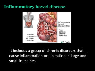
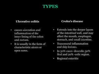
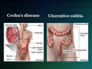
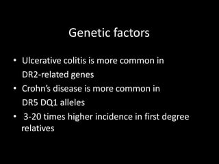
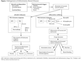
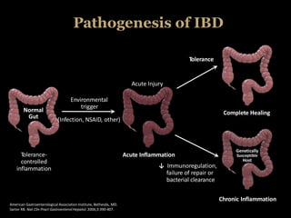
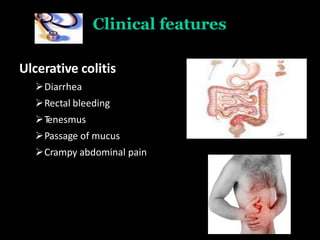
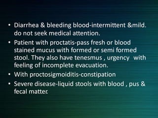
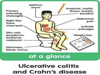
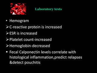
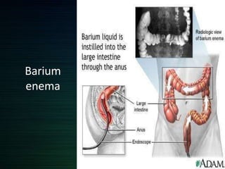
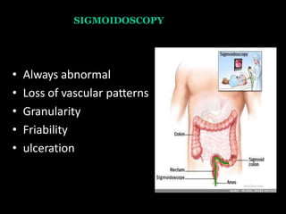
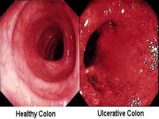
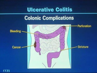
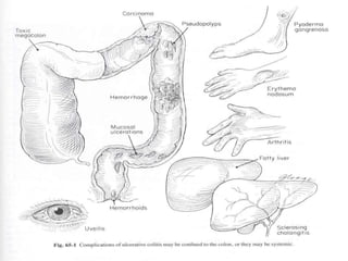
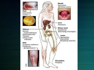
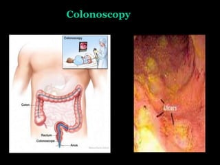
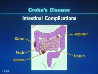
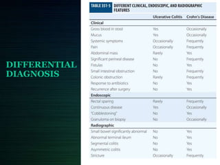
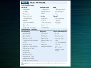
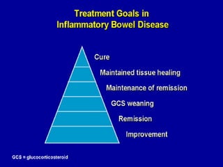
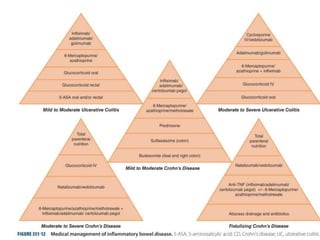
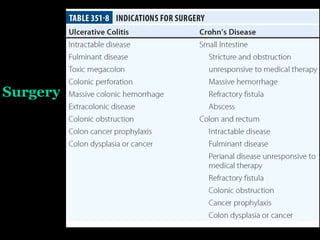
INFLAMMATORY
BOWEL DISEASES
DR. DURGESH MAHAJAN
MD MEDICINE
ASSISTANT PROFESSOR
SVNGMC, YAVATMAL
Inflammatory bowel disease
It includes a group of chronic disorders that
cause inflammation or ulceration in large and
small intestines.
TYPES
Ulcerative colitis
• causes ulceration and
inflammation of the
inner lining of the colon
and rectum.
• It is usually in the form of
characteristic ulcers or
open sores.
Crohn’s disease
• Extends into the deeper layers
of the intestinal wall, and may
affect the mouth, esophagus,
stomach, and small intestine.
• Transmural inflammation
and skip lesions.
• In 50% cases -ileocolic,30%
ileal and 20% -colic region.
• Regional enteritis
Ulcerative colitisCrohn’s disease
OTHER FORMS OF IBD
• Collagenous colitis
• Lymphocytic colitis
• Ischemic colitis
• Behcet’s syndrome
• Infective colitis
• Intermediate colitis
Epidemiology
Etiopathogenesis
• Exact cause is unknown.
• Genetic factors
• Immunological factors
• Microbial factors
• Psychosocial factors
Genetic factors
• Ulcerative colitis is more common in
DR2-related genes
• Crohn’s disease is more common in
DR5 DQ1 alleles
• 3-20 times higher incidence in first degree
relatives
Immunologic factors
• Defective regulation of immunesuppresion
• Activated CD+4 cells activate other
inflammatory cells like macrophages & B-cells
or recruit more inflammatory cells by
stimulation of homing receptor on leucocytes
& vascular epithelium.
Pathogenesis of IBD
American Gastroenterological Association Institute, Bethesda, MD.
Sartor RB. Nat Clin Pract Gastroenterol Hepatol. 2006;3:390-407.
Normal
Gut
Tolerance-
controlled
inflammation
Tolerance
Acute Injury
Environmental
trigger
(Infection, NSAID, other)
Complete Healing
Chronic Inflammation
Genetically
Susceptible
Host
Acute Inflammation
↓ Immunoregulation,
failure of repair or
bacterial clearance
Ulcerative colitis
Pathology
Macrocopic features
Usually involves rectum & extends proximally to involve all
or part of colon.
Spread is in continuity.
May be limited colitis( proctitis &
proctosigmoiditis)
in total colitis there is back wash ileitis (lumpy- bumpy
appearance)
• Mild disease- erythema & sand paper
appearance(fine granularity)
• Moderate-marked erythema,coarse granularity,contact
bleeding & no ulceration
• Severe- spontaneous bleeding, edematous &
ulcerated(collar button ulcer).
• Long standing-epithelial regeneration so
pseudopolyps , mucosal atrophy &
disorientation leads to a precancerous condition.
• Eventually can lead to shortening and narrowing
of colon.
• Fulminant disease-Toxic colitis/megacolon
Ulcerative colitis
Ulcer
pseudopolyps
Microscopic features
 Crypts atrophy & irregularity
 Superficial erosion
 Diffuse mixed inflammation
 Basal lymphoplasmacytosis
Clinical features
Ulcerative colitis
Diarrhea
Rectal bleeding
Tenesmus
Passage of mucus
Crampy abdominal pain
• Diarrhea & bleeding blood-intermittent &mild.
do not seek medical attention.
• Patient with proctatis-pass fresh or blood
stained mucus with formed or semi formed
stool. They also have tenesmus , urgency with
feeling of incomplete evacuation.
• With proctosigmoiditis-constipation
• Severe disease-liquid stools with blood , pus &
fecal matter.
Physical signs
Proctitis – Tender anal canal & blood on rectal
examination
Extensive disease-tenderness on palpation of
colon
Toxic colitis-severe pain &bleeding
If perforation-signs of peritonitis
Clinical Severity of UC
Diagnosis
• Laboratory tests
• Endoscopy
• Radiography
• Biopsy
Laboratory tests
• Hemogram
C-reactive protein is increased
ESR is increased
Platelet count-increased
Hemoglobin-decreased
 Fecal Calponectin levels correlate with
histological inflammation,predict relapses
&detect pouchitis
Barium
enema
BARIUM ENEMA
• Fine mucosal granularity
• Superficial ulcers seen
• Collar button ulcers
• Pipe stem appearance-
loss of haustrations
• Narrow & short colon-
ribbon contour colon
SIGMOIDOSCOPY
• Always abnormal
• Loss of vascular patterns
• Granularity
• Friability
• ulceration
Extra intestinal manifestations
Crohn’s disease
Macroscopic features
Can affect any part of GIT
Transmural
Segmental with skip lesions
Cobblestone appearance
Creeping fat- adhesions & fistula
Microscopic features
• Aphthous ulcerations
• Focal crypt abscesses
• Granuloma-pathognomic
• Submucosal or subserosal lymphoid aggregates
• Transmural with fissure formation
Clinical features
Ileal Crohn’s Disease
Abdominal pain
 Diarrhea
 Weight loss
Low grade fever
Jejunoileitis disease
 Malabsorption
 Steatorrhea
Colitis and perianal disease
 Bloody diarrohea
 Passage of mucus
 Lethargy
 Malaise
 Anorexia
 Weight loss
Diagnosis
• Laboratory tests
• Endoscopy
• Radiography
• Biopsy
• CT enterography
Laboratory tests
• CRP-elevated
• ESR-elevated
• Anemia
• Leukocytosis
• hypoalbuminemia
Barium enema
String
sign
Colonoscopy
CT enterography
• Mural hyperenhancement
• Stratification
• Engorged vasa recta
• Perienteric inflammatory
changes
DIFFERENTIAL
DIAGNOSIS
Treatment
Treatment
Lifestyle changes
Drugs
• 5-ASA agents
• Glucocorticoids
• Antibiotics
• Immunosuppresants
• Biological therapy
5-ASA Agents
•Sulfasalazine (5-aminosalicylic
acid and sulfapyridine as carrier
substance)
•Mesalazine (5-ASA), e.g. Asacol,
Pentasa
•Balsalazide (prodrug of 5-ASA)
• Olsalazine (5-ASA dimer cleaves
in colon)
Distribution of 5-ASA Preparations
Oral
• Varies by agent: may be released in the distal/terminal
ileum, or colon1
Suppositories
• Reach the upper rectum2,5
(15-20 cm beyond the anal verge)
Liquid Enemas
• May reach the splenic flexure2-4
• Do not frequently concentrate in the rectum3
Topical Action of 5-ASA: Extent of Disease
Impacts Formulation Choice
1. Sandborn WJ, et al. Aliment Pharmacol Ther. 2003;17:29-42; 2. Regueiro M, et al. Inflamm Bowel Dis. 2006;12:972–978; 3. Van Bodegraven AA,
et al. Aliment Pharmacol Ther. 1996; 10:327-332; 4. Chapman NJ, et al. Mayo Clin Proc. 1992;62:245-248; 5. Williams CN, et al. Dig Dis Sci. 1987;32:71S-75S.
• Use
 In mild to moderate UC & crohn’s colitis
 Maintaining remission
 May reduce risk of colorectal cancer
• Adverse effects
 Nausea, headache, epigastric pain, diarrhoea,
hypersensitivity, pancreatitis
 Caution in renal impairment, pregnancy, breast feeding
Glucocorticoids
• Anti inflammatory agents for moderate to
severe relapses.
• Inhibition of inflammatory pathways
• Budesonide- 9mg/dl used for 2-3 months &
then tapered.
• Prednisone-40-60mg/day
• No role in maintainence therapy
Antibiotics
• No role in active/quienscent UC
• Metronidazole is effective in active
inflammatory,fistulous & perianal CD.
• Dose-15-20mg/kg/day in 3 divided doses.
• Ciprofloxacin
• Rifaximin
Immunosuppresants
 Thiopurines
Azathioprine
6-mercaptopurin
 Methotrexate
 Cyclosporine
Cyclosporine
• Preventing clonal expansion of T cell subsets
• Used in Steroid sparing Active and chronic disease
• Side effects
Tremor, paraesthesiae, malaise, headache, gingival
hyperplasia, hirsutism Major: renal impairment,
infections, neurotoxicity
Biological therapy
• Infliximab
Anti TNF monoclonal antibody
Infliximab binds to TNF trimers with high affinity, preventing cytokine
from binding to its receptors
It also binds to membrane-bound TNF- a and neutralizes its activity &
also reduces serum TNF levels.
• Use
Fistulizing CD
Severe active CD
Refractory/intolerant of steroids or immunosuppression
• Side effects
Infusion reactions, Sepsis, Reactivation of Tb, Increased risk of Tb
Other medications
Anti- diarrheals - Loperamide (Imodium)
Laxatives - senna, bisacodyl
Pain relievers. acetaminophen (Tylenol).
Iron supplements
 Nutrition
Surgery
• Reconstructive proctocolectomy with illeoanal pouch
• Proctocolectomy & ileostomy
• Rectal &anal dissection
• Colectomy with ileorectal anastomosis
• Ileostomy with intraabdominal pouch
SURGERIES IN ULCERATIVE COLITIS
SURGERIES IN Crohn’s disease
 Ileocaecal resection
 Segmental resection
 Colectomy & ileorectal anastamosis
 Temporary loop ileostomy
 Proctocolectomy
 Stricturoplasty
Strictureplasty
THANK YOU

Inflammatory Bowel Diseases

  • 1.
    INFLAMMATORY BOWEL DISEASES DR. DURGESHMAHAJAN MD MEDICINE ASSISTANT PROFESSOR SVNGMC, YAVATMAL
  • 2.
    Inflammatory bowel disease Itincludes a group of chronic disorders that cause inflammation or ulceration in large and small intestines.
  • 3.
    TYPES Ulcerative colitis • causesulceration and inflammation of the inner lining of the colon and rectum. • It is usually in the form of characteristic ulcers or open sores. Crohn’s disease • Extends into the deeper layers of the intestinal wall, and may affect the mouth, esophagus, stomach, and small intestine. • Transmural inflammation and skip lesions. • In 50% cases -ileocolic,30% ileal and 20% -colic region. • Regional enteritis
  • 4.
  • 5.
    OTHER FORMS OFIBD • Collagenous colitis • Lymphocytic colitis • Ischemic colitis • Behcet’s syndrome • Infective colitis • Intermediate colitis
  • 6.
  • 7.
    Etiopathogenesis • Exact causeis unknown. • Genetic factors • Immunological factors • Microbial factors • Psychosocial factors
  • 8.
    Genetic factors • Ulcerativecolitis is more common in DR2-related genes • Crohn’s disease is more common in DR5 DQ1 alleles • 3-20 times higher incidence in first degree relatives
  • 9.
    Immunologic factors • Defectiveregulation of immunesuppresion • Activated CD+4 cells activate other inflammatory cells like macrophages & B-cells or recruit more inflammatory cells by stimulation of homing receptor on leucocytes & vascular epithelium.
  • 11.
    Pathogenesis of IBD AmericanGastroenterological Association Institute, Bethesda, MD. Sartor RB. Nat Clin Pract Gastroenterol Hepatol. 2006;3:390-407. Normal Gut Tolerance- controlled inflammation Tolerance Acute Injury Environmental trigger (Infection, NSAID, other) Complete Healing Chronic Inflammation Genetically Susceptible Host Acute Inflammation ↓ Immunoregulation, failure of repair or bacterial clearance
  • 12.
  • 13.
    Pathology Macrocopic features Usually involvesrectum & extends proximally to involve all or part of colon. Spread is in continuity. May be limited colitis( proctitis & proctosigmoiditis) in total colitis there is back wash ileitis (lumpy- bumpy appearance)
  • 15.
    • Mild disease-erythema & sand paper appearance(fine granularity) • Moderate-marked erythema,coarse granularity,contact bleeding & no ulceration • Severe- spontaneous bleeding, edematous & ulcerated(collar button ulcer). • Long standing-epithelial regeneration so pseudopolyps , mucosal atrophy & disorientation leads to a precancerous condition. • Eventually can lead to shortening and narrowing of colon. • Fulminant disease-Toxic colitis/megacolon
  • 16.
  • 17.
    Microscopic features  Cryptsatrophy & irregularity  Superficial erosion  Diffuse mixed inflammation  Basal lymphoplasmacytosis
  • 18.
    Clinical features Ulcerative colitis Diarrhea Rectalbleeding Tenesmus Passage of mucus Crampy abdominal pain
  • 19.
    • Diarrhea &bleeding blood-intermittent &mild. do not seek medical attention. • Patient with proctatis-pass fresh or blood stained mucus with formed or semi formed stool. They also have tenesmus , urgency with feeling of incomplete evacuation. • With proctosigmoiditis-constipation • Severe disease-liquid stools with blood , pus & fecal matter.
  • 20.
    Physical signs Proctitis –Tender anal canal & blood on rectal examination Extensive disease-tenderness on palpation of colon Toxic colitis-severe pain &bleeding If perforation-signs of peritonitis
  • 21.
  • 23.
    Diagnosis • Laboratory tests •Endoscopy • Radiography • Biopsy
  • 24.
    Laboratory tests • Hemogram C-reactiveprotein is increased ESR is increased Platelet count-increased Hemoglobin-decreased  Fecal Calponectin levels correlate with histological inflammation,predict relapses &detect pouchitis
  • 25.
  • 26.
    BARIUM ENEMA • Finemucosal granularity • Superficial ulcers seen • Collar button ulcers • Pipe stem appearance- loss of haustrations • Narrow & short colon- ribbon contour colon
  • 27.
    SIGMOIDOSCOPY • Always abnormal •Loss of vascular patterns • Granularity • Friability • ulceration
  • 31.
  • 33.
  • 34.
    Macroscopic features Can affectany part of GIT Transmural Segmental with skip lesions Cobblestone appearance Creeping fat- adhesions & fistula
  • 37.
    Microscopic features • Aphthousulcerations • Focal crypt abscesses • Granuloma-pathognomic • Submucosal or subserosal lymphoid aggregates • Transmural with fissure formation
  • 38.
    Clinical features Ileal Crohn’sDisease Abdominal pain  Diarrhea  Weight loss Low grade fever Jejunoileitis disease  Malabsorption  Steatorrhea Colitis and perianal disease  Bloody diarrohea  Passage of mucus  Lethargy  Malaise  Anorexia  Weight loss
  • 39.
    Diagnosis • Laboratory tests •Endoscopy • Radiography • Biopsy • CT enterography
  • 40.
    Laboratory tests • CRP-elevated •ESR-elevated • Anemia • Leukocytosis • hypoalbuminemia
  • 41.
  • 42.
  • 43.
    CT enterography • Muralhyperenhancement • Stratification • Engorged vasa recta • Perienteric inflammatory changes
  • 45.
  • 47.
  • 48.
  • 49.
  • 50.
    Drugs • 5-ASA agents •Glucocorticoids • Antibiotics • Immunosuppresants • Biological therapy
  • 52.
    5-ASA Agents •Sulfasalazine (5-aminosalicylic acidand sulfapyridine as carrier substance) •Mesalazine (5-ASA), e.g. Asacol, Pentasa •Balsalazide (prodrug of 5-ASA) • Olsalazine (5-ASA dimer cleaves in colon)
  • 53.
    Distribution of 5-ASAPreparations Oral • Varies by agent: may be released in the distal/terminal ileum, or colon1 Suppositories • Reach the upper rectum2,5 (15-20 cm beyond the anal verge) Liquid Enemas • May reach the splenic flexure2-4 • Do not frequently concentrate in the rectum3 Topical Action of 5-ASA: Extent of Disease Impacts Formulation Choice 1. Sandborn WJ, et al. Aliment Pharmacol Ther. 2003;17:29-42; 2. Regueiro M, et al. Inflamm Bowel Dis. 2006;12:972–978; 3. Van Bodegraven AA, et al. Aliment Pharmacol Ther. 1996; 10:327-332; 4. Chapman NJ, et al. Mayo Clin Proc. 1992;62:245-248; 5. Williams CN, et al. Dig Dis Sci. 1987;32:71S-75S.
  • 54.
    • Use  Inmild to moderate UC & crohn’s colitis  Maintaining remission  May reduce risk of colorectal cancer • Adverse effects  Nausea, headache, epigastric pain, diarrhoea, hypersensitivity, pancreatitis  Caution in renal impairment, pregnancy, breast feeding
  • 55.
    Glucocorticoids • Anti inflammatoryagents for moderate to severe relapses. • Inhibition of inflammatory pathways • Budesonide- 9mg/dl used for 2-3 months & then tapered. • Prednisone-40-60mg/day • No role in maintainence therapy
  • 56.
    Antibiotics • No rolein active/quienscent UC • Metronidazole is effective in active inflammatory,fistulous & perianal CD. • Dose-15-20mg/kg/day in 3 divided doses. • Ciprofloxacin • Rifaximin
  • 57.
  • 58.
    Cyclosporine • Preventing clonalexpansion of T cell subsets • Used in Steroid sparing Active and chronic disease • Side effects Tremor, paraesthesiae, malaise, headache, gingival hyperplasia, hirsutism Major: renal impairment, infections, neurotoxicity
  • 59.
    Biological therapy • Infliximab AntiTNF monoclonal antibody Infliximab binds to TNF trimers with high affinity, preventing cytokine from binding to its receptors It also binds to membrane-bound TNF- a and neutralizes its activity & also reduces serum TNF levels. • Use Fistulizing CD Severe active CD Refractory/intolerant of steroids or immunosuppression • Side effects Infusion reactions, Sepsis, Reactivation of Tb, Increased risk of Tb
  • 60.
    Other medications Anti- diarrheals- Loperamide (Imodium) Laxatives - senna, bisacodyl Pain relievers. acetaminophen (Tylenol). Iron supplements  Nutrition
  • 61.
  • 62.
    • Reconstructive proctocolectomywith illeoanal pouch • Proctocolectomy & ileostomy • Rectal &anal dissection • Colectomy with ileorectal anastomosis • Ileostomy with intraabdominal pouch SURGERIES IN ULCERATIVE COLITIS
  • 64.
    SURGERIES IN Crohn’sdisease  Ileocaecal resection  Segmental resection  Colectomy & ileorectal anastamosis  Temporary loop ileostomy  Proctocolectomy  Stricturoplasty
  • 65.
  • 66.